Introduction
Plant life in the continental Antarctic is scarce and only in some favourable oases does vegetation cover more than a few square metres. In the harsh climate severe environmental constraints threaten plant survival at all times. The terrestrial vegetation, composed mainly of lichens and mosses, is regularly exposed to very low temperatures (Kappen et al. Reference Kappen, Schroeter, Green and Seppelt1998) but, when active, thallus temperatures are apparently close to, or above freezing point (Schroeter et al. Reference Schroeter, Green, Pannewitz, Schlensog and Sancho2010, Seppelt et al. Reference Seppelt, Tuerk, Green, Moser, Pannewitz, Sancho and Schroeter2010, Schroeter et al. Reference Schroeter, Green, Pannewitz, Schlensog and Sancho2011). Low temperatures are suggested to increase the sensitivity to damage by high light (Björkman Reference Björkman1981) because photosynthetic pathways and repair mechanisms are slowed or inhibited (Öquist et al. Reference Öquist, Greer and Ögren1987, Krause Reference Krause1994). One strategy in Antarctica that is often suggested for lichens is to avoid this problem through their poikilohydric nature, i.e. they just dry out at high light intensities (Schlensog & Schroeter Reference Schlensog and Schroeter2000, Schlensog et al. Reference Schlensog, Schroeter, Pannewitz and Green2003, Kappen & Valladares Reference Kappen and Valladares2007). However, whilst this strategy may operate in the Maritime Antarctic, in the continental Antarctic the lichens are often active during times of snowmelt and, therefore receive high Photosynthetic Photon Flux Density (PPFD). Umbilicaria aprina Nyl. at Botany Bay, 77°S, southern Victoria Land, when active, received maximal PPFD of > 2500 μmol photons m-2 s-1 and a mean level in January of 880 μmol photons m-2 s-1 (Schroeter et al. Reference Schroeter, Green, Pannewitz, Schlensog and Sancho2011). The contrast with the Maritime Antarctic is immense: at Botany Bay U. aprina received a mean level of 247 mol photons m-2 month-1 for the four summer months compared to just 12 mol photons m-2 month-1 for Usnea aurantiaco-atra (Jacq.) Bory at Livingston Island, 62°S, South Shetland Islands (Schroeter et al. Reference Schroeter, Green, Pannewitz, Schlensog and Sancho2010).
Similarly, for mosses, especially those growing in melt stream areas, the light environment of continental Antarctica is especially severe. They are also poikilohydric and are only wet and active when temperatures are high enough for substantial meltwater to occur. The result is that they remain active at times of high PPFD (Pannewitz et al. Reference Pannewitz, Green, Scheidegger, Schlensog and Schroeter2003, Green et al. Reference Green, Schroeter and Sancho2007).
Mosses, however, are suggested to be constitutive shade plants (Green & Lange Reference Green and Lange1995, Marschall & Proctor Reference Marschall and Proctor2004, Glime Reference Glime2007). Chlorophyll a/b ratios for 39 mosses had a mean value of 2.29 compared to 3.0 and 4.0 for C3 and C4 vascular plants, respectively (Glime Reference Glime2007) and it would be expected that substantial protection must be present in order to maintain the photosynthetic apparatus in a healthy state. It has been previously demonstrated (Green et al. Reference Green, Schroeter and Seppelt2000) that Bryum argenteum var. muticum Brid. (Bryum argenteum var. muticum has previously been referred to as Bryum subrotundifolium, fide Ochyra et al. Reference Ochyra, Lewis Smith and Bednarek-Ochyra2008) is not deleteriously affected when growing in full ambient sunlight. Physiological indicators such as stable net photosynthesis (NP) and maximal quantum efficiency of photosystem II ( ), remained unaffected under full sunlight and there was no difference if ambient levels of UVA and UVB were present or not. This contrasts with studies on another species, Schistidium antarctici (Card.) L. Savic. & Smirn., which showed clear signs of photoinhibition (Lovelock et al. Reference Lovelock, Osmond and Seppelt1995a, Reference Lovelock, Jackson, Melick and Seppelt1995b). It was also demonstrated with B. argenteum that samples kept shaded for several days changed their colour from a creamy white to a clear green and that these samples only showed transient photoinhibition under ambient PPFD with
 declining in the presence of high PPFD but recovering if held in darkness (Green et al. Reference Green, Schroeter and Seppelt2000).
Plants can regulate the strain caused by excess light in several ways (Björkman & Demmig-Adams Reference Björkman and Demmig-Adams1995) including controlling excess excitation energy by the xanthophyll cycle. It has been shown that the state of the xanthophyll cycle, especially the concentrations of zeaxanthin and antheraxanthin, is closely related to the amount of non-photochemical quenching and that the protection system is ubiquitous throughout plants but with considerable variation according to plant type and environment (Demmig-Adams & Adams Reference Demmig-Adams and Adams III2006). Information available about the pigments present in Antarctic mosses is growing but is not yet extensive (Post Reference Post1990, Lovelock & Robinson Reference Lovelock and Robinson2002, Robinson et al. Reference Robinson, Turnbull and Lovelock2005). Studies on Bryum pseudotriquetrum (Hedw.) Gaertn., Meyer et Scherb., Ceratodon purpureus (Hedw.) Brid. and Schistidium antarctici at different sites suggest that high light stress exists and that this leads to high xanthophyll cycle pigment levels that differed between plants in different light environments (Lovelock & Robinson Reference Lovelock and Robinson2002). We do not know in any detail what changes occur when the mosses first become hydrated at the start of summer or how rapidly the plants can respond to changes in their PPFD environment and how such changes are achieved. However, studies on the response of Antarctic bryophytes to changes in UV radiation suggest that rapid responses can occur (Newsham et al. Reference Newsham, Hodgson, Murray, Peat and Smith2002, Snell et al. Reference Snell, Convey and Newsham2007) and that unprotected plants can regain normal protection levels within a week (Green et al. Reference Green, Kulle, Pannewitz, Sancho and Schroeter2005). Also, the liverwort Cephaloziella varians (Gottsche) Steph. showed only a small increase in protective pigments as it became active during snow melt (Snell et al. Reference Snell, Convey and Newsham2007).
Surprisingly there is also only a small amount of data for temperate mosses but there are indications that mosses generally can have high levels of protection against high light. Marschall & Proctor (Reference Marschall and Proctor2004) noted that non-photochemical quenching (NPQ) levels in vascular plants reached values of around 4.5 in full sunlight whereas levels of 10–15 are often measured for mosses. Also, Proctor & Smirnoff (Reference Proctor and Smirnoff2011) have presented good evidence that mosses can use alternative electron sinks, such as oxygen, under high light. Certainly, although not yet well studied, bryophytes appear to have some fascinating abilities to adapt to, and survive, high light stress. The performance of mosses in important geographical areas, such as alpine zones, seems to have received little attention as yet.
We studied samples of B. argenteum during the first hydrated exposure to sun during snowmelt in the early Antarctic summer, and we also adapted other samples to deep shade and then compared their performance to untreated individuals at the same site. By this means we were not only able to analyse the differences between the shaded and normal forms but also to study the recovery of the treated samples. The results suggest that B. argenteum is always well protected against high light but can also rapidly adapt to changes in its radiation environment.
Material and methods
Site description and samples
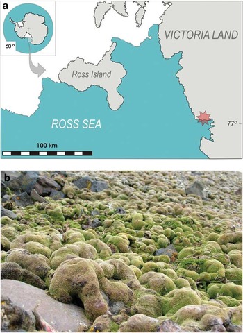
The research was carried out in the field during January 2001 at Botany Bay, southern Victoria Land, 77°01′S, 162°34′E (Fig. 1). The area consists of a narrow beach, backed by a small cliff with NNE exposure, above which there is a plateau ending at the foot of a small glacier at about 200 m above sea level. The beach is covered with granite boulders of various sizes and melt streams run to the sea from December–February with flow determined by the weather (Schroeter et al. Reference Schroeter, Green, Pannewitz, Schlensog and Sancho2011). The climate of Botany Bay is unusually warm for the latitude with temperatures often above freezing point in late December and January (Schroeter et al. Reference Schroeter, Green, Pannewitz, Schlensog and Sancho2011). The low occurrence of cloud, coupled with reflection from the sea ice lying in front of the bay means that when winter snowmelt occurs the lichens and mosses can receive very high light levels, with PPFD often in excess of 2000 μmol photons m-2 s-1. Small to large patches of moss grow amongst the rocks with the dominant species being Ceratodon purpureus (wettest sites), Hennediella heimii (Hedw.) Zand. (drier sites) and Bryum argenteum var. muticum (wet, intermediate sites) (Seppelt et al. Reference Seppelt, Tuerk, Green, Moser, Pannewitz, Sancho and Schroeter2010).
Location map for Botany Bay (marked with a star), southern Victoria Land, continental Antarctica (77°00′14′′S, 162°32′52′′E). Botany Bay lies in a protected site within Granite Harbour and has an exceptional vegetation for the latitude (Seppelt et al. Reference Seppelt, Tuerk, Green, Moser, Pannewitz, Sancho and Schroeter2010).

The appearance of B. argenteum varies depending on its exposure to light and it forms so-called ‘sun’ and ‘shade’ forms (Green et al. Reference Green, Schroeter and Seppelt2000). The sun form grows in a densely packed cushion that appears yellowish to whitish green (Fig. 1). The photosynthetically active part of the shoot is short, about 1 mm long with only around five green leaves. The shade form, on the other hand, is a clear, dark green colour, less closely packed with longer active shoots which are up to 7 mm long and with around ten green leaves. The two forms occur naturally and grade into one another with the shade form being less common and found typically close to, or under the edge of, rocks or on the sides of large hummocks.
Experimental procedures
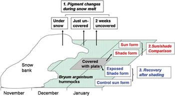
A schematic version of the experimental plan is shown in Fig. 2.
Diagrammatic representation of the research plan investigating the performance and pigments of Bryum argenteum in the field at Botany Bay. Three main comparisons were made: 1) between sun and shade forms (CO2 exchange, chlorophyll fluorescence and pigment content), 2) between samples at the start of the season from snow covered to exposed (pigment content), 3) between sun and shade forms that were exposed to normal ambient sunlight - dynamics of stress reaction (CO2 exchange, chlorophyll fluorescence and pigment content). Full details in Methods.

1. Pigment changes during snowmelt: samples were collected at the same time (at midday, approximately one hour before solar noon) on three occasions on 6 November, 3 December and 6 December 2000, in order to investigate changes in pigment content following the winter. At the first sampling the mosses were dry and still covered with snow, at the second they were moist and had just become uncovered from the snow, and at the third they had been uncovered for about two weeks during which time they had been continuously moist and active (note, this is a different site to that of the second collection). On each occasion five samples, c. 1 cm diameter with the lower brown parts removed, were taken from an area of about 1 m2 of the B. argenteum carpet and immediately placed in small tubes and stored in liquid nitrogen until further preparation and analysis in the laboratory at Waikato University, New Zealand.
2. Comparison of sun and shade forms: samples were collected in mid-January to compare sun forms and shade forms. The sun forms were abundant and samples were taken from the middle of moss hummocks. Shade forms were much less in extent and, because they graded into surrounding sun forms, they proved to be highly variable. It was known from previous studies that B. argenteum could rapidly change from the sun form to the shade form if shaded so six areas of B. argenteum were placed in deep shade. This was achieved by laying opaque plastic plates (about 5 mm thick and 15 cm on each side) over areas of B. argenteum with the sheet supported just above the moss by small stones. The shading was put in place in early December and when removed for sample collection in early January (after about six weeks) the shaded areas of moss had become a clear dark green colour typical of deeply shaded moss in natural conditions. Shade form, therefore, always refers to B. argenteum that had been subjected to deep shade under the plastic plates.
For CO2 gas exchange measurements, samples, square pieces of moss carpet about 8–10 cm2, were collected and taken straight to the laboratory tent and prepared for use. Damaged and discoloured parts of the moss were removed and the older parts cut away to leave a thickness of about 10 mm predominantly composed of green, photosynthetic tissue (Green et al. Reference Green, Schroeter and Seppelt2000). The samples were then moistened and placed in small, foil trays.
Five samples were taken from sun and shade forms for pigment analysis as described above.
3. Recovery after shading: the recovery of the deeply shaded moss (shade form) to full sunlight exposure was analysed in mid-January. Five samples were collected immediately after removal of the shading plastic plate, placed in individual aluminium trays exposed to ambient PPFD for five days. All samples were square and about 8–10 cm2 in area and were kept moist whilst exposed. A set of samples of normal sun forms were treated identically. CO2 gas exchange and chlorophyll fluorescence measurements were made on all samples at 0, 1, 6, 24, 48, 96 and 120 h after removal of the shading plastic plate. Samples were taken for pigment analysis (as detailed above) at the beginning (these were the same samples used in the previous sun/shade comparison) and the end (after 120 h) of the treatment.
Experimental techniques
Microclimate recording
A Squirrel data logger (SQ1021, Grant Instruments Ltd, UK) recorded the microclimate of the moss area at five minute intervals. Photosynthetic Photon Flux Density was measured with GaAsP-photodiodes (Hamamatsu, Japan) equipped with filter and cosine correction according to Pontailler (Reference Pontailler1990) and calibrated against a quantum sensor (190 SB, Licor, USA) using an Optical Radiation Calibrator (1800-02, Licor, USA). The sensors were mounted level on a rock adjacent to the mosses. Air-temperature was measured at 1 m above the ground with shielded thermocouples and thallus-temperature was measured in the top layer (5 mm) of the moss turf.
CO2 gas exchange
The CO2 gas exchange was measured at a field camp close to Botany Bay using a differential open measuring system, consisting of the Compact Minicuvette System CMS4P (Walz GmbH, Germany) with a climatized cuvette and an infrared gas analyser BINOS100 (Rosemount Analytical, Germany) (Schroeter et al. Reference Schroeter, Green, Kappen and Seppelt1994). All measurements were carried out at 10°C, a temperature expected to be close to optimal for the species. Light was provided by a fibre optic lamp (Kaltlicht-Fiberleuchte FL-400 and Spezial Fiberoptik 400-F, Walz GmbH, Germany) with the intensity regulated by adjustment of lamp power and neutral filters, so that spectral distribution was not altered. CO2 gas exchange rates were related to projected surface area of the sample. Three replicates of each treatment were measured.
Chlorophyll a fluorescence
A pulse amplitude modulated fluorometer (MINI-PAM, Walz GmbH, Germany) was used with the fibre optic probes fixed about 10 mm from the sample. Standard routines programmed within the MINI-PAM allowed the determination of maximal and minimal fluorescence in dark adapted samples (Fm and F0) or maximal and ground fluorescence in the light (Fm’ and Ft). The quantum yield for photosystem II (ΦPSII) could then be calculated either after darkening (maximal quantum efficiency of PSII =  = Fv/Fm = (Fm - F0)/Fm) or in the light (effective quantum efficiency of PSII = yield = ΦPSII = ΔF/Fm’ = (Fm’ - Ft)/Fm’). The relative electron transport rate (ETR) was calculated from ΦPSII and incident light (PPFD = photosynthetic photon flux density) as ETR = PPFD * ΦPSII (Schreiber et al. Reference Schreiber, Bilger and Neubauer1994). Non-photochemical quenching (NPQ) was calculated as NPQ = Fm/Fm’ - 1 (Bilger & Björkman Reference Bilger and Björkman1990, Schreiber et al. Reference Schreiber, Bilger and Neubauer1994).
Determination of pigments
The samples were ground under liquid nitrogen, using CaCO3 as a buffer. After the nitrogen had evaporated, the samples were dissolved in methanol, ground again, and centrifuged for two minutes at 200 g. The supernatant was decanted, while the pellet was again dissolved in methanol and centrifuged. This process was repeated until the supernatant was colourless. Nitrogen gas was blown through the extract to remove oxygen. Before HPLC, the extracts were filtered with a 0.2 μm filter.
The separation of carotenoids followed the method of Martínez-Ferri et al. (Reference Martínez-Ferri, Balaguer, Valladares, Chico and Manrique2000) on a HPLC system (Waters 717plus autosampler, Waters 600S controller and Waters 996PAD photodiode array detector with a 5 μm ODS2, Waters Spherisorb, 4.6 x 250 mm column with a mean pore size of 80Å and surface area of 220 m2 g-1). Pigments were detected at 400 nm and contents calculated from calibration curves. Chlorophyll absorption of the extracts was separately measured with an Uvikon 930 spectrometer (Kontron Instruments, Japan) at 652 nm, 665.2 nm and 750 nm. The chlorophyll content was then calculated using the formula by Porra (Reference Porra1991).
Total carotenoid content of the xanthophyll cycle (VAZ) was calculated as (V + A + Z) and de-epoxidation status (DPS) was calculated according to Demmig-Adams & Adams (Reference Demmig-Adams and Adams III1996) as DPS = (A + Z)/(V + A + Z) where, in both cases, V is violaxanthin, A is antheraxanthin and Z is zeaxanthin. It has been reported that β-carotene is not completely extracted by methanol solvents and is, therefore, probably under-reported here (Dunn et al. Reference Dunn, Turnbull and Robinson2004).
Results
Microclimate
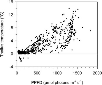
Photosynthetic Photon Flux Density consistently reached about 1500 μmol photons m-2 s-1 on days without cloud and, for c. 25% of the day, PPFD were greater than 1000 μmol photons m-2 s-1 (Fig. 3). Moss thallus temperatures were buffered by the effects of the meltwater running around the cushions. The protective effect of freezing meant that only 1.5% of records were below freezing point but 52% were between 0 and 1.0°C. The cooling effect of the water meant that temperatures were 1–4°C, 4–8°C and > 8°C for 18.7, 19.1 and 8.6% of the time, respectively. Absolute maximum was 14°C (Fig. 3). The temperature record was not complete because of damage to probes from marauding skua gulls but it does cover the period when the shaded plants were exposed to full sunlight.
Distribution of thallus temperature (°C, y-axis) and incident PPFD (photosynthetic photon flux density, μmol photons m-2 s-1, x-axis) measured simultaneously every five minutes and averaged to 20 minute intervals. Approximately 52% of the values lie between 0 and 1°C, and only 1.5% below zero. The upper boundary of the data distribution equates to a 1°C rise with each 100 μmol photons m-2 s-1 increase in PPFD.

Pigment changes during snowmelt under natural conditions
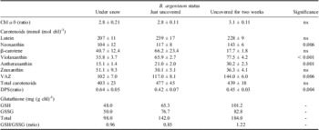
There were only slight changes in pigment content of sun forms at the beginning of the season as the plants became rehydrated and exposed after melting of covering snow. There was a small but non-significant increase in total carotenoids (on a molar chlorophyll basis) but there was a significant (P = 0.006) increase in the VAZ pool which rose from 25 to 32% of total carotenoids in the first two weeks (Table I). There were significant rises in violaxanthin and antheraxanthin and a non-significant fall in zeaxanthin. This was accompanied by a lowering in the de-epoxidation status from 0.6 to c. 0.45 (Table I) and an increase in glutathione content from 98 to 184 mg m-2 but little change in the GSH/GSSG ratio (reduced glutathione/oxidised glutathione ratio; Table I). Chlorophyll a/b ratio was unchanged (Table I) while loss of data in the field meant that chlorophyll could not be presented on an area basis.
Comparison of chl a/b ratio, carotenoid content and glutathione content in Bryum argenteum sampled at three times in the early summer, first whilst still covered by snow, second immediately after being uncovered by snow melt and third, two weeks after being uncovered. In all cases samples were taken in the field and stored in liquid nitrogen until later analysis. Carotenoid contents in mmol (mol chl)-1, glutathione contents in mg (g chl)-1. Statistical significance by ANOVA on groups of five replicates, ns = P > 0.05, - = not calculated.

VAZ = total carotenoid content of the xanthophyll cycle, DPS = de-epoxidation status, GSH = reduced glutathione, GSSG = oxidised glutathione.
Sun and shade form comparison
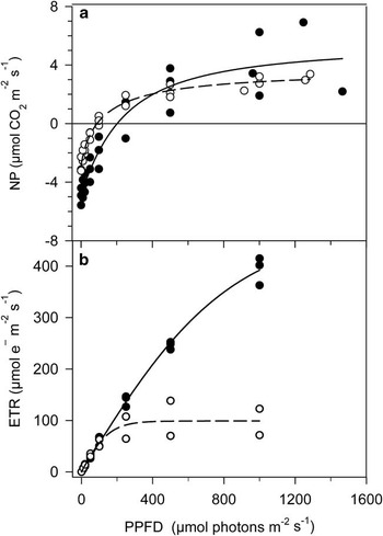
The response of CO2 exchange to PPFD at 10°C for the sun (normal sun form) and shade (produced by artificial shading) forms of B. argenteum (Fig. 4a) showed several features that might be expected as adaptations to different light climates. The shade form had a lower light compensation point (90 compared to 200 μmol photons m-2 s-1), lower dark respiration (2.8 compared to 4.8 μmol CO2 m-2 s-1) and higher quantum efficiency for CO2 uptake (0.058 compared to 0.036). There was little difference in maximal NP between the two forms but gross photosynthesis of the shade form was also much lower. The lower compensation point of the shade form is also typical of shade adapted higher plants but is normally produced by a much smaller dark respiration (DR) rather than a combination of slightly lower DR and a lower quantum efficiency as found here. Quantum efficiencies in higher plants are usually very similar in shade and sun forms and the differences in quantum efficiencies found here for the sun and shade forms almost certainly represents differential absorption of light or internal filtering but we have no measurements that allow this to be confirmed.
Relationship between incident PPFD (x-axis, μmol photons m-2 s-1) and a. net photosynthesis (NP, μmol m-2 s-1, y-axis), and b. relative electron transport rate (ETR, μmol e- m-2 s-1, y-axis) for shade form (○) and sun form (●) of Bryum argenteum measured in the field.

The response of ETR to PPFD (Fig. 4b) was almost identical for both forms at PPFD up to about 150 μmol photons m-2 s-1 (slopes of 0.671 shade, 0.645 sun) above which the shade form showed a clear saturation at around 300 μmol photons m-2 s-1 whereas the sun form did not saturate at all under the conditions used in this study. There was only a small difference in maximal quantum yield (Fv/Fm) between the two forms (0.744 shade, compared to 0.689 sun) which contrasts with the large difference in the quantum efficiency of CO2 uptake (Fig. 4a).
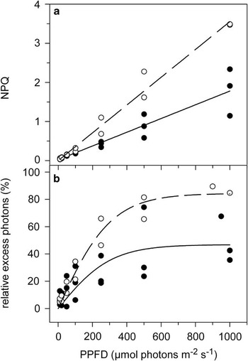
Non-photochemical quenching was linearly dependent on light intensity for both sun and shade forms (Fig. 5a) and, as might be expected from the NP responses, both NPQ (Fig. 5a) and relative excess photons (Fig. 5b) were higher for the shade form at all PPFD.
Relationship between incident PPFD (x-axis, μmol photons m-2 s-1) and a. non-photochemical quenching (NPQ, y-axis) and b. relative excess photons (%, y-axis) for shade form (○) and sun form (●) of Bryum argenteum measured in the field.

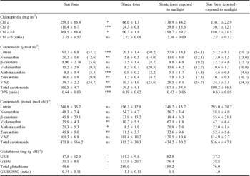
Pigment content differed between sun and shade forms with chl a, chl b and chl a+b contents significantly higher in sun plants although their photosynthetically active layer was much thinner (about 4–5 vs > 10 leaves, 1 vs 5–7 mm, sun and shade forms, respectively) (Table II). The chl a/b ratio was lower, but not significantly, in sun forms than in shade forms (2.35 and 2.72, respectively, Table II). The absolute pigment contents on an area basis showed the same pattern as the chlorophyll content. All carotenoids were higher in the sun plant, including lutein, neoxanthin, β-carotene, and members of the xanthophyll cycle (VAZ, violaxanthin, and especially antheraxanthin and zeaxanthin) and all differences were significant except for violaxanthin and β-carotene (Table II). However, when calculated on a chlorophyll base lutein, neoxanthin, β-carotene, VAZ and total carotenoids were similar in sun and shade samples (Table II) although changes in the individual carotenoids contributing to the xanthophyll cycle were very different. Violaxanthin was much higher in the shade plant, 20.5% of all carotenoids, compared to only 9.5% in the sun plant. Antheraxanthin and zeaxanthin, on the other hand, were higher in the sun plant, 8.5% and 16.0% respectively compared to 0.9% and 4.7% in the shade form. As expected, the de-epoxidation status of the sun forms was, therefore, much higher at 0.64 (Table II) compared to 0.19 in the shade form that was sampled in darkness. Total glutathione levels and GSH/GSSG ratio were substantially higher in the shade plants (289.0 vs 48.6 mg (mg chl)-1, 1.1 versus 0.34, respectively). This suggests the presence or potential presence of increased oxidative stress in shade plants, but we are not able to identify what this is.
Comparison of chlorophyll, carotenoid and glutathione contents of B. argenteum at Botany Bay, southern Victoria Land. Sun and shade forms were sampled at the same time in January whilst samples of sun and shade forms were also exposed to natural full sunlight conditions for five days (see text for description). In all cases samples were taken in the field and stored in liquid nitrogen until later analysis. Chlorophyll contents in mg m-2; carotenoid contents in μmol m-2 (upper values) and mmol (mol chl)-1 (lower values), glutathione contents in mg (g chl)-1. Values in brackets in the section on carotenoids on a mmol m-2 basis are percentage of the total carotenoids. Statistical significances were calculated only for the sun form/shade form comparison and are shown as: ns = non-significant, P > 0.05, *P = 0.05–0.01, **P < 0.01 > 0.001, ***P < 0.001, calculated for five replicates by unpaired t-test; - = not calculated. It should be noted that the thickness of the chlorophyll containing zone was very different between the sun (1 mm, five leaves) and shade (7 mm, ten leaves) forms. Dry weights are not available for any samples. The shade samples exposed to sunlight contain a mixture of old and new shoots as these were not separated.

VAZ = total carotenoid content of the xanthophyll cycle, DPS = de-epoxidation status, GSH = reduced glutathione, GSSG = oxidised glutathione.
Recovery after shading
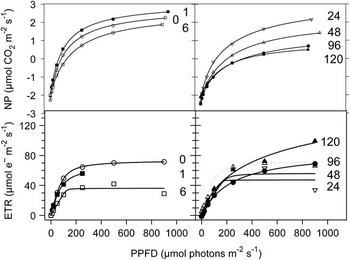
Samples of both sun and shade forms were exposed to ambient PPFD that ranged between 100 and 1500 μmol photons m-2 s-1 every day for five days. The shade form (produced by artificial shading, see methods) showed a decline in both maximal NP and the initial slope of the response to PPFD (quantum efficiency of CO2 fixation), a change that had already started after six hours (Fig. 6). The initial slope of the response of ETR to PPFD also showed a fall after six hours but then remained almost constant for the remainder of the time. The ETR response to PPFD saturated at about 200 μmol photons m-2 s-1 for the first 48 hours and then changed, first seen after 96 hours, to showing no saturation at 1000 μmol photons m-2 s-1 (Fig. 6). All changes in quantum efficiency of CO2 fixation and ETR response to PPFD were towards that of the sun form of the moss whilst maximal NP declined rather than increased (Fig. 4).
Change in photosynthetic performance of shade form of Bryum argenteum after exposure to ambient incident PPFD. Upper panels: net photosynthesis (NP, μmol m-2 s-1, y-axis), lower panels: relative electron transport rate (ETR, μmol e- m-2 s-1, y-axis), over a six day period. Numbers at end of each fitted line is hours since the start of the exposure of the shade form to ambient light.

Interpretation of changes in pigments is made difficult because a replication of three was used and this makes statistical testing highly subject to the slightest variability. Total chlorophyll increased by 220% in the shade form as a consequence of large increases in both chl a and b (Table II) whilst the sun form showed a decline. The chl a/b ratio remained almost constant for both forms (Table II). On a chlorophyll basis all individual carotenoids and total carotenoids for both sun and shade forms remained almost constant but, on an area basis because of the large rise in chlorophyll content, the shade form had sharp increases in all carotenoids, but especially in lutein, VAZ and all components of the xanthophyll cycle (Table II). The sun form also showed some changes with total carotenoids falling on an area basis but rising per molecule chlorophyll because of the decline in total chlorophyll content (Table II). The de-epoxidation status of the shade form rose whilst that of the sun forms remained constant (Table II). Glutathione rose from 43 to 76 mg m-2 in the sun form but almost halved from 289 to 159 mg m-2 in the shade form (Table II).
The changes in the shaded moss were not due to acclimation of the existing shoots. There was, instead, a rapid change in morphology of the moss as the original, long green shoots were replaced by short compact, sun shoots, which grew rapidly from axillary cells.
Discussion
These results from continental Antarctica show that B. argenteum does not avoid high PPFD by drying out as suggested for poikilohydric lichens and mosses (Kappen & Valledares 2007). Activation is typically by melting of snow or ice and this occurs during periods of high ambient PPFD that consistently exceeded 1500 μmol photons m-2 s-1 and averaged six hours over 1000 μmol photons m-2 s-1 every day. A similar situation has been reported for the lichen U. aprina at the same site (Schroeter et al. Reference Schroeter, Green, Pannewitz, Schlensog and Sancho2011). The desiccation strategy is probably most applicable to mosses and lichens that are activated by precipitation but this is a rare event in the continental Antarctic although normal in the Maritime Antarctic (Kappen et al. Reference Kappen, Schroeter, Green and Seppelt1998).
Bryum argenteum was found to be well able to tolerate the high PPFD of its habitat and it achieved this by protection of its photosystems using the xanthophyll cycle. The amount of xanthophyll cycle pigments was very different in sun (higher) and shade forms when presented on an area basis, however, there were no differences when xanthophyll cycle pigments were related to chlorophyll content. Bryum argenteum, therefore, does not appear to alter the xanthophyll cycle pigment content with respect to the chlorophyll of its antennas to any great extent in response to the light environment so protection must be regarded as being constitutive.
Higher plants typically have a lower NP in shade leaves and the lack of difference in maximal NP found here has also been reported by Green et al. (Reference Green, Schroeter and Seppelt2000) at about 4 μmol CO2 m-2 s-1 for both forms. Comparing sun and shade leaves of the same species or between species, sun leaves coped with an enhanced necessity for non-photochemical quenching by having a much higher amount of VAZ than shade leaves (Bilger & Björkman 1990, Pfündel & Bilger Reference Pfündel and Bilger1994, Demmig-Adams Reference Demmig-Adams1998). This was not the situation for B. argenteum where sun and shade forms, and plants under snow all showed high levels of protective pigments. Total VAZ was always high, over 100 mmol (mol chl)-1, similar levels to those found by Robinson et al. (Reference Robinson, Turnbull and Lovelock2005) for the Antarctic moss Schistidium antarctici, which are levels equivalent to those of the sun leaves of higher plants and well above those of shade leaves (104 and 39 mmol (mol chl)-1, respectively; Demmig-Adams Reference Demmig-Adams1998). In B. argenteum the VAZ pool seems to be large enough at all times to not limit non-photochemical quenching and this produces the obvious difference found in NPQ between the two forms. A similar relatively constant level of VAZ in plants from different light environments has also been shown for the Antarctic mosses Bryum pseudotriquetrum, Ceratodon purpureus and Schistidium antarctici (Lovelock & Robinson Reference Lovelock and Robinson2002). Although the general state of the xanthophyll cycle, i.e. the large amount of antheraxanthin and zeaxanthin in contrast to violaxanthin, did reflect the strain from excess light energy, conversion was not complete. Violaxanthin still made up about a third of the total amount of VAZ in the sun forms, an effect that has also been reported by Pfündel & Bilger (Reference Pfündel and Bilger1994) and Horton et al. (Reference Horton, Ruban and Walters1996). Some antheraxanthin and zeaxanthin also always remained, 5% of total pigments, even in the shade plant, which is quite unusual as these pigments normally amount to less than 0.5% in shade grown plants (Demmig-Adams Reference Demmig-Adams1998). A maintained level of zeaxanthin also appears to be important during the winter in evergreen plants (Adams et al. Reference Adams III, Zarter, Ebbert and Demmig-Adams2004) and a similar ‘persistent zeaxanthin protection’ appears also to exist in Antarctic B. argenteum. Unlike in the winter in evergreen trees we do not yet have any evidence of down-regulation of photosynthetic rates in Antarctic mosses.
The protection by the xanthophyll cycle was maintained despite low environmental temperatures which are often suggested to negatively impact photoprotection (Krause Reference Krause1994). High PPFD, however, tended to coincide with higher moss temperatures. The 52.1% of the active time at 0–1°C was almost entirely at PPFD < 400 μmol photons m-2 s-1. Subzero temperatures were rare (1.5% of the time) and only occurred at low light levels. This is a similar temperature regime to those reported for the same moss at Botany Bay (Green et al. Reference Green, Schroeter and Seppelt2000) and Hennediella heimii in the McMurdo Dry Valleys (Pannewitz et al. Reference Pannewitz, Green, Scheidegger, Schlensog and Schroeter2003) where subzero temperatures were absent at midsummer due to the protective effect of latent heat release during freezing at night.
This maintained level of protection is the probable explanation for the absence of photodamage in shade forms exposed to high light. Photoinhibition occurs during high light periods in the daytime but recovery is complete overnight (Green et al. Reference Green, Schroeter and Seppelt2000). Lovelock et al. (Reference Lovelock, Osmond and Seppelt1995a, Reference Lovelock, Jackson, Melick and Seppelt1995b) found a similar result for Schistidium antarctici exposed to freeze/thaw cycles. The moss Sanionia uncinata (Hedw.) Loeske in the Maritime Antarctic accumulated DNA damage in the day which was repaired at night and there was no apparent effect of UV radiation on photosynthesis (Lud et al. Reference Lud, Schlensog, Schroeter and Huiskes2003, Reference Lud, Moerdijik, van De Poll, Buma and Huiskes2002). Sun plants becoming exposed to full sunlight as snow cover melted also showed few changes in pigment content. The contribution of the VAZ pool increased from 25 to 32% of total carotenoids but the overall totals remained constant as also did the chl a/b ratio. It appears that the protection against light is maintained when frozen and dehydrated through the winter, and is constitutive. Snell et al. (Reference Snell, Convey and Newsham2007) also found that total carotenoids in the liverwort Cephaloziella varians (Gottsche) Steph. increased by a similar amount within one day after snowmelt.
The “shade” form proved to be remarkably robust and, within the five day period exposure to ambient PPFD, had regained almost all the features of the sun form. In particular, ETR after an initial collapse had increased and had changed its response from a saturation curve to no saturation at the maximal PPFD tested. The quantum efficiency of CO2 exchange declined but this seems to also be typical of the sun form. Levels of all pigments moved towards the sun form including large increases in chlorophyll content and VAZ. Somewhat of a surprise was that these changes were achieved not by acclimation of the existing moss shoots but by their replacement after exposure to full sunlight by large numbers of new, compact sun shoots that seem to have originated from axillary cells. In this manner the mosses behaved like higher plants which also replace shade leaves by new organs. However, the result is a strong contrast to the reported growth rates of mosses in this area which are very low with replacement of removed sections of turf taking several decades (Green, unpublished data). If the plants can respond so quickly to changes in PPFD environment by initiating new shoots then it seems that the overall growth rate is being controlled by some other, at the moment unknown, factor and not solely by the environment.
Chlorophyll a/b ratios (2.72 and 2.35 for shade and sun forms, respectively, Table II), were similar to those of higher plant shade leaves (2.42 and 3.40 for shade and sun leaves, respectively; Demmig-Adams Reference Demmig-Adams1998) and this, together with changes in CO2 exchange response to PPFD, shows that photosynthetic acclimation occurred independently of the level of photosystem protection. Not all of the changes shown by the moss are similar to those found in the leaves of higher plants. The maximal NP, for example, was very similar in both sun and shade forms and certainly did not show the level of difference that are typically found between sun and shade leaves. The sun form of the moss also consistently had lower quantum efficiency for CO2 fixation, whereas no difference is typically found for higher plant leaves. Most different is the much higher chlorophyll content in the sun form, the opposite of the situation in leaves. The results generally support the suggestion by Green & Lange (Reference Green and Lange1995) that, with respect to CO2 fixation, mosses are constitutively shade plants and that adaptation to high light is by some form of light attenuation with compensation by higher chlorophyll levels. Sancho et al. (Reference Sancho, Pintado, Green, Pannewitz and Schroeter2003) reported a similar situation for the lichen Umbilicaria aprina from Botany Bay.
The results give an apparently contradictory picture of B. argenteum: first, as having a constitutive pigment protection system with levels similar to those of higher plant sun leaves but, second, being dynamic and taking only about six days to change from shade to sun form. The plants show stability combined with agility. The results do not support suggestions that the high PPFD in conjunction with low temperatures is a problem for B. argenteum. In part, this was because the temperatures were not that low when the light levels were high. The monitoring results of Schroeter et al. (Reference Schroeter, Green, Pannewitz, Schlensog and Sancho2010, Reference Schroeter, Green, Pannewitz, Schlensog and Sancho2011) on lichens showed that the conditions under which the organisms were active bore little resemblance to the standard meteorological data and this shows the necessity of having field data for performance and environmental conditions. The results also show that it is not possible to predict the behaviour of these mosses by extrapolation from results obtained with higher plants. Bryophytes appear to have many of the features of shade plants but these are combined with a high ability to tolerate high light stress (Glime Reference Glime2007) and probably also relatively novel methods to remove excess electrons in the photosynthtic pathways (Proctor & Smirnoff Reference Proctor and Smirnoff2011). Despite these interesting features the group remains relatively unstudied outside the polar regions.
Acknowledgements
We thank Antarctica New Zealand for provision of logistic support and the University of Waikato, through their Antarctic Research Program, for their continuing support for Antarctic research over many years. BS, DK, SP and MS gratefully acknowledge financial support from the Deutsche Forschungsgemeinschaft (SCHR 473/4-3). LGS and TGAG were supported by the Spanish Ministry of Science (POL2006-08405 and CTM2009-12838-C04-01). TGAG was supported by a Ramon y Cajal Fellowship at Vegetal II, Farmacia, Universidad Complutense, Madrid, Spain, and by FRST grant: Understanding, valuing and protecting Antarctica's unique terrestrial ecosystems: predicting biocomplexity in Dry Valley ecosystems, during the writing of this paper. A special thank you goes to Prof Ute Harms, IPN, Kiel, Germany for support to BS. We thank three anonymous reviewers for their comments which helped to substantially improve the manuscript.