Introduction
With 52 valid species (Boyko et al., Reference Boyko, Bruce, Hadfield, Merrin, Ota and Poore2025), Mesanthura Barnard, Reference Barnard1914 in the isopod family Anthuridae is characterized by having a body with conspicuous dorsal pigmentation (the chief character; Poore, Reference Poore2001), the tri-articulate maxillipedal palp with the terminal article at least one-third as long as the penultimate article, the pereopod 1 with stepped palm and the carpus not fused with the propodus, the upper margin on the carpus of pereopods 4–7 shorter than the lower margin, the pereopods 4–7 with ventrodistal spiniform seta, and the leaf-like uropodal exopod (Chew et al., Reference Chew, Abdul Rahim and Haji Ross2014; Poore, Reference Poore2001). Within species, the pigmentation pattern is roughly similar in females across ontogenetic stages from post-manca to ovigerous (Shiraki and Kakui, Reference Shiraki and Kakui2024) and is the primary taxonomic character (Wägele, Reference Wägele1984a). Although some studies have used the male pigmentation pattern in species discrimination, the pattern in males generally differs from that in females (Müller, Reference Müller1993); this was confirmed with molecular evidence in Mesanthura miyakoensis Nunomura, Reference Nunomura1979 (Shiraki and Kakui, Reference Shiraki and Kakui2024). Mesanthura isopods commonly occur in shallow waters in tropical or temperate seas (Müller, Reference Müller1993).
To date, five Mesanthura species have been reported from Japan (Figure 1). Mesanthura nigrodorsalis Nunomura, Reference Nunomura1977, with females almost completely pigmented, was described from shallow water at Amakusa, Kumamoto (Nunomura, Reference Nunomura1977). Mesanthura miyakoensis, with females having a roughly square zone of black pigmentation dorsally on each pereonite, was described from a depth of 3 m off Miyako Island, Okinawa (Nunomura, Reference Nunomura1979) and was subsequently reported from Tatsukushi, Kochi (Shiraki and Kakui, Reference Shiraki and Kakui2024); in addition, Nunomura (Reference Nunomura1992) reported M. sp. aff. miyakoensis from Kuroshima, Yaeyama Islands, Okinawa. Mesanthura atrata Nunomura, Reference Nunomura1985, for which information on female pigmentation is lacking, was described from a single male from a rocky shore at a depth of 1 m in Toyama (Nunomura, Reference Nunomura1985). Mesanthura cinctula Nunomura, Reference Nunomura2006, in which females have a dark square zone dorsally on each pereonite, was described from depths of 247–269 m in Sagami Bay (Nunomura, Reference Nunomura2006). Finally, Mesanthura saikaiensis Nunomura, Reference Nunomura2016, for which the original description did not mention the female pigmentation pattern in detail, was described from the subtidal zone in Shijiki Bay, Nagasaki (Nunomura, Reference Nunomura2016); an illustration in Nunomura (Reference Nunomura2016) shows the female to have a single large pigmented patch on pereonite 1 and a pair of patches on each of pereonites 2–7.
Distribution of six Mesanthura species in Japan: (A) map of Japan; (B) map of Sagami Bay; (C) map of southwestern Japan. Symbols: purple circles, M. nigrodorsalis; blue hexagons, M. miyakoensis; pink triangle, M. atrata; red squares, M. cinctula; green diamond, M. saikaiensis; orange stars, M. sol sp. nov.; solid symbols, type locality; open symbols, locality reported after original description.

Here we describe a new species of Mesanthura having orange pigmentation, collected from coral rubble at 10–15 m depth on the north reef of Irabu Island, Miyako Islands, Okinawa, Japan and at 1 m depth of Onna Village, Okinawa main island, Okinawa, and present a description of M. miyakoensis based on specimens collected from Tatsukushi, Kochi, Shikoku, Japan. We also provide the first records for M. cinctula and M. nigrodorsalis after their original descriptions. For four of the species mentioned above, we present partial sequences of the 16S rRNA gene (16S) and a phylogenetic tree based on them.
Materials and methods
Mesanthura individuals were collected around Japan from 2018 to 2023: from intertidally by hand, from subtidally by free or SCUBA diving, and from deeper water by biological dredge (Figure 1; Table 1). Animals were photographed alive to record the pigmentation pattern and then fixed and preserved in 70–99% ethanol. The ontogenetic stage of each specimen was determined according to Shiraki and Kakui (Reference Shiraki and Kakui2024). Some specimens were dissected, mounted on glass slides, and drawn as described in Shiraki et al. (Reference Shiraki, Shimomura and Kakui2021). Appendages were mounted on slides, observed with an Olympus BX53 microscope, and sealed with Canada balsam. Body length was measured from the tip of the anterolateral lobe of the head to the tip of the telson, and body width at the widest portion of pereonite 5. Telson length was measured from the posterior tip to the narrowest point in the anterior half. Measurements were made axially from digital images by using Adobe Illustrator CC or ImageJ (Schneider et al., Reference Schneider, Rasband and Eliceiri2012). The specimens examined are deposited in the Invertebrate Collection of the Hokkaido University Museum (ICHUM), Sapporo, Japan.
Specimens used in this study and accession numbers for DNA sequence data; bold font, novel sequences obtained in this study; *, sequences from Shiraki and Kakui (Reference Shiraki and Kakui2024)

1 Specimen ICHUM8946 was an ovigerous female when collected but molted and became a female lacking oostegites through rearing; in this study, we treated it as a female lacking oostegites, whereas Shiraki and Kakui (Reference Shiraki and Kakui2024: p. 324, right column, lines 31–32) treated it as an ovigerous female.
DNA was extracted from a pereopod of selected specimens by using a NucleoSpin Tissue XS Kit (Macherey-Nagel, Germany). Primers Copepod16S_F and Copepod16S_R (Kakui et al., Reference Kakui, Fukuchi and Ohta2023) were used for PCR amplification and cycle sequencing. Amplification conditions for 16S from M. cinctula with TaKaRa Ex Taq DNA polymerase (TaKaRa Bio, Japan) were 94°C for 1 min; 35 cycles of 98°C for 10 s, 45°C for 30 s, and 72°C for 50 s; and 72°C for 2 min, and those for 16S from the remaining three species (M. miyakoensis, M. nigrodorsalis, and our new Mesanthura species) with KOD One (Toyobo, Japan) were as described by Kakui et al. (Reference Kakui, Fukuchi and Ohta2023). Nucleotide sequences were determined with a BigDye Terminator Kit ver. 3.1 and a 3730 DNA Analyzer (Life Technologies, USA). Sequences obtained were deposited in the International Nucleotide Sequence Database (INSD) through the DNA Data Bank of Japan.
Our 16S dataset for phylogenetic analysis (Table 1) comprised eight new sequences from four Mesanthura species, three sequences from M. miyakoensis (accession numbers LC787697, LC787698, LC795631; Shiraki and Kakui, Reference Shiraki and Kakui2024), and one sequence from the paranthurid Paranthura japonica Richardson, 1909 as an outgroup taxon (accession number GQ302694; unpublished). Sequences ranged in length from 421 to 439 bp. The sequences were aligned by using the MAFFT online platform version 7 (Katoh et al., Reference Katoh, Rozewicki and Yamada2019; Katoh and Standley, Reference Katoh and Standley2013) with the ‘Q-INS-i’ strategy (Katoh and Toh, Reference Katoh and Toh2008), and ambiguous sites were removed with Gblocks version 0.91b (Castresana, Reference Castresana2000); the final alignment was 396 bp long. The optimal phylogenetic tree was determined with the maximum likelihood method implemented in IQtree version 2.1.2 (Minh et al., Reference Minh, Schmidt, Chernomor, Schrempf, Woodhams, von Haeseler A and Lanfear2020), with nodal support values obtained through 1000 Shimodaira-Hasegawa-like approximate likelihood ratio tests (SH-aLRT; Guindon et al., Reference Guindon, Dufayard, Lefort, Anisimova, Hordijk and Gascuel2010) and analysis of 1000 ultrafast bootstrap (UFBoot; Hoang et al., Reference Hoang, Chernomor, von Haeseler, Minh and Vinh2018) pseudoreplicates. Clades with SH-aLRT ≥80 and UFBoot ≥95 were considered to be well supported. The optimal substitution model determined under the corrected AIC (Akaike information criterion) option in ModelFinder (Kalyaanamoorthy et al., Reference Kalyaanamoorthy, Minh, Wong, von Haeseler A and Jermiin2017) was TIM2 + F + G4. The resulting phylogenetic tree was drawn with FigTree software version 1.4.4 (https://github.com/rambaut/figtree/releases/tag/v1.4.4) and edited with Adobe Illustrator CC. P-distances and Kimura (Reference Kimura1980) 2-parameter (K2P) distances among the eleven aligned Mesanthura sequences were calculated with MEGA7 (Kumar et al., Reference Kumar, Stecher and Tamura2016).
Results and discussion
Systematics
Superfamily Anthuroidea Leach, Reference Leach and Brewster1814
Family Anthuridae Leach, Reference Leach and Brewster1814
Genus Mesanthura Barnard, Reference Barnard1914
Diagnosis
See Poore (Reference Poore2001).
Remarks
Mesanthura atrata and Mesanthura javensis Wägele, 1984, were described from only a single male specimen each and are distinguished from congeners mainly by differences in pigmentation pattern (Nunomura, Reference Nunomura1985; Wägele, Reference Wägele1984b). As species in Mesanthura show sexual dimorphism in pigmentation pattern, the male pattern in one species cannot be compared to the female pattern in other species (e.g., Shiraki and Kakui, Reference Shiraki and Kakui2024; this study). Morphological information of their females that are molecularly linked to the males are needed to fix their taxonomic status.
Among the five Mesanthura species known from Japan, M. saikaiensis may not be a true member of the genus because it shows the following characters which contradict Poore’s (Reference Poore2001) generic diagnosis: short article 3 of maxillipedal palp; pereopod-1 palm not stepped; and the carpus of pereopods 4–7 without ventrodistal spiniform seta. In this study, however, we refrain from transferring the species to another genus because we did not observe M. saikaiensis specimens in this study.
Mesanthura cinctula Nunomura, Reference Nunomura2006
(Japanese name: Futairo-uminanafushi)
(Figure 2)
Mesanthura cinctula Nunomura, Reference Nunomura2006, female lacking oostegites, living individual (A) and fixed specimen (B–F; ICHUM8940): (A) dorsal view (photographed by Hisanori Kohtsuka; no museum deposition), scale unavailable; (B, C) lateral and dorsal views; (D–F) head in dorsal and lateral views and pleon in lateral view. Scale bars: 1 mm.

Mesanthura cinctula Nunomura, Reference Nunomura2006, p. 14, figure 4.
Material examined
Female lacking oostegites (ICHUM8940, 1 vial), body length 10.78 mm; sandy mud bottom, 321–224 m depth off Jogashima (35°07′13.0″N 139°33′46.0″E to 35°07′35.9″N 139°33′07.7″E), Kanagawa, Japan, 23 May 2018, collected by Hisanori Kohtsuka. Manca (ICHUM8941, 1 vial), body length 5.71 mm; 109–144 m depth off Jogashima (35°08′30.3″N 139°32′56.9″E to 35°08′34.3″N 139°32′43.1″E), Kanagawa, Japan, 22 February 2019, collected by Keiichi Kakui; LC849816 (16S).
Distribution
Mesanthura cinctula is known from Sagami Bay, Japan, at depths of 109–321 m (Nunomura, Reference Nunomura2006; this study) (Figure 1).
Remarks
We assigned our specimens to M. cinctula based on (1) the square or rectangular pigmentation zone occupying half or more of each pereonite and located posteriorly in pereonites 4–6 and (2) the pleonites 1–5 dorsally fused but laterally articulated (Figure 2A–C, F; Nunomura, Reference Nunomura2006). In the original description, the pigmentation zones on M. cinctula were simply described as ‘dark’ (Nunomura, Reference Nunomura2006: p. 14), with no mention of the hue. We observed the pigmentation to be reddish orange in both fresh and ethanol-preserved specimens. This species is unique in its depth of occurrence (200 m or deeper); all other Mesanthura species are known only from much shallower depths (Poore, Reference Poore2001).
Mesanthura miyakoensis Nunomura, Reference Nunomura1979
(Japanese name: Moyou-uminanafushi)
Mesanthura miyakoensis Nunomura, Reference Nunomura1979, dorsal views of living individuals: (A) female lacking oostegites (ICHUM8944) from Tatsukushi (photographed by Yuki Oya); (B) female lacking oostegites (ICHUM8943) from Irabu island; (C) subadult male (ICHUM8945) from Tatsukushi (photographed by Yuki Oya). Scale bar: 1 mm.

Mesanthura miyakoensis Nunomura, Reference Nunomura1979, female lacking oostegites (ICHUM8944): (A) head and pereonite 1, dorsal view; (B) pereonites 2–4, dorsal view; (C) pereonites 5–7 and pleon, dorsal view; (D) body, lateral view; (E) left antennula; (F) right antenna; (G) flagellum of right antenna; (H) left mandible; (I) left maxilla; (J) maxilliped. Scale bars: A–D, 1 mm; E–J, 100 µm.

Mesanthura miyakoensis Nunomura, Reference Nunomura1979, female lacking oostegites (ICHUM8944): (A–G) left pereopods 1–7. Scale bar: 100 µm.

Mesanthura miyakoensis Nunomura, Reference Nunomura1979, female lacking oostegites (ICHUM8944): (A–C) left pleopods 1–3; (D) right uropodal endopod; (E) right uropodal exopod; (F) telson. Scale bar: 100 µm.

Mesanthura miyakoensis Nunomura, Reference Nunomura1979, subadult male (ICHUM8945): (A) lateral view; (B) left antennula; (C) right mandible; (D) right maxilliped; (E) right pereopod 1; (F) right pleopod 2. Scale bars: A, 1 mm; B–F, 100 µm.

Mesanthura miyakoensis Nunomura, Reference Nunomura1979, p. 31, figures 1, 2; Shiraki and Kakui, Reference Shiraki and Kakui2024, p. 3, figures 2, 3, 5, 6.
Mesanthura sp. (aff miyakoensis, Nunomura, Reference Nunomura1979): Nunomura, Reference Nunomura1992, p. 53, figure 4.
Material examined
Post-manca (ICHUM8942, 1 vial), body length 2.75 mm; coral rubble, 10–15 m depth, north reef of Irabu Island (24°51′55.5″N 125°08′39.6″E), Miyako Islands, Okinawa, Japan, 24 April 2022, collected by Shoki Shiraki; LC849817 (16S). Post-manca (ICHUM8943, 1 vial), body length 3.21 mm; same collection data as for ICHUM8942. Female lacking oostegites (ICHUM8944, 10 slides and 1 vial), body length 3.76 mm; coral rubble, intertidal zone, Tatsukushi (32°47′09.3″N 132°51′22.2″E), Kochi, Shikoku, Japan, 26 July 2021, collected by Yuki Oya. Subadult male (ICHUM8945, 6 slides and 1 vial), body length 3.79 mm; same locality as for ICHUM8944, 13 June 2021, collected by Yuki Oya; LC787697 (16S; Shiraki and Kakui, Reference Shiraki and Kakui2024). Female lacking oostegites (see the footnote for Table 1) (ICHUM8946, 1 vial), body length 4.59 mm; coral rubble, 1 m depth, same locality as for ICHUM8944, 10 October 2022, collected by Shoki Shiraki; LC795631 (16S; Shiraki and Kakui, Reference Shiraki and Kakui2024). Post-manca (ICHUM8947, 1 vial), body length 2.92 mm; same collection data as for ICHUM8946; LC787698 (16S; Shiraki and Kakui, Reference Shiraki and Kakui2024).
Description of female lacking oostegites based on ICHUM8944
Body (Figures 3A, 4A–D) length 12.49 times body width, slender. Dark brown pigmentation pattern as shown in Figure 4A–D, 6D, F. Head (Figure 4A) length 1.21 times head width, with several lateral simple setae; rostrum protruding, slightly shorter than anterolateral lobes; eyes dorsolateral, relatively large, length 0.19 times head length. Pereonites 1–7 (Figure 4A–D) with length ratio 1.17: 1.15: 1.05: 1.21: 1.49: 1.32: 0.84: 0.90 relative to head length; each pereonite with several lateral simple setae. Pleonites 1–5 (Figure 4C, D) completely fused, 0.08 times body length, with several simple setae. Pleonite 6 free from pleonites 1–5, fused dorsally to telson. Telson (Figure 6F) linguliform, length 2.11 times width; with two indentations in apical edge comprising three protrusions, two posterior plumose sensory setae, one pair of posterior short, 28 dorsal short, and two pairs of posterior long simple setae, patches of dark brown pigmentation, and two statocysts anteriorly. Pigmentation pattern as described by Shiraki and Kakui (Reference Shiraki and Kakui2024).
Antennula (Figure 4E) with three peduncular articles and three flagellar articles. Peduncular article 1 longest, with plumose sensory seta, two simple setae, and seta (tip broken); article 2 with two plumose sensory setae; article 3 with four simple setae. Flagellar article 1 with plumose sensory seta; article 2 naked; article 3 with three aesthetascs and six simple setae.
Antenna (Figure 4F, G) with five peduncular articles and five flagellar articles. Peduncular article 1 with outer simple seta; article 2 with one outer and two distal simple setae, and inner seta (tip broken); article 3 with three inner simple setae; article 4 with two distal plumose sensory setae, four simple setae, and two inner setae (each tip broken); article 5 with four distal plumose sensory setae and eleven simple setae. Flagellar articles 1–5 with numerous distal simple setae.
Mandible (Figure 4H) with tri-articulate palp. Palp article 1 with distal simple seta; article 2 longest, without simple setae; article 3 with six spiniform setae. Incisor with three-toothed cusp and two-toothed cusp. Lamina dentata with three denticles. Molar process rounded.
Maxilla (Figure 4I) bent inward, with six distal teeth.
Maxilliped (Figure 4J) with tri-articulate palp. Palp article 1 with two inner simple setae; article 2 with three distal simple setae; article 3 nearly as long as broad, 0.63 times as long as article 2, with two inner plumose setae and three inner distal simple setae. Endite lacking. Epipod oval.
Pereopod 1 (Figure 5A) subchelate. Basis with one dorsal and one ventrodistal simple setae. Ischium with ventrodistal simple seta. Merus with two dorsal and two ventral simple setae. Carpus triangular, with three membranous processes and four ventral simple setae. Propodus oval, with one dorsodistal and one outer simple setae. Palm with middle rounded process, several transparent membranous processes, and nine simple setae. Dactylus with ventral transparent membranous process and one ventral, three ventrodistal, and four inner simple setae. Unguis as long as dactylus, naked.
Pereopod 2 (Figure 5B) narrower than pereopod 1. Basis with three dorsal plumose sensory setae, and two dorsal and one ventrodistal simple setae. Ischium with two dorsal simple setae. Merus with two dorsal and four ventral simple setae. Carpus triangular, narrower than merus, with three ventral simple setae. Propodus rectangular, with ventrodistal transparent membranous process, ventrodistal spiniform seta with sensory bristle sensu Negoescu (Reference Negoescu1994), dorsal plumose sensory seta, and two dorsodistal and six ventral simple setae. Dactylus with ventrodistal spiniform seta, two ventrodistal plumose sensory setae, and one ventral, two ventrodistal, and three inner simple setae. Unguis about half length of dactylus.
Pereopod 3 (Figure 5C) similar to pereopod 2 except in number of plumose sensory setae and simple setae.
Pereopod 4 (Figure 5D) nearly as narrow as pereopods 2 and 3. Basis with four dorsal plumose sensory setae and five simple setae. Ischium with one dorsal and two ventral simple setae. Merus with two dorsal and two ventral simple setae. Carpus trapezoidal, with ventrodistal spiniform seta having sensory bristle, dorsal plumose sensory seta, and one dorsal and two ventral simple setae. Propodus rectangular, with ventrodistal spiniform seta having sensory bristle, dorsodistal plumose sensory seta, and two dorsodistal and five ventral simple setae. Dactylus with ventrodistal spiniform seta and one mid-ventral, four ventrodistal, and four outer simple setae. Unguis about half length of dactylus, naked.
Pereopods 5–7 (Figure 5E–G) similar to pereopod 4, except in numbers of spiniform setae, plumose sensory setae on basis, and simple setae. Propodus of pereopod 7 with two ventrodistal trifurcate spiniform setae in addition to ventrodistal spiniform seta with sensory bristle.
Pleopod 1 (Figure 6A) protopod with three inner spiniform setae. Exopod operculiform, with twelve plumose setae and seven short simple setae on surface near distal margin. Endopod shorter than exopod, with three plumose setae.
Pleopods 2 and 3 (Figure 6B, C) with protopod bearing one and two simple setae, respectively. Exopods with six distal plumose setae and two short outer simple setae. Endopods with four plumose setae.
Uropodal (Figure 6D, E) protopod shaped like triangular prism, with network of stellate patches of dark brown pigmentation and eight plumose setae. Endopod (Figure 6D) rounded, pigmented similarly to protopod, with two distal plumose sensory setae, five plumose setae, and 31 simple setae. Exopod (Figure 6E) finely sinuate along dorsodistal margin, with 13 plumose sensory setae, 12 simple setae, and four setae (each tip broken).
Description of subadult male ICHUM8945
Body (Figures 3C, 7A) slender. Head length 1.59 times head width; rostrum overlapping anterolateral lobes; dorsolateral eyes large, 0.35 times as long as head. Pleonites 1–5 (Figure 7A) completely fused, 0.10 times body length. Pleonite 6 free from pleonites 1–5, fused dorsally to telson. Telson linguliform, similar to that in females, with two indentations in apical edge comprising three protrusions; with two statocysts anteriorly. Pigmentation pattern as described by Shiraki and Kakui (Reference Shiraki and Kakui2024).
Antennula (Figure 7B) swollen, with three peduncular articles and eleven flagellar articles. Peduncular articles 1–3 naked. Flagellar article 1 with three inner simple setae; articles 2–10 naked; article 11 with four distal simple setae.
Antenna similar to that in females, with five peduncular articles and five flagellar articles.
Mandible (Figure 7C) with tri-articulate palp. Palp article 1 naked; article 2 longest, with zero (right) or one (left) simple seta; article 3 with six spiniform setae on both mandibles. Incisor, lamina dentata, and molar process reduced.
Maxilliped (Figure 7D) similar to that in females, with tri-articulate palp. Palp article 1 with inner simple seta; article 2 with three distal simple setae; article 3 nearly as long as broad, with two inner plumose setae and two inner simple setae. Endite lacking.
Pereopod 1 (Figure 7E) subchelate, slightly slenderer than and similar to that in females. Basis with one dorsal and two ventral simple setae. Ischium with one dorsal and one ventral simple setae. Merus with two dorsal and two ventral simple setae. Carpus triangular, with four ventral simple setae. Propodus oval, with inner distal spiniform seta and one dorsal, two dorsodistal, and one inner simple setae. Palm with middle weak rounded process and ten simple setae. Dactylus with one ventral, two ventrodisal, and five inner distal simple setae. Unguis slightly shorter than dactylus, naked.
Pereopods 3 and 7 slightly slenderer than and similar to those in females.
Pleopod 1 similar to that in females. Exopod operculiform, with 19 (right) or 17 (left) plumose setae. Endopod shorter than exopod, with six (right) or five (left) plumose setae.
Pleopod-2 (Figure 7F) exopod with six distal plumose setae and outer simple seta. Endopod with four plumose setae and appendix masculina. Appendix masculina 0.83 times as long as endopod, slightly curved inward.
Uropodal exopod and endopod similar to those in females.
Distribution
Mesanthura miyakoensis is widely distributed in southwestern Japan (Figure 1). It has been reported from Tatsukushi, Kochi, at a depth of 1 m (Shiraki and Kakui, Reference Shiraki and Kakui2024; this study); Miyako Island, Miyako Islands, Okinawa, at a depth of 3 m (type locality; Nunomura, Reference Nunomura1979); Irabu Island, Miyako Islands, Okinawa, at a depth of 10–15 m (this study); and Kuroshima, Yaeyama Islands, Okinawa, depth unknown (Nunomura, Reference Nunomura1992).
Remarks
We identified non-male (i.e., post-manca to ovigerous female) specimens from Irabu and Tatsukushi as M. miyakoensis by having a pigmentation pattern (Figures 3A, B, 4A–D) identical to that described by Nunomura (Reference Nunomura1979). However, some differences in morphological characters are evident between our description and the original description (character state of the latter in parentheses). Our specimen have the pereopod-2 carpus narrower than the merus (as wide as the merus), one spiniform seta on the pereopod-3 propodus (two), a rounded uropodal endopod (weakly tapering; this apparent difference might be due to having drawn the uropod at a different angle), and two pairs of long simple setae on the posterior edge of the telson (one pair).
The pigmentation pattern in M. miyakoensis males differs from that in conspecific non-males (Shiraki and Kakui, Reference Shiraki and Kakui2024). Other sexually dimorphic characters in this species are typical for Mesanthura: males have larger eyes; longer, swollen antennulae; longer pereopods; an appendix masculina; and a longer pleon.
We concluded that Mesanthura sp. aff. miyakoensis sensu Nunomura (Reference Nunomura1992) is conspecific with M. miyakoensis. Although Nunomura (Reference Nunomura1992) listed four morphological differences between the two, we judged none of these to be interspecific differences: (1) The eyes (= dark pigmentation of the ommatidia) being scattered or not is likely an effect of fixation in alcohol; pigmentation has been reported to shrink after ethanol fixation (e.g., Shiraki and Kakui, Reference Shiraki and Kakui2024). (2), (3) Differences in the number of articles in the antennule and antenna; these numbers can vary among conspecific specimens in anthuroids (e.g., Shiraki et al., Reference Shiraki, Shimomura and Kakui2021: table 1). (4) The longer pereopodal merus in M. sp. aff. miyakoensis: only pereopod-1 merus looks longer in M. sp. aff. miyakoensis than in M. miyakoensis sensu stricto, but by measuring from the illustrations, the length ratio of the merus relative to the carpus was almost same in M. sp. aff. miyakoensis and M. miyakoensis sensu stricto, both 0.79 (Nunomura, Reference Nunomura1979, Reference Nunomura1992).
Mesanthura nigrodorsalis Nunomura, Reference Nunomura1977
(Japanese name: Seguro-uminanafushi)
(Figure 8)
Mesanthura nigrodorsalis Nunomura, Reference Nunomura1977, females lacking oostegites, living individual (A; ICHUM8949) and fixed specimen (B–E; ICHUM8948): (A) dorsal view of individual from Araihama (photographed by Aoi Tsuyuki); (B) lateral view; (C) head, dorsal view; (D, E) pleon, dorsal and lateral views. Scale bars: A, B, 1 mm; C–E, 500 µm.

Mesanthura nigrodorsalis Nunomura, Reference Nunomura1977, p. 77, figures 5, 6.
Material examined
Female lacking oostegites (ICHUM8948, 1 vial), body length 6.74 mm; sea algae, 0.2 m depth, Nagamatsu Beach (34°19′51.4″N 135°09′10.4″E), Osaka, Japan, 10 September 2022, collected by Shoki Shiraki. Female lacking oostegites (ICHUM8949, 1 vial), body length 3.91 mm; calcareous algae, 1 m depth, Araihama (35°09′37.4″N 139°36′38.9″E), Kanagawa, Japan, 25 July 2021, collected by Hisanori Kohtsuka and Aoi Tsuyuki; LC849812 (16S). Female lacking oostegites (ICHUM8950, 1 vial), body length 5.19 mm; sea algae, 1 m depth, Jogashima (35°08′11.4″N 139°36′39.3″E), Kanagawa, Japan, 12 July 2022, collected by Shoki Shiraki; LC849811 (16S).
Distribution
Mesanthura nigrodorsalis is known in the middle Japan (Figure 1): from Araihama and Jogashima, Kanagawa, at a depth of 1 m (this study); Nagamatsu Kaigan, Osaka, at a depth of 0.2 m (this study); and Amakusa, Kumamoto, in shallow water (type locality; Nunomura, Reference Nunomura1977).
Remarks
We assigned our females lacking oostegites to M. nigrodorsalis due to their body entirely chestnut-coloured dorsally (Figure 8; Nunomura, Reference Nunomura1977), which likely acts as concealing coloration, as we collected M. nigrodorsalis from brown (Sargassum sp.) and calcareous algae that were similar in colour. Another dark-bodied species, Mesanthura nigra Müller Reference Müller1993, was also reported from Sargassum sp. (Müller, Reference Müller1993).
Mesanthura sol sp. nov.
(New Japanese name: Taiyo-uminanafushi)
Mesanthura sol sp. nov., living individuals: (A, B) dorsal and lateral views of holotype ovigerous female from Irabu (ICHUM8951); (C, D) dorsal views of paratype females lacking oostegites from Onna Village (ICHUM8952 and ICHUM8953, respectively); (E, F) dorsal views of paratype adult males from Onna Village (ICHUM8954 and ICHUM8955, respectively). Scale bars: 1 mm.

Mesanthura sol sp. nov., holotype ovigerous female from Irabu (ICHUM8951): (A) head and pereonites 1, 2, dorsal view; (B) pereonites 3–7 and pleon, dorsal view; (C) lateral view; (D) left antennula; (E) right antenna; (F) left mandible; (G) maxilla; (H) maxilliped. Scale bars: A–C, 1 mm; D–H, 100 µm.

Mesanthura sol sp. nov., holotype ovigerous female from Irabu (ICHUM8951): (A–C, E–G) left pereopods 1–3 and 5–7; (D) right pereopod 4. Scale bar: 100 µm.

Mesanthura sol sp. nov., holotype ovigerous female from Irabu (ICHUM8951): (A–E) right pleopods 1–5; (F) left uropodal exopod, pigmentation omitted; (G) right uropodal endopod, pigmentation omitted; (H) telson, pigmentation omitted. Scale bars: 100 µm.

Mesanthura sol sp. nov., paratype adult male (ICHUM8955) from Onna Village: (A) head and pereonite 1, dorsal view; (B) pereonites 2, 3, dorsal view; (C) pereonites 4–6, dorsal view; (D) pereonite 7 and pleon, dorsal view; (E) lateral view, (F) left antennula, (G) right antenna; (H) right mandible; (I) left mandibular palp; (J) maxilliped. Scale bars: A–E, 1 mm; F–J, 100 µm.

Mesanthura sol sp. nov., paratype adult male (ICHUM8955) from Onna Village: (A) left pereopod 1; (B) right pereopod 2; (C) left pereopod 7; (D, E) right pleopods 1, 2; (F) tip of appendix masculina on right pleopod 2; (G) right uropodal exopod; (H) right uropodal endopod; (I) telson. Scale bars: A–E, G–I = 100 µm; F = 50 µm.

Zoobank: urn:lsid:zoobank.org:act:76BE9F43-F488-4C19-A4F4-BB28B87CFC10
Material examined
Holotype. Ovigerous female (ICHUM8951, 14 slides and 1 vial), body length 5.53 mm; coral rubble, 10–15 m depth, north reef of Irabu Island (24°51′55.5″N 125°08′39.6″E), Miyako Islands, Okinawa, Japan, 24 April 2022, collected by Shoki Shiraki; LC849814 (16S). Paratypes. All from coral rubble, 1 m depth, Onna Village (26°29′57.4″N 127°50′30.8″E), Okinawa main island, Okinawa, Japan, 23 April 2023, collected by Shoki Shiraki. Female lacking oostegites (ICHUM8952, 1 vial), body length 5.39 mm; LC849810 (16S). Female lacking oostegites (ICHUM8953, 1 vial), body length 4.21 mm. Adult male (ICHUM8954, 1 vial), body length 6.09 mm; LC849815 (16S). Adult male (ICHUM8955, 6 slides and 1 vial), body length 4.54 mm; LC849813 (16S).
Etymology
The specific name is a noun in apposition derived from the Latin noun sōl (sun) referring to the orange colour of the species.
Diagnosis
Female. Head with hollowed-out rectangular zone of pigmentation composed of distinct patches of orange. Pereonites and fused pleonites with distinct patches of orange pigmentation along margins, with non-pigmented elliptical ‘window’ located mid-segment and occupying almost half zone of each segment. Telson 2.28 times as long as wide. Uropodal exopod oval, sinuous along dorsodistal margin. Male. Telson with two indentations at apical edge. Uropodal exopod tapering.
Description of holotype ovigerous female
Body (Figures 9A, B, 10A–C) length 12.52 times body width, slender. Head (Figure 10A) length 1.31 times head width, with hollowed-out rectangular pattern composed of distinct patches of orange pigmentation; rostrum protruding, as long as anterolateral lobes; dorsolateral eyes relatively large, 0.20 times head length. Pereonites 1–7 (Figure 10A–C) with length ratio 1.48: 1.32: 1.19: 1.51: 1.47: 1.33: 0.94 relative to head length; each pereonite with several lateral simple setae, distinct patches of orange pigmentation along the margin of each pereonite, with pigmentation lacking in oval-shaped ‘window’ in middle; yellow pigmentation along anterior margin of pereonite 1 and posterior part of other pereonites evident only in living animals. Pleonites 1–5 (Figure 10B, C) completely fused, 0.08 times body length, with trapezoidal pattern of distinct patches of orange pigmentation but without pigmentation in oval-shaped ‘window’ in the middle. Pleonite 6 (Figure 10B) free from pleonites 1–5, fused dorsally to telson, with yellow pigmentation along posterior margin only in living animal. Telson (Figure 12H) linguliform, length 2.28 times width, with two posterior plumose setae and 36 simple setae; pigmentation weak; with two statocysts anteriorly.
Antennula (Figure 10D) with three peduncular and two flagellar articles. Peduncular article 1 longest, with three outer plumose sensory setae; article 2 with four distal plumose sensory setae and outer seta (tip broken); article 3 slightly shorter than article 1, with outer distal plumose sensory seta and four outer distal simple setae. Flagellar article 1 naked; article 2 with three distal aesthetascs and seven distal simple setae.
Antenna (Figure 10E) with five peduncular and four flagellar articles. Peduncular article 1 triangular, with two outer simple setae; article 2 with one outer and two distal simple setae; article 3 with two inner simple setae and inner seta (tip broken); article 4 with two distal plumose sensory setae and one inner and four distal simple setae; article 5 with four distal plumose sensory setae, nine distal simple setae, and inner seta (tip broken). Flagellar articles 1–4 with numerous distal simple setae.
Mandible (Figure 10F) with tri-articulate palp. Palp article 1 with distal simple seta; article 2 longest, with two simple setae; article 3 slightly shorter than article 1, with nine spiniform setae. Incisor with two three-toothed cusps. Lamina dentata with five denticles. Molar process weakly rounded.
Maxilla (Figure 10G) bent inward, with six distal teeth.
Maxilliped (Figure 10H) with tri-articulate palp. Palp article 1 with inner simple seta; article 2 with four inner and one outer simple setae; article 3 nearly as long as broad, length 0.68 times that of article 2, with two inner plumose setae and two inner simple setae. Endite lacking. Epipod oval.
Pereopod 1 (Figure 11A) subchelate. Basis with three dorsal plumose sensory setae and four simple setae. Ischium with one dorsodistal and two ventral simple setae and ventrodistal seta (tip broken). Merus with one dorsal and two ventral simple setae. Carpus triangular, with one inner, one outer, and three distal simple setae, and two setae (each tip broken). Propodus oval, with two dorsal, two dorsodistal, one inner, and 13 outer simple setae. Palm denticulate, with middle rounded process bearing seven simple setae and three distal simple setae. Dactylus with one ventral, five ventrodistal, and three inner simple setae. Unguis as long as dactylus, naked.
Pereopod 2 (Figure 11B) narrower than pereopod 1. Basis with dorsal plumose sensory seta, two dorsal and two ventral simple setae, and two setae (each tip broken). Ischium with two dorsal and three ventral simple setae. Merus with two dorsal and three ventral simple setae and ventral seta (tip broken). Carpus triangular, with six simple setae. Propodus rectangular, with ventro-subdistal transparent membranous process, ventrodistal spiniform seta with sensory bristle sensu Negoescu (Reference Negoescu1994), dorsal plumose sensory seta, and four dorsal and ten ventral simple setae. Dactylus with ventrodistal spiniform seta and one ventral, five ventrodistal, and five inner simple setae. Unguis about half as long as dactylus.
Pereopod 3 (Figure 11C) similar to pereopod 2 except in numbers of plumose sensory setae on basis and simple setae.
Pereopod 4 (Figure 11D) slightly narrower than pereopods 2 and 3. Basis with four dorsal plumose sensory setae and seven simple setae. Ischium with two dorsal and three ventral simple setae. Merus with two dorsal and four ventral simple setae. Carpus trapezoidal, with ventral triangular transparent membranous process, ventrodistal spiniform seta with sensory bristle, dorsal plumose sensory seta, and one dorsal and five ventral simple setae. Propodus slightly narrower than pereopods 2 and 3, with six ventral triangular transparent membranous processes, ventrodistal spiniform seta with sensory bristle, dorsodistal plumose sensory seta, and five dorsal and 10 ventral simple setae. Dactylus with ventrodistal spiniform seta and one mid-ventral, four ventrodistal, and five outer simple setae. Unguis about two-fifths as long as dactylus.
Pereopods 5–7 (Figure 11E–G) similar to pereopod 4, except for numbers of triangular transparent membranous processes, spiniform setae, plumose sensory setae, and simple setae. Propodus of pereopod 7 with two ventrodistal spiniform setae with sensory bristle.
Pleopod 1 (Figure 12A) protopod with outer spiniform seta. Exopod operculiform, with 20 plumose setae and ten simple setae on surface near distal margin. Endopod slightly shorter than exopod, with five plumose setae.
Pleopod 2 (Figure 12B) protopod with two inner spiniform setae and outer simple seta. Exopod with seven distal and one short outer plumose setae, and outer simple seta. Endopod with five plumose setae.
Pleopod 3 (Figure 12C) protopod with outer plumose sensory seta and two inner simple setae. Exopod with slight slit in the middle, with eight distal and one short outer plumose setae. Endopod with five plumose setae.
Pleopod 4 (Figure 12D) protopod with outer plumose sensory seta. Exopod with slit in middle, with seven plumose setae and two outer and one proximal simple setae. Endopod with five plumose setae.
Pleopod 5 (Figure 12E) protopod with outer plumose sensory seta and two inner simple setae. Exopod with slit in middle, with seven plumose setae (or eight, one possibly lost by damage to exopod) and outer simple seta. Endopod with three plumose setae.
Uropodal (Figure 12F, G) protopod shaped like triangular prism, with seven plumose setae and two simple setae. Exopod (Figure 12F) oval, sinuous along dorsodistal margin, with 21 plumose setae, 35 simple setae, and six setae (each tip broken). Endopod (Figure 12G) rounded, length 1.41 times width, with four plumose sensory setae, eight plumose setae, and numerous simple setae.
Description of adult male ICHUM8955
Body (Figures 9F, 13A–E) slender, length 14.28 times width. Head (Figure 13A) length 1.70 times head width, entirely pigmented reddish brown dorsally; rostrum overlapping anterolateral lobes; dorsolateral eyes large, 0.33 times as long as head. Pereonites 1–7 (Figure 13A–D) with length ratio 0.84: 0.91: 0.92: 1.04: 1.10: 0.89: 0.68 relative to head length; each pereonite with brown pigmentation around margin but lacking pigmentation in oval-shaped ‘window’ in centre. Pleonites 1–5 (Figure 13D) completely fused, 0.10 times body length, with segment in posterior half of pleon outlined with marginal pigmentation, leaving paired, circular or oval unpigmented ‘windows’ medially and laterally. Pleonite 6 (Figure 13D) free from pleonites 1–5, fused dorsally to telson. Telson (Figure 14I) linguliform, length 2.19 times width, with two indentations in apical edge comprising three protrusions, with two posterior plumose setae, 41 simple setae, and two setae (each tip broken); pigmented primarily in anterior half; with two statocysts anteriorly.
Antennula (Figure 13F) swollen, with three peduncular and ten flagellar articles. Peduncular article 1 naked; article 2 with two plumose sensory setae; article 3 with three inner distal simple setae. Flagellar articles 1–10 with numerous aesthetascs on distal edge.
Antenna (Figure 13G) with five peduncular and five flagellar articles. Peduncular article 1 triangular, naked; article 2 with two simple setae; article 3 with three simple setae; article 4 with two distal plumose sensory setae and 12 simple setae; article 5 with three distal plumose sensory setae and 13 simple setae. Flagellar articles 1–5 with numerous distal simple setae; article 5 tiny.
Mandible (Figure 13H, I) with tri-articulate palp. Palp article 1 with distal simple seta; article 2 longest, with simple seta; article 3 naked (right) or with seven spiniform setae (left). Incisor, lamina dentata, and molar process reduced.
Maxilliped (Figure 13J) with tri-articulate palp. Palp article 1 with inner simple seta; article 2 with two inner and one outer simple setae; article 3 nearly as long as broad, length 0.65 times that of article 2, with three inner plumose and two inner simple setae. Endite lacking. Epipod oval.
Pereopod 1 (Figure 14A) subchelate, slenderer than in females. Basis with three plumose sensory setae and two simple setae. Ischium with four simple setae. Merus with two dorsal and two ventral simple setae. Carpus triangular, with five simple setae. Propodus narrow, with 27 inner spiniform setae and four dorsal, two dorsodistal, and three outer simple setae. Palm denticulate, with slight process in middle, with three outer distal and five ventral simple setae. Dactylus with ventral membranous process, ventrodistal spiniform seta, and one ventral and eight distal simple setae. Unguis as long as dactylus, naked.
Pereopod 2 (Figure 14B) narrower than pereopod 1. Basis with two dorsal plumose sensory setae, two dorsal and three ventral simple setae, and seta (tip broken). Ischium with one dorsal and three ventral simple setae. Merus with two dorsal and five ventral simple setae. Carpus triangular, with four simple setae. Propodus rectangular, with ventrodistal spiniform seta with sensory bristle, dorsal plumose sensory seta, and two dorsal and eight ventral simple setae. Dactylus with ventral membranous process, ventrodistal spiniform seta, and nine simple setae. Unguis about half length of dactylus.
Pereopod 7 (Figure 14C) slightly narrower than pereopod 2. Basis with five dorsal plumose sensory setae and five simple setae. Ischium with one dorsal and three ventral simple setae. Merus with ventral plumose seta, and two dorsodistal and two ventral simple setae. Carpus trapezoidal, with two ventral triangular transparent membranous processes, ventrodistal spiniform seta with sensory bristle, dorsal plumose sensory seta, and one dorsal and three ventral simple setae. Propodus slightly narrower than pereopod 2, with three ventral triangular transparent membranous processes, three ventrodistal spiniform setae with sensory bristle, dorsodistal plumose sensory seta, and four dorsal and four ventral simple setae. Dactylus with ventral membranous process, ventrodistal spiniform seta, and eight distal simple setae. Unguis about two-fifths as long as dactylus.
Pleopod 1 (Figure 14D) protopod with four inner spiniform setae and outer plumose seta. Exopod operculiform, with 15 plumose and seven simple setae. Endopod with six plumose setae and distal simple seta.
Pleopod 2 (Figure 14E) protopod with two inner spiniform setae and outer simple seta. Exopod with nine distal and one short outer plumose setae, and outer simple seta. Endopod with three plumose setae and appendix masculina bearing six small triangular processes. Appendix masculina (Figure 14E, F) 1.04 times as long as endopod, slightly curved inward.
Uropodal (Figure 14G, H) protopod triangular-prism-shaped, with three plumose setae. Exopod (Figure 14G) narrower than in female, tapering, not sinuous along dorsodistal margin, with 15 plumose and 21 simple setae. Endopod (Figure 14H) rounded, 1.81 times as long as wide, with five plumose sensory setae, four plumose setae, and numerous simple setae.
Distribution
Mesanthura sol sp. nov. is known from Onna Village, Okinawa main island, Okinawa, Japan, at a depth of 1 m, and Irabu Island, Miyako Islands, Okinawa, Japan, at a depth of 10–15 m (type locality; this study).
Remarks
We determined the 16S sequences (LC849810, LC849813–LC849815) for two adult males and one female lacking oostegites from Onna Village and one ovigerous female from Irabu. The three sequences from Onna specimens were identical but were 3.2% divergent (p-distance) from the sequence from Irabu. Based on the low p-distance and the identical pigmentation pattern between Onna and Irabu females, we concluded that our males and females from the two sites are conspecific.
In M. sol sp. nov., the pigmentation pattern is identical between ovigerous females (Figures 9A, B, 10A, B) and females lacking oostegites (Figure 9C, D). The pigmentation pattern in males and females is similar for the pereonites, but differs in the head and pleon. Males have the head entirely pigmented (Figures 9E, F, 13A), and a rectangular zone of pigmentation occupying the posterior half of the pleon and showing median and laterally paired unpigmented ‘windows’ (Figures 9E, F, 13D), while females have only the anterior two-thirds of the head pigmented (Figures 9A–D, 10A), and only a large, elliptical non-pigmented ‘window’ medially (Figures 9A–D, 10B). Yellow pigmentation was evident along anterior margin of pereonite 1 and posterior part of the other pereonites and pleonite 6 in females but absent in males. It should be noted that, in M. miyakoensis, the yellow pigmentation is present in adult males just after the molt but gradually faded and disappeared eventually (Shiraki and Kakui, Reference Shiraki and Kakui2024: Figure 5B–E).
The female pigmentation pattern differentiates M. sol sp. nov. from all the other Mesanthura species for which the female pattern is known. The female pattern is unknown for Mesanthura crucis Barnard, Reference Barnard1925; Barnard (Reference Barnard1925) did not describe it, and Kensley (Reference Kensley1987: p. 114) mentioned only that the female syntypes had ‘lost all pigmentation’. However, female M. sol sp. nov. differs from female M. crucis in the shape of the telson (length 2.28 times width in M. sol sp. nov.; 1.62 times in M. crucis) and the uropodal exopod (sinuous along dorsodistal margin; not sinuous) (Kensley, Reference Kensley1987).
Males in M. sol sp. nov. differ from the two species M. atrata and M. javensis in which only a male is known: from M. atrata in the shape of the uropodal exopod (tapering in M. sol sp. nov.; rounded in M. atrata) and from M. javensis in the number of indentations at the apical edge of the telson (two in M. sol sp. nov.; one in M. javensis) (Nunomura, Reference Nunomura1985; Wägele, Reference Wägele1984b).
In addition to the pigmentation pattern, the following characters were sexually dimorphic in M. sol sp. nov.: males have larger eyes; swollen and longer antennulae; longer pereopods; an appendix masculina; a longer pleon; reduced incisor, lamina dentata, and molar process (toothed incisor, denticulate lamina dentata, and weakly rounded molar process in females); an oval pereopod-1 propodus with the palm lacking spiniform setae (narrow propodus with the palm bearing 27 spiniform setae in females); and oval uropodal exopods with a sinuous dorsodistal margin (narrower and tapering exopods with a smooth dorsodistal margin in females).
Molecular phylogeny
In our phylogenetic tree (Figure 15), M. sol sp. nov. forms a clade with M. miyakoensis, with high nodal support (SH-aLRT = 95.6% and UFBoot = 95%). The relationship between M. cinctula and the other taxa included is unclear due to low support values.
Maximum likelihood tree for Mesanthura species based on 16S rRNA gene sequences (396 bp), with the information on their depth range (deep or shallow) and substrate (sandy mud, sea algae, or coral rubble). Numbers near nodes are SH-aLRT/UFBoot support values (clades having SH-aLRT ≥80% and UFBoot ≥95% were considered to be well supported).

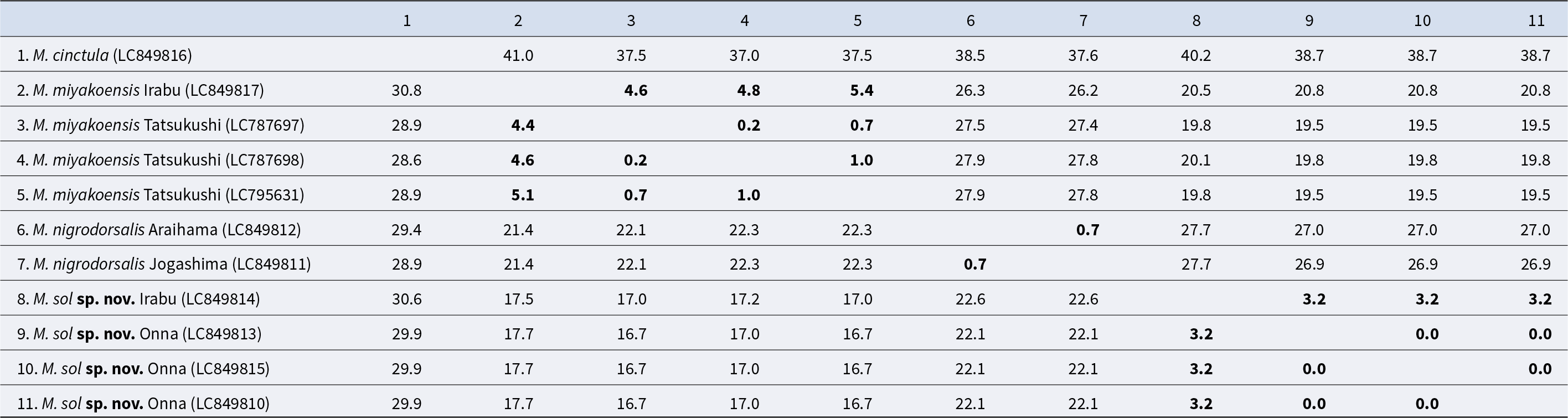
Genetic distances (Table 2) among the four species were 16.7–30.8% (p-distance) and 19.5–40.2% (K2P). These high values suggest that 16S may be evolving too rapidly for reliable inference of interspecific relationships in Mesanthura; sequencing more slowly evolving genes such as 18S rRNA or 28S rRNA may be necessary. Intraspecific distances were much lower, ranging 0.0–5.1% (p-distance) or 0.0–5.4% (K2P), and were lower for single localities (0.2–0.7% for M. miyakoensis at Tatsukushi; 0.0% for M. sol sp. nov. at Onna) than between localities (M. miyakoensis, 4.4–5.1% p-distance, 4.6–5.4% K2P; M. sol sp. nov., both distances 3.2%). These inter-population values fall within the ‘gray zone’ (Riehl et al., Reference Riehl, Lins and Brandt2018) of 3–10% in p-distance, which can encompass both intraspecific and interspecific variation in Isopoda.
Genetic distances (as percentages) among partial 16S rRNA sequences from Mesanthura specimens; above diagonal, K2P distances; below diagonal, p-distances; intraspecific distances are in bold font

Acknowledgements
We thank the following people for assistance. Hisanori Kohtsuka (The University of Tokyo); Yuki Oya (J. F. Oberlin University); Aoi Tsuyuki (University of the Ryukyus); Naohiro Hasegawa (Hiroshima Shudo University); Yuki Kita, Hiroki Matsushita, Kentaro Ageishi, Yoshinobu Matsushima, Maho Matsushita, and Shogo Sekiguchi (Hokkaido University); the staff at the Usa Marine Biological Institute (Kochi University); and Yosuke Kamata, Junko Watanabe, and Manami Kimura (Miyakojima diving shop ‘AQUA STAR’) helped in sampling. Hiroaki Nakano (University of Tsukuba) and Hisanori Kohtsuka for conducting the 19th JAMBIO (Japanese Association for Marine Biology) Coastal Organism Joint Survey (ICHUM8941 was collected). Hiroshi Kajihara and Kevin Wakeman (Hokkaido University) commented on our results. Hisanori Kohtsuka, Yuki Oya, and Aoi Tsuyuki provided photos of living specimens. The staffs of the Miyako, Ikema, and Irabu fisheries cooperative associations permitted sampling in the Miyako Islands. Takashi Akamine, Reijiro Haga, and Hidetaka Shibao (Okinawa Prefecture) handled the documentation necessary for permission to collect coral rubble. Matthew H. Dick (Hokkaido University) reviewed the manuscript and edited the English.
Author contributions
SS conceived and designed the study, conducted the faunal survey, made morphological observations and did the molecular work, wrote a first draft, and reviewed and edited the draft. KK supervised the study and critically reviewed and edited the draft.
Funding
This study was supported by the Research Institute of Marine Invertebrates (KO2022-04) and a Grant-in-Aid for JSPS Fellows (JP23KJ0063) from the Japan Society for the Promotion of Science (JSPS).
Competing interests
The authors declare none.
Data availability
The data supporting this study are available in the article and INSD.
Ethical standards
Not applicable.