Introduction
The black inch looper Hyposidra talaca (Walker) (Lepidoptera: Geometridae) attacks and defoliates mature tea Camellia sinensis (L.) O. Kuntze in the foothills of Darjeeling and Terai and the Dooars of northeastern India (26° to 27°13′ North latitude and 87°59′ to 88° 53′ East longitude). In recent years, the looper activities have largely increased. Although Buzura suppressaria Guen. (Lepidoptera: Geometridae) has been recorded in tea plantations of Darjeeling and Terai and the Dooars in the last six decades (Anonymous, 1994), recent studies have revealed the incidence of other looper species such as H. talaca as a common defoliating insect in tea plantations of Darjeeling Terai and the Dooars (Basu Majumdar and Ghosh, Reference Basu Majumdar and Ghosh2004; Das et al., Reference Das, Mukhopadhyay and Roy2010a). H. talaca usually undergoes four or more overlapping generations throughout the year. Maximum damage occurs during the pre-monsoon period (March–June) followed by a reduction in population during the peak monsoon months (Das and Mukhopadhyay, Reference Das, Mukhopadhyay, Ramamurthy and Subrahmanyam2009). A residual population was also recorded during the winter months (Das et al., Reference Das, Mukhopadhyay, Roy and Biswa2010b).
Populations of looper pests of tea have so far been controlled by regular application of synthetic insecticides especially organophosphates and pyrethroids, but with time the pests have become less susceptible, often resulting in control failures (Sannigrahi and Talukdar, Reference Sannigrahi and Talukdar2003; Sarker and Mukhopadhyay, Reference Sarker and Mukhopadhyay2006). Since the use of synthetic pesticides is being discouraged in tea due to residue problems and safety concerns to human health and the environment, a search for a baculoviral pathogen was conducted by screening natural populations of H. talaca that show symptoms of viral infection. Once isolated, characterized and bioassayed, the virus can be formulated, developed and applied as a biopesticide in future. Baculoviruses are also well suited for use in integrated pest management strategies because of their compatibility with other control agents, chemical or biological. They have a proven safety track record with minimal effects on non-target insects, such as honeybees and silkworms, they do not appear to cause health hazards to humans and other vertebrates, and lastly, they do not invoke resistance in insects (Persley, Reference Persley1996).
Among the various alternatives to synthetic pesticides, microbial control with nucleopoly-hedrovirus (NPV) is a favoured tactic for Helicoverpa armigera (Hübner) (Lepidoptera: Noctuidae) management in different crops (Jayaraj et al., Reference Jayaraj, Rabindra, Narayana, King and Jackson1985; Chandel et al., Reference Chandel, Ahmad and Singh2004). NPV of Perigonia lusca (Fab.) (Lepidoptera: Sphingidae), a pest of Paraguayan tea, was isolated and used for controlling the insect on 900 ha of tea (Sosa-Gómez et al., Reference Sosa-Gómez, Kitajima and Rolon1994). NPV incorporated in occlusion bodies (OBs) was isolated from B. suppressaria and used for controlling B. suppressaria on tea and tung oil trees in over 20,000 ha in China (Yi and Li, Reference Yi and Li1989). In India, preliminary reports on the occurrence of baculoviruses in B. suppressaria and H. talaca from Terai and Darjeeling foothills are available (Mukhopadhyay et al., Reference Mukhopadhyay, De, Sarker and Bambawale2007, Reference Mukhopadhyay, De and Khewa2010). Virulence of NPV against B. suppressaria was subsequently proved in laboratory testing (Mukhopadhyay and De, Reference Mukhopadhyay, De, Ignacimuthu and Jayaraj2008). Granulovirus isolated from Arctornis submarginata (Walker) (Lepidoptera: Lymantriidae), another defoliator in Terai tea plantations, has been added to the list of baculoviruses of tea pests (Mukhopadhyay and Roy, Reference Mukhopadhyay, Roy, Ramamurthy and Gupta2008; Mukhopadhyay and De, Reference Mukhopadhyay and De2009). The objective of this study was to isolate naturally occurring NPVs of H. talaca, characterize them and determine their virulence by establishing the lethal concentration (LC50) and lethal time (LT50) using laboratory bioassays.
Materials and methods
Preparation of polyhedral OBs
The dead cadavers and moribund larvae showing typical symptoms of NPV infections were collected from different tea plantations of Darjeeling foothills, Terai and the Dooars. An individual cadaver was stored in 1 ml water in a stoppered flask and was left to putrefy for about 15 days to enable the release of OBs (polyhedra) from the infected tissue. The putrefied suspension of ten infected larvae was homogenized using a tissue homogenizer and left undisturbed for 2–3 days enabling the OBs to settle at the bottom of the flask. Then, the dead tissue on the upper layer of the flask was removed gently so that the bottom should not be disturbed. The remaining suspension at the bottom was filtered through four layers of cheesecloth, and the filtrate was immediately centrifuged at 1000 g for 20 min. The supernatant was removed by suction, and the sedimented OBs were suspended in 25% (w/v) sucrose dissolved in water. The suspension was again centrifuged at 1000 g for 20 min, the supernatant was discarded and the sedimented OBs were re-suspended in 10 ml of a 25% sucrose solution. The suspended OBs (10 ml) were layered on 30 ml of a 50% sucrose solution and centrifuged at 1800 g for 40 min in a 50 ml centrifuge tube. The differential centrifugation was repeated twice, and the OBs were washed seven to eight times with deionized distilled water following the method of Kawarabata and Matsumoto (Reference Kawarabata and Matsumoto1973). The pellets containing pure OBs were re-suspended in distilled water and stored at 4°C until further use.
The number of OBs/ml was determined by using a Neubauer haemocytometer (0.1 mm depth, 0.0025 mm2 area, Brand GmbH & Co., Wertheim, Germany). Appropriate dilutions were made from the stock with a concentration of 1.12 × 10¹⁸ OBs/ml.
Characterization of the virus
Phase contrast and transmission electron microscopy
The crystalline white precipitate (OBs) isolated from each cadaver was examined at × 1000 magnification in a phase contrast microscope (Olympus CX31 Biological microscope, made in the Phillipines) and photographed with Motic image plus camera attachment. For the transmission electron microscopic (TEM) study, the OBs were fixed with 2.5% glutaraldehyde in 0.01 m Sørenson's buffer, placed on carbon-coated copper grid, dried overnight, and observed and photographed under TEM of FEI-Tecnai G2 Spirit BioTWIN (FEI company, principal HQ in USA, made in Czech Republic) at 80 kV with magnification of × 16,500 at the Indian Institute of Chemical Biology (IICB, Kolkata, India).
Sodium dodecyl sulphate-polyacrylamide gel electrophoresis (SDS-PAGE) of major polyhedrin protein of occlusion bodies
SDS-PAGE of the polyhedrin protein was performed following the method of Mitsuhashi et al. (Reference Mitsuhashi, Kawakita, Watanabe and Sato1995) to determine its molecular weight. The OBs were incubated for 2 h at 70°C to inactivate their protease. A large concentration of the polyhedral suspension (1.7 × 10¹⁰ OBs/ml) was added to an equal volume of SDS protein solubilizing buffer (Laemmli, Reference Laemmli1970) and submerged in boiling water for 3 min. The treated suspension was centrifuged at 7200 g for 3 min to remove the undissolved OBs. Polyhedral protein, thus obtained, was loaded on 10% polyacrylamide gel overlaid with 4% stacking gel and molecular weight marker of protein as reference (Broad range; Genei, Bangalore, India), and was electrophoresed at 100 V for 2 h.
Multiplication of virus
Late second instar larvae were infected with the virus (1 × 10⁶ OBs/ml) and reared for 2 days at 30 °C. Within 2–4 days, third instar larvae showed typical symptoms of viral infection (pale, glossy, flaccid appearance and hanging head down). Fat body cells were dissected out of third instar larvae after 2 days and smeared onto a glass slide for observation under a phase contrast microscope for the baculovirus in vivo.
Bioassay
Various instars of H. talaca were collected from tea bushes (high-yielding Tocklai vegetative clone, TV-25) from Terai plantations. These were reared in the laboratory for two generations at 27 ± 2°C and 72 ± 2% relative humidity with a photoperiod of 11 h light-13 h dark in aseptic conditions. Fresh tea twigs (TV-25) collected from an organically maintained tea garden at the North Bengal University campus were supplied as food after surface sterilizing with 1% sodium hypochloride by dipping the leaves in the solution for 3 s and then washing them with distilled water. Laboratory-reared early second instar larvae of the second generation were used for bioassay in the laboratory.
The viral suspension was diluted decimally with distilled water (Kawarabata and Matsumoto, Reference Kawarabata and Matsumoto1973). The leaf dip-feeding method was used to treat the larvae (n = 60) with five concentrations (1 × 10⁶, 1 × 10⁵, 1 × 10⁴ 1 × 10³ and 1 × 10² OBs/ml). For each concentration, three replicates of 20 larvae each were used, and three replicates of double distilled water-treated leaves served as control. Before treating with NPV, the larvae were starved for 2 h. After 24 h treatment with NPV, the larvae were provided with fresh tea leaves, and the mortality was recorded every day. After Abbott's (Reference Abbott1925) correction of mortality, the data were subjected to Probit analysis (Finney, Reference Finney1954) to determine the median lethal concentration (LC50) of NPV. Median lethal time (LT50) was also determined using the following equation (Biever and Hostetter, Reference Biever and Hostetter1971):

where a is the number of hours from the initiation of the test until the reading made immediately before the 50% value was recorded, b is the total number of larvae dead at the reading immediately before the 50% value was recorded, c is 50% of the total number tested, D is the number of larvae dying in 24 h during which the 50% mortality was reached and e is the number of hours between mortality counts.
Results and Discussion
Baculoviruses are host specific for insects and can cause epizootics in nature resulting in the control of insect populations. These viruses are, therefore, attractive biological control agents and offer an alternative to broad-spectrum synthetic insecticides. Hu (Reference Hu1998) listed at least six baculoviruses that had been developed and used as biopesticides against defoliating caterpillars of tea.
Naturally occurring wild-type baculoviruses, NPV in particular, are able to spread rapidly and independently from an epicentre of infection into the surrounding crop habitat, where new hosts may encounter them, either immediately or during subsequent generations of the pests. Most baculoviruses so far employed for biological control of insect pests are natural entities, which occur in nature as a result of insect–pathogen co-evolution (Speight et al., Reference Speight, Hunter and Watt1999).
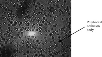
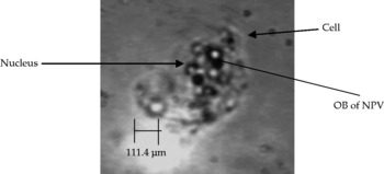
In tea bushes of Darjeeling foothills, virus-infected larvae of H. talaca hung head down and turned pale and flaccid with putrefied and liquefied tissues. The mean OBs isolated from a cadaver of fifth instars were at a concentration of 3.36 × 10¹⁷ OBs/ml. The OBs were evident as bright particles under phase contrast microscopy (Fig. 1). The virus was present in the nucleus of the fat body cell (Fig. 2), which is the principal multiplying site of NPVs (Asayama, Reference Asayama1975). The OBs were dissolved in 5% 1 N NaOH (Lacey and Brooks, Reference Lacey, Brooks and Lacey1997). TEM showed a distinct polyhedral shape of the viral OBs (Fig. 3).
Smear showing polyhedral occlusion bodies of nucleopolyhedrovirus of Hyposidra talaca under phase contrast microscope

Occlusion bodies (OBs) of nucleopolyhedroviruses (NPVs) formed within fat body cell nucleus of Hyposidra talaca

Transmission electron micrographs ( × 16,500) of polyhedral bodies of nucleopolyhedrovirus of looper pest Hyposidra talaca

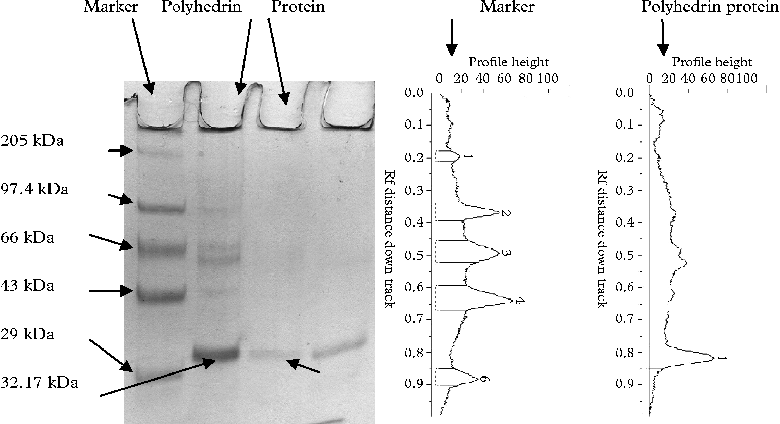
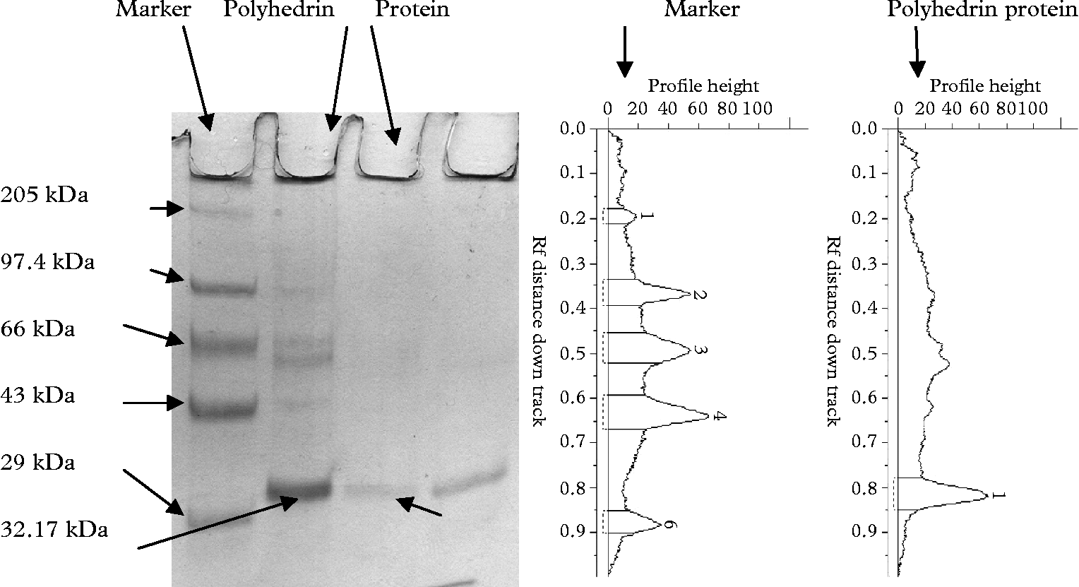
The SDS-PAGE analysis showed that the molecular weight of the polyhedrin is 32.17 kDa (Fig. 4). The molecular weights of polyhedrin of some other NPVs reported are as follows: Autographa californica multiple-nucleocapsid NPV (MNPV) 30 kDa, Anticarsia gemmatalis MNPV 29 kDa, Heliothis armigera MNPV 28 kDa, Trichoplusia ni single-nucleocapsid NPV 31 kDa, Bombyx mori NPV 31 kDa, Spodoptera exigua NPV about 30 kDa and Cyclophragma yamadai NPV 33 kDa (Summers and Smith, Reference Summers and Smith1978; Taniai and Inoue, Reference Taniai and Inoue1988; Kondo et al., Reference Kondo, Yamamoto, Takashi and Maeda1994; Mitsuhashi et al., Reference Mitsuhashi, Kawakita, Watanabe and Sato1995).
Sodium dodecyl sulphate-polyacrylamide gel electrophoresis profile of polyhedrin protein of occlusion bodies of nucleopolyhedrovirus obtained from Hyposidra talaca along with graphical representation
Application of the isolated virus (NPV) of H. talaca in various doses through food (tea leaves) was effective in causing dose-dependent mortality in the laboratory. The mortality ranged from 25 to 83.3%, and the LC50 value was estimated as 2.8 × 10³ OBs/ml, with 1.16 × 10³ and 6.75 × 10³ OBs/ml as the lower and upper limits, respectively. The LT50 values were 5.45 days for 1 × 10⁴ OBs/ml, 4.15 days for 1 × 10⁵ OBs/ml and 4.05 days for 1 × 10⁶ OBs/ml concentrations (Table 1).
Dose–mortality values of nucleopolyhedrovirus on second instar Hyposidra talaca for determining LC50 and LT50 within 7 days of inoculation

A major limitation to the wider use of baculoviruses in insect pest control is their relatively slow speed of action leading to feeding inhibition in larvae and their consequent death when immediate insecticidal effect is often desired. Another drawback is their relative low virulence for the older instars, which cause the majority of damage in crops. Finally, baculoviruses are often too specific to meet the commercial demands for broad-spectrum activity (Hu, Reference Hu1998).
Thus to improve the insecticidal properties of baculoviruses, one approach is to select the natural variants by in vitro cloning with cell culture technique or in vivo cloning with larvae. Such clones have proved to be 100-fold more lethal for A. gemmatalis NPV (Ribiero et al., Reference Ribiero, Pavan and Muotri1997) and seven times more lethal and speedy for Panolis flammea NPV (Hodgson et al., Reference Hodgson, Vanbergen, Watt, Hail and Cory2001).
Introduction and establishment of baculoviruses in a crop environment is intended to result in permanent suppression of the target pest (Moscardi, Reference Moscardi1999). Wild-type baculoviruses are an integral component of the natural biological control of many pest species, and in several cases application of wild-type viruses has been very effective for pest management (Bonning and Hammock, Reference Bonning and Hammock1996; Sun, Reference Sun2005).
Large-scale use of baculoviruses, wild-type or genetically modified ones, is hampered not so much by biology and ecology or technology, but more by public and even scientific distrust of the imagined danger of ‘germ warfare’ in our crops. Nonetheless, insect pest populations can be regulated very efficiently by baculoviruses (Speight et al., Reference Speight, Hunter and Watt1999). The efficacy has been showcased by the NPV-based successful control of the soya bean looper A. gemmatalis Hübner (Beach et al., Reference Beach, Carner and Turnipseed1984). Clearly, the system works most efficiently and should be considered as a model for other crop pest management including the tea loopers in India, where environmentally benign control systems are required as alternatives to the large-scale use of synthetic insecticides.
The present finding on the virulence of the H. talaca NPV (Hyta NPV) at a reasonably low concentration holds promise of its future use as a microbial pesticide against the tea-attacking black inch loopers.
Acknowledgements
Thanks are due to Entomology Division, IARI, New Delhi, for identifying Hyposidra; Indian Institute of Chemical Biology, Kolkata, for extending the TEM facility; and the University Grants Commission for funding the project. We thank Dr A. Raman, Charles Stuart University, Australia, for reviewing the manuscript in addition to two anonymous reviewers. We are also thankful to our fellow researcher Dr Somnath Roy for his kind help in the statistical analysis.