Introduction
Wild boar populations occur throughout the steppe and forest regions of the world (Oliver and Leus, Reference Oliver and Leus2008). They remain widely distributed and often locally abundant but are also increasing in numbers in several countries, spreading into northern regions. As a result of their depredation on crops, wild boar are not appreciated in many countries, where they are managed as a game animal. In addition, they harbour many infectious agents (bacteria, viruses and parasites) transmissible to other animal species and humans, through contaminated food and/or the environment (Fredriksson-Ahomaa, Reference Fredriksson-Ahomaa2018). Changes of human habitation to suburban areas, increased use of land for agricultural purposes and deforestation have all increased chances of wild boar contact with domestic animals and humans (Meng et al., Reference Meng, Lindsay and Sriranganathan2009). Wild boar, Sus scrofa algira and the Barbary wild boar, Sus scrofa barbarus are two species native to North African countries including Morocco, Algeria and Tunisia (http://fr.wikipedia.org). Wild boar is among the most abundant and economically important wildlife species in Tunisia. Despite their ubiquitous presence across most of Tunisia, accurate figures of wild boar population are not currently available. However, their observed numbers during the hunting season (September–April) for the period between 1991 and 2009 was estimated to be approximately 10 293 animals and the mean annual number hunted during the same period was 2437 wild boar (Karem et al., Reference Karem, Triplet and Lartiges2009). Studies on parasitic infections of wildlife species are important to evaluate the extent to which wild animals may potentially serve as source of disease transmission to livestock and humans. Information on the helminth parasitic fauna of wild boar in Tunisia remains limited. Being omnivorous, wild boar feed on small mammals, invertebrates, carrions and plants and could therefore potentially play an important role in the transmission of zoonotic parasites (Solaymani-Mohammadi et al., Reference Solaymani-Mohammadi, Mobedi, Rezaian, Massoud, Mohebali, Hooshyar, Ashrafi and Rokni2003). Wild boar from Tunisia were molecularly confirmed to harbour Echinococcus granulosus sensu stricto (s.s), although their epidemiological role in the spread of cystic echinococcosis (CE) is unknown (Boufana et al., Reference Boufana, Lahmar, Rebaï, Ben Safta, Jebabli, Ammar, Kachti, Aouadi and Craig2014). Due to their encroachment on human habitation, where they have been observed in Tunisia to roam freely around houses and suburban areas, wild boar may serve as reservoirs for human helminthiases.
The aims of this survey were to describe the helminth fauna of wild boar from northeastern and northwestern Tunisia; to determine parasite prevalence and intensity, and analyse their association with gender and age with special reference to CE infection pressure.
Materials and methods
A total of 591 North African wild boar (S. scrofa) shot during the 2008–2013 hunting seasons in northeast (Ariana, Mabtouh, Bou Argoub, Djebel Ressas) and northwest (Bou Salem, Bou Arada, Gaâfour, Fernana, Touaref) Tunisia were included in this study. The type of organs recovered from each wild boar carcass was determined by the hunters and thus the investigated sample was conveniently divided into three groups. Wild Boar Group 1 (WBG1) included 297 wild boar carcasses collected between 2008 and 2011, which were used to examine the lungs, liver and abdominal cavity for the presence of helminth species. Wild Boar Group 2 (WBG2) consisted of 264 animals obtained during 2009–2013 hunting seasons which were used to identify gastrointestinal helminths. Wild Boar Group 3 (WBG3) consisted of 30 wild boar shot between 2010 and 2012 which were specifically used for the detection of Trichinella species. Age was estimated for animals within WBG1 and WBG2 using the dental formula described by Oroian et al. (Reference Oroian, Oroian, Paşca, Oroian and Covrig2010). Animals were classified into three age classes, <2 (n = 212), 2–3 (n = 208) and ⩾4 years old (n = 141). Among the WBG1 and WBG2 animals, 305 and 256 originated from the northeast and the northwest regions of Tunisia, respectively. Host gender was registered only for WBG1 which included 163 males and 134 females. Wild boar carcasses and/or organs were transported to the Parasitology Laboratory of the Veterinary School, Sidi Thabet, Tunisia where analysis was carried out.
Lung and hepatic helminths
Individual samples of lungs and livers from 297 necropsied wild boar (WBG1) were examined for lungworms, E. granulosus metacestodes and Taenia hydatigena larvae. The trachea, large bronchi and bronchioles were opened using a pair of scissors and inspected for the presence of metastrongylids. Lungs were then cut into small pieces and soaked in 0.9% NaCl. After discarding large pieces of tissue, lungworms were collected in the sediment and mounted in chloral lactophenol solution (44%). The shape and length of spicules as well as the size and ray arrangement of the copulatory bursa were used to identify metastrongylid species as previously described (Skrjabin et al., Reference Skrjabin, Shikhobalova, Shults, Popova, Boev and Delyamure1952; Gassó et al., Reference Gassó, Rossi, Mentaberre, Casas, Velarde, Nosal, Serrano, Segales, Fernandez-Llario and Feliu2014).
Echinococcus granulosus hydatid cysts were identified by gross examination and palpation of the lungs and livers. Livers and lungs were then cut into thin slices and the number, size, localization and type of cysts were recorded. Hydatid fluid was aspirated from each pulmonary and hepatic viable cyst and examined to determine the protoscoleces fertility rate. The mean fertility rate corresponded to the total number of the extrapolated protoscoleces/ml in each cyst divided by the total number of fertile cysts. Viability of protoscoleces was assessed for each fertile cyst per animal and organ using the method described by Smyth and Barrett (Reference Smyth and Barrett1980). A drop of 0.2% aqueous eosin solution was added to an equal volume of protoscoleces retrieved from a drop of cyst fluid sediment. A total of 100 protoscoleces in the sample was then scored as stained (indicating that the protoscoleces were dead) or unstained (indicating that the protoscoleces were viable). The mean percentage of protoscoleces viability was calculated by dividing the total number of viable protoscoleces of each cyst by the total examined protoscoleces in all fertile cysts multiplied by 100. Caseous hydatid cysts were considered infertile.
Gastro-intestinal helminths
Abdominal viscera were removed from 264 necropsied wild boar (WBG2). The oesophagus, stomach and intestinal tract of each animal were separated then opened longitudinally and carefully inspected for the presence of worms; the contents were washed in 0.9% NaCl and repeatedly rinsed through 100 mesh standard sieves which allowed the collection of helminths. Nematodes and trematodes were cleared using chloral lactophenol solution (44%) and identified under a light microscope based on figures and descriptions provided by Anderson (Reference Anderson1992); they were then counted and stored in 70% alcohol. Identification of cestodes was based on the size and morphology of the proglottids following staining with 1% acetocarmine solution (Khalil et al., Reference Khalil, Jones and Bray1994). Worm size, shape and the arrangement of the proboscis hooks were used in the identification of acanthocephalans.
Collection and examination of faecal samples
Of the 264 wild boar within WBG2, 232 faecal samples were collected directly from the rectum of each animal and examined for the presence of parasite eggs and larvae using the qualitative flotation method. From each sample, 10 g of faeces were weighted and mixed with 75 mL of saturated MgSO4 solution. Eggs were isolated onto glass cover slips superimposed on convex meniscus for 15 min. Recovered eggs were identified based on their morphology (shape, envelope structure, number and size of blastomeres or presence of larvae) and biometrics (Thienpont et al., Reference Thienpont, Rochette and Vanparijs1986; Georgi and McCulloch, Reference Georgi and McCulloch1989).
Examination of wild boar muscle for larvae of Trichinella
A total of 120 muscle samples from 30 wild boar (WBG3) (four muscle samples per wild boar) were used to investigate the presence of Trichinella larvae. This was carried out using trichinoscopy and by artificial digestion (artificial gastric juice) using 5 g of the tongue, masseter, pillar of diaphragm and inter-costal muscles. About 80 g of muscle tissue was collected from each animal including 20 g from the tongue, 20 g from the masseter, 20 g from the diaphragm and 20 g from the inter-costal muscle. The trichinoscopy was performed by compressing 28 small pieces of muscle tissue, about 2 mm by 10 mm, with a total weight of approximately 0.5 g from each sample. Muscle pieces were compressed between two glass plates until they become translucent, and examined using a stereomicroscope (Nöckler et al., Reference Nöckler, Pozio, Voigt and Heidrich2000). Artificial digestion for the detection of muscle larvae was performed using the magnetic stirrer method (Gamble et al., Reference Gamble, Bessonov, Cuperlovic, Gajadhar, van Knapen, Noeckler, Schenone and Zhu2000; Mayer-Scholl et al., Reference Mayer-Scholl, Pozio, Gayda, Thaben, Bahn and Nöckler2017).
Data analysis
Data were analysed in R (R Core Team, Reference R Core2017). The probability distribution of the abundance of each parasite in its host was analysed through maximum likelihood. Count models analysed included Poisson, negative binomial and zero inflated models. The most parsimonious model was used for further analysis. The abundance of each parasite was analysed according to age and geographic origin of the host (northeast or northwest Tunisia). Generalized linear models (GLM) (with or without zero inflation) were utilized to explore these relationships. Generalized additive models (GAM) were also used to model the variations of abundance and prevalence of E. granulosus with age in wild boar. Finally, variations in abundance and prevalence of echinococcosis was examined according to existing models and explored for evidence of parasite-induced immunity or variations in infection pressure. Thus, the variation in abundance m (t) with age of the host t could be modelled as follows
 $$m\,(t)\, = \,\displaystyle{{ght} \over {g + ah}}\, + \,\displaystyle{{ah^2} \over {{(g + ah)}^2}}\,\lsqb {1-\,{\rm exp}\,{\rm \{} -\,(g + ah)t{\rm \}}} \rsqb $$
where h is the infection pressure in cysts per year, a is the probability of inducing immunity and g is the rate of loss of immunity (Roberts et al., Reference Roberts, Lawson and Gemmell1986; Torgerson and Heath, Reference Torgerson and Heath2003). Once infected, an animal is assumed to be infected for life. In the absence of parasite-induced host immunity (i.e. a = 0), this can be expressed simply as:
 $$m\,(t)\, = \,ht$$
Likewise, the prevalence p (t) can be expressed as:
 $$p\,(t)\, = \,k\,(1-{\rm exp}\,{\rm \{} -bt{\rm \}} )$$
where b is the infection pressure in exposures per year and k is a constant that reduces the asymptotic prevalence below 1 in the presence of parasite-induced host immunity. The data were fitted to the models using maximum likelihood techniques to find the most likely parameter values and assess the goodness of fit to the model.
Statistical analysis of the prevalence of infection used Fisher's exact and χ 2 tests (Schwartz, Reference Schwartz1993), with a P value of <0.05 considered indicative of a statistically significant difference.
Results
Global infection
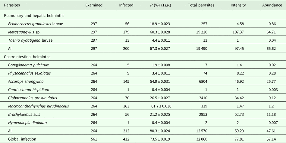
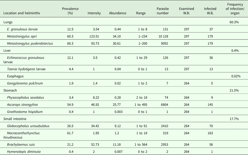
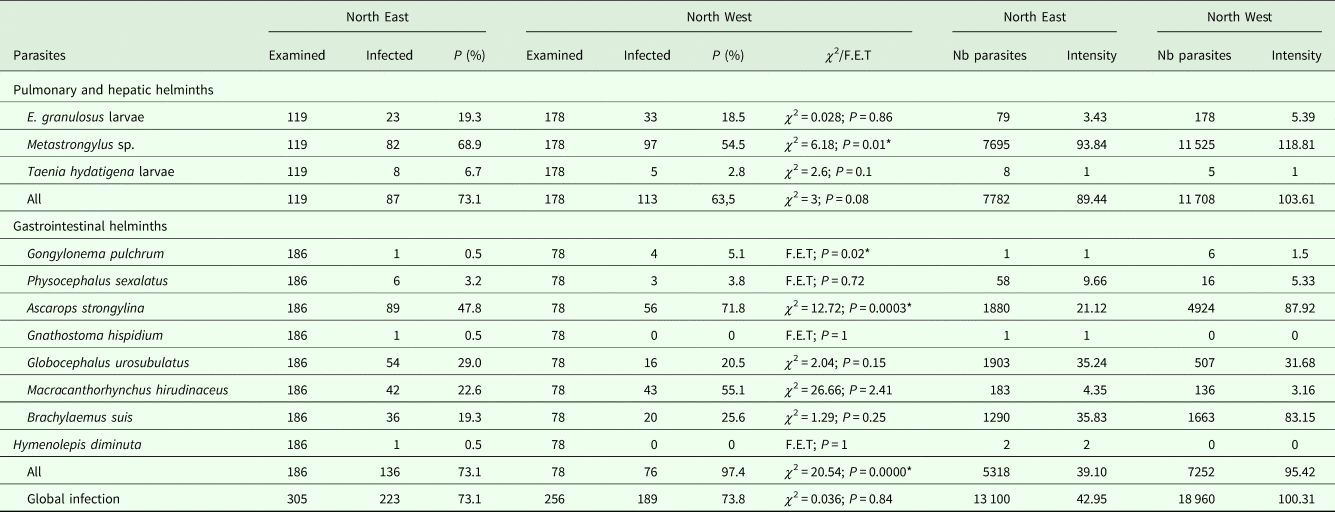
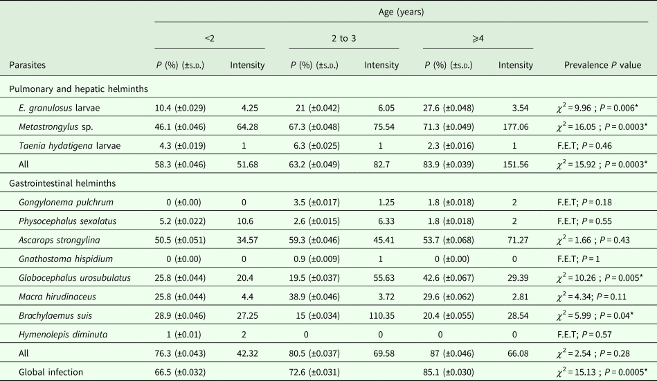
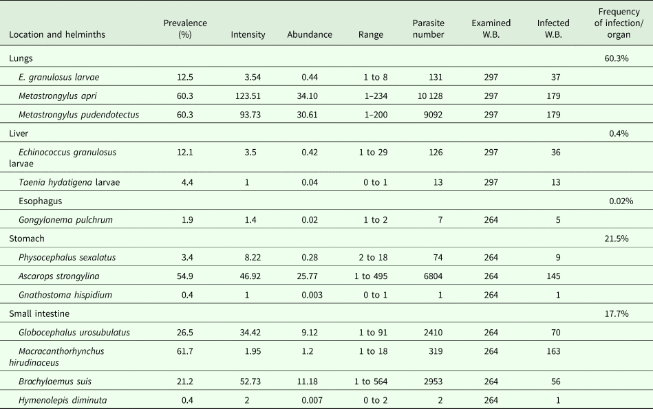
A total of 32 060 recovered helminths belonging to fourteen species (adults of Brachylaemus suis, Hymenolepis diminuta, Metastrongylus apri, Metastrongylus pudendotectus, Ascarops strongylina, Globocephalus urosubulatus, Physocephalus sexalatus, Gnathostoma hispidum, Gongylonema pulchrum and Macracanthorhynchus hirudinaceus; larval stages of E. granulosus and T. hydatigena; eggs of Strongyloides ransoni and Capillaria spp.) were recovered in this study. The visceral necropsy of the examined wild boar (n = 561) revealed a global infection prevalence of 73.5% (±0.019) (412/561) with a global mean intensity and mean abundance of 77.81 helminths per infected animal and 57.14 parasites per examined wild boar, respectively (Table 1). The most prevalent helminth was M. hirudinaceus while lung nematodes (Metastrongylus sp.) were the most abundant. The most frequently infected tissues were the lungs (60.3%), followed by the stomach (21.5%), small intestine (17.7%), liver (0.4%) and oesophagus (0.02%) (Table 2). Coproscopic examination of faecal samples collected from WBG2 revealed the presence of two other nematode species not detected by necropsy namely, Capillaria spp. and S. ransoni in one (1/232) and two wild boar (2/232), respectively. Trichinella larvae were not detected in the muscular tissues of wild boar within WBG3. Wild boar from the northwest (73.8%) were similarly infected with visceral helminths as those from the northeast (73.1%) regions (χ 2 = 0.036; P = 0.84) (Table 3). The global prevalence of infection with visceral helminths relative to host age was significantly different in the examined three age groups, increasing with age from 66.5% (±0.032) to 85.1% (±0.03) for wild boar <2 and ⩾4 years old, respectively (χ 2 = 15.13; P = 0.0005) (Table 4).
Prevalence, intensity and abundance of pulmonary, hepatic and gastrointestinal helminth species in Tunisian wild boar

s.d., standard deviation.
Prevalence, intensity, range and abundance of visceral helminth species per organ in Tunisian wild boar
Prevalence of visceral helminths in wild boar from northeast and northwest Tunisia

P, prevalence; Nb, number.
*Significant.
Prevalence and intensity of helminth species in Tunisian wild boar relative to host age

P, prevalence; s.d., standard deviation.
*Significant.
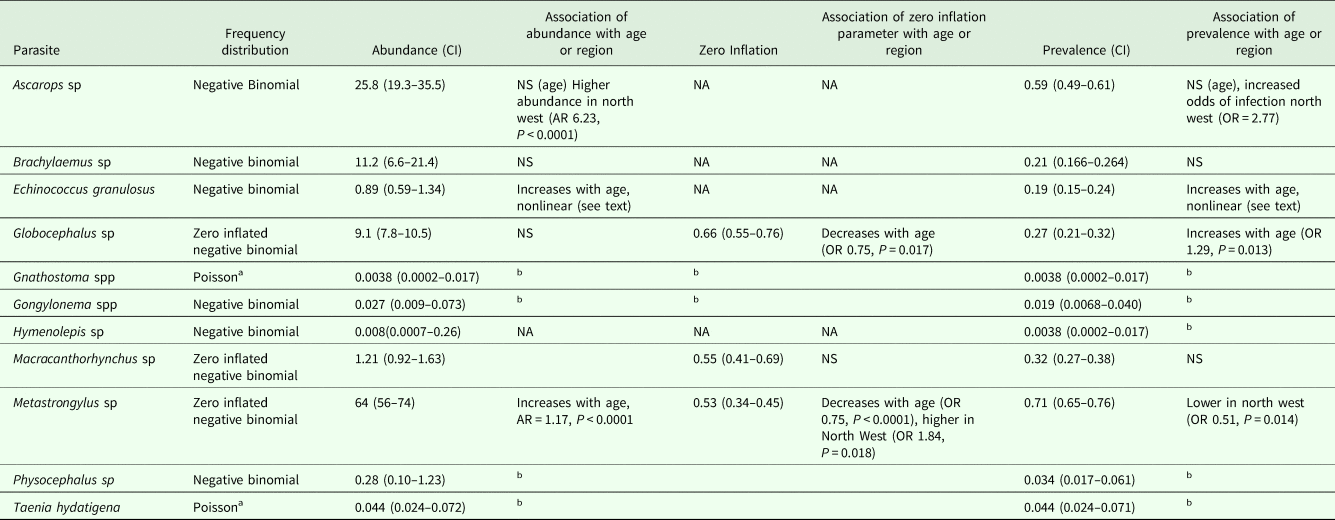
Most parasites had an over dispersed distribution which could be best modelled by either a negative binomial or zero inflated negative binomial distribution. For T. hydatigena and Gnathostoma species, the small number of positive counts were single parasites, thus it was not possible to conclude that these parasites had an over dispersed distribution and a Poisson distribution was assumed (Table 5).
Frequency distributions of wild boar parasites and association with age or geographical region

AR, abundance ratio; OR, odds ratio.
a Positive animals only had single parasites, so no meaningful analysis of other frequency distributions was possible.
b Insufficient positive animals to evaluate.
Prevalence and intensity of lung, liver and abdominal cavity helminths
Among the 297 animals in WBG1 investigated for lung and hepatic helminth infections, 200 wild boar were infected (67.3% ± 0.027) with a mean intensity of 97.45 and a mean abundance of 65.62 worms per host (Table 1). Animals from the northeast (73.1%) were more infected than those from the northwest (63.5%) (χ 2 = 3; P = 0.08) (Table 3). Prevalence of infection with pulmonary and hepatic helminths increased significantly with age from 58.3 to 63.2 and 83.9% in animals aged <2, 2–3 and ⩾4 years old, respectively (χ 2 = 15.92; P = 0.0003) (Table 4). No significant difference was observed between prevalence in female (80.6 ± 0.034) and male wild boar (73 ± 0.035) (χ 2 = 2.35; P = 0.125) nor between the abundance of infection with lung and hepatic helminth species (P = 0.128) (Table 6).
Prevalence, intensity and abundance of pulmonary and hepatic helminths in Tunisian wild boar relative to host gender

Exam, examined; Inf, infected; I, Intensity; A, Abundance.
Three parasite species were recovered from the lungs, liver and abdominal cavity namely, Metastrongylus spp., E. granulosus metacestodes and larvae of T. hydatigena. Metastrongylus spp. (M. apri and M. pudendotectus) were the most frequent nematode species (60.3% ± 0.028) isolated from the bronchi and bronchioles. Prevalence was similar for both species (60.3% ± 0.028). However, M. apri showed a higher intensity of infection (123.51 worms) than M. pudendotectus (93.73 worms per infected animal) (Table 2). A statistically significant difference in the prevalence was observed between the two studied regions for Metastrongylus spp. infection (χ 2 = 6.18; P = 0.01) and the mean intensity was higher in the northwest (118.81) than in the northeast (93.84) of Tunisia (Table 3). Prevalence of these respiratory strongyles was higher in female 61.9% (±0.042) than male wild boar 58.9% (±0.039) (χ 2 = 0.28; P = 0.59) as well as the abundance (73.4 for females; 57.28 for males) (P = 0.12) (Table 6). Prevalence of metastrongylids increased significantly with age from 46.1% (±0.046) to 71.3% (±0.049), in wild boar <2 and >4 years old, respectively (χ 2 = 16.05; P = 0.0003) (Table 4). Also, the intensity of Metastrongylus increased with age. Metastrongylus had a zero-inflation which decreased with age and was higher in the northwest (Table 5).
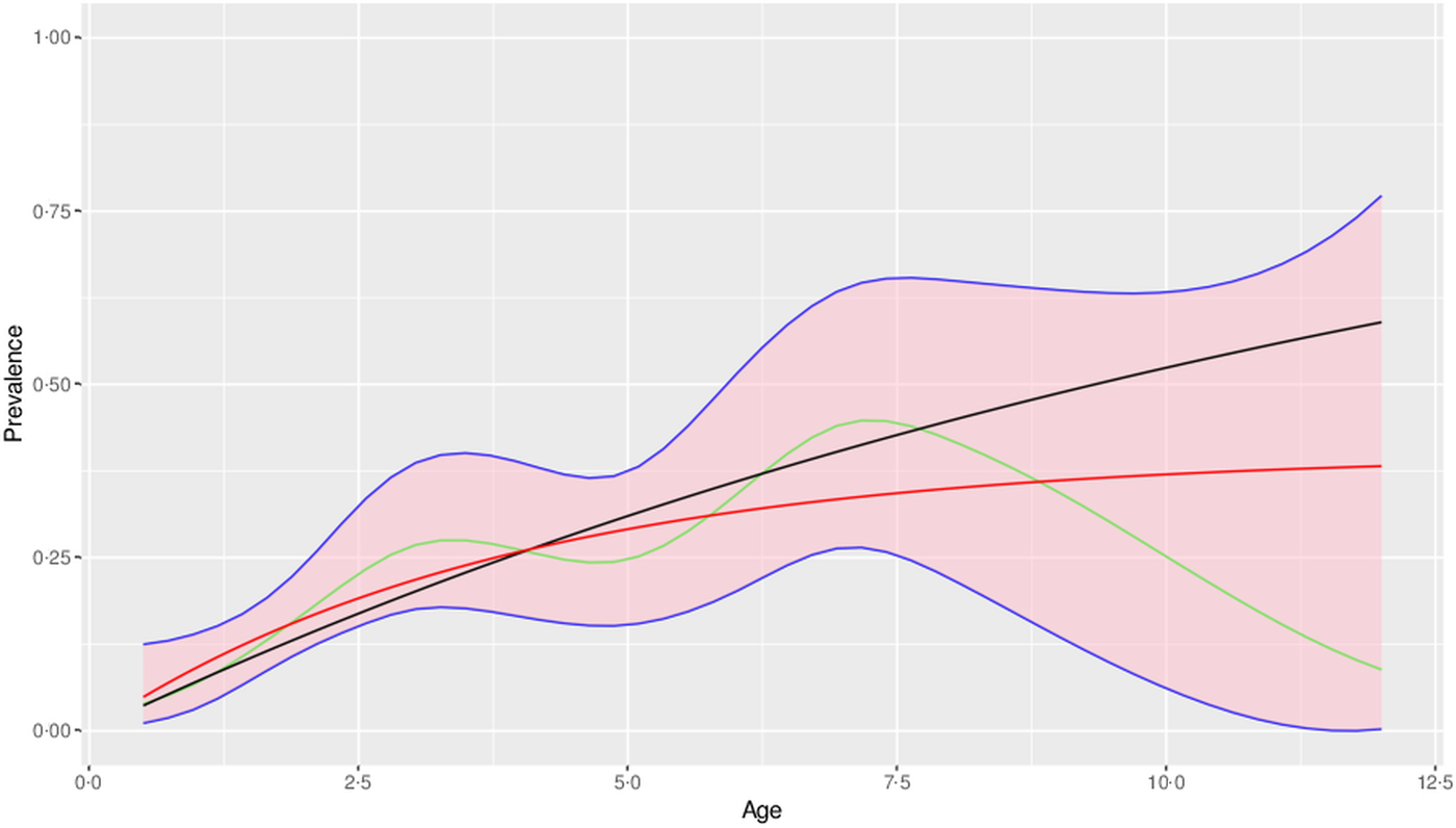
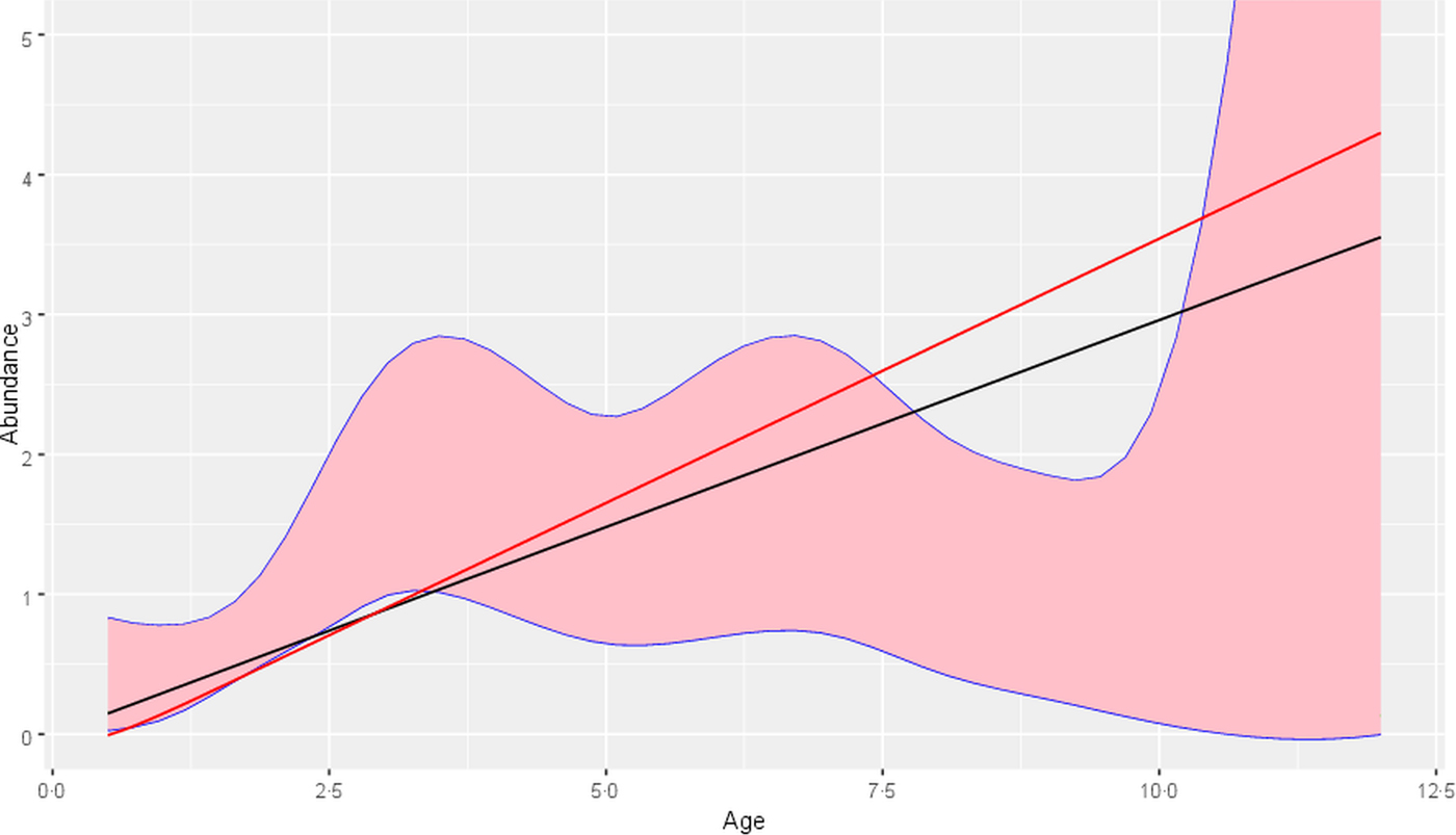
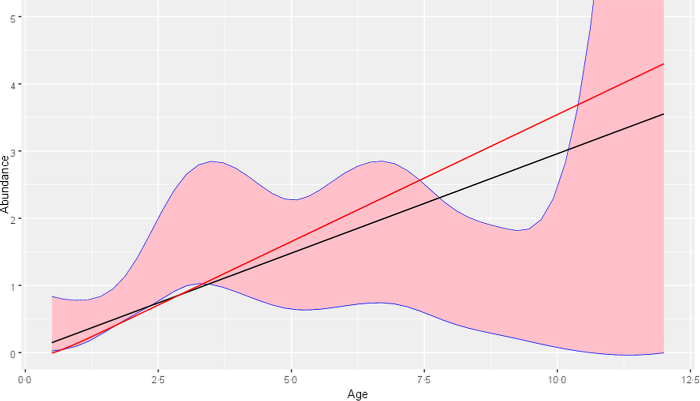
Echinococcus granulosus was the most prevalent cestode recorded in 18.9% (±0.023) of the 297 examined wild boar with a mean intensity of 4.58 worms per infected host and an abundance of 0.86 worms per examined host (Table 1). The liver and lungs were the only infected organs with co-infection of hepatic and pulmonary tissues encountered in 5.7% (17/297) of cases, and observed in all age-groups with 2.6, 5.3 and 10.3% infection recorded for <2, 2–3 and ⩾4 years old, respectively. There were no significant differences in prevalence rates between hepatic hydatid cysts (12.1%) and pulmonary cysts (12.5%) (χ 2 = 0.015; P = 0.9) nor in the abundance of E. granulosus larvae in the two organs (0.42 and 0.44 cyst per examined animal) (P = 0.93) (Table 2). There was a nonlinear relationship with age both in prevalence and abundance of E. granulosus hydatid cysts (Figs 1 and 2). The variation in age prevalence did not give a good fit to the standard mathematical model, with a reduction in prevalence between ages 3 and 5 and a decrease in older animals (Fig. 1). Likewise, the age abundance variation gave a poor fit (Fig. 2). The fit given by the GAM model suggested that hydatid cyst abundance peaked at approximately 3 years and decreased as the animals became older.
Variation of prevalence with age for Echinococcus granulosus. Best fit generalized additive model (GAM) (green) with 95% confidence limits (pink). Predicted models in the absence (black line) and presence (red line) of parasite-induced immunity.

Variation of abundance with age for Echinococcus granulosus. Best fit generalized additive model (GAM) (green) with 95% confidence limits (pink). Predicted models in the absence (black line) and presence (red line) of parasite-induced immunity.
No significant difference was noted in CE rates between males (16.6% ± 0.029) and females (21.6% ± 0.036) (χ 2 = 1.23; P = 0.26) neither in abundance (0.9 for males; 0.81 for females) (P = 0.83) (Table 6). Differences in CE prevalence between regions of the northeast (19.3%) and the northwest (18.5%) were not statistically significant (χ 2 = 0.028; P = 0.86) (Table 3). The mean number of cysts per infected wild boar as well as abundance were highest (6.05; 1.27) in animals aged between 2 and 3 years old and lower in the two other age groups (<2 and ⩾4 years) varying between 4.25–0.97 and 3.54–0.44 cysts per infected animal, respectively (Table 4). Cyst size varied between 5 and 60 mm in diameter for each animal. The diameter of hepatic and pulmonary cysts ranged from 11 to 20 mm for 53 cysts (20.6%), 21 to 60 mm for 39 cysts (15.2%) while 165 cysts (64.2%) were less 10 mm wide.
Of the 257 recovered hydatid cysts, 238 (92.6%) were viable including 6 (2.3%) fertile cysts while 19 (7.4%) cysts were caseous. Animals aged between 2 and 3 years had the highest proportion of viable (43.2%) and dead (3.9%) cysts. The percentage of viable cysts followed a positive linear correlation with age (R 2 = 0.2892; y = 0.0681x + 0.1724). Lung hydatid cysts (3.8%) were more fertile than their hepatic counterparts (0.8%). Mean fertility rates of the six fertile hydatid cysts were estimated to be 850, 950, 1450, 1600 and 1750 for the five pulmonary fertile cysts and 200 protoscoleces per ml of hydatid cyst fluid for the unique fertile hepatic cyst. The fertility rate was highest in cysts measuring between 21 and 60 mm in diameter. No fertile cyst was detected in wild boar <2 years old whereas the mean fertility rate increased with age from 1950 to 4850 protoscoleces per ml in 2–3 and ⩾4 years age groups, respectively. Of the total viable hydatid cysts, germinal layers and protoscoleces of 27 hydatid cysts (12 hepatic and 15 pulmonary) removed from 23 wild boar originating equally from the northwest and the northeast studied areas, were characterized at the molecular level and reported to be those of E. granulosus s.s (Boufana et al., Reference Boufana, Lahmar, Rebaï, Ben Safta, Jebabli, Ammar, Kachti, Aouadi and Craig2014).
Taenia hydatigena larvae were encountered in the liver and the abdominal cavity of 4.4% (±0.011) of the examined wild boar with a mean intensity and a mean abundance of 1 and 0.04 larvae, respectively (Tables 1 and 5). Infection in wild boar originating from the northeast was higher (6.7%) than that observed in animals from the northwest (2.8%) (Table 3) (χ 2 = 2.6; P = 0.1). With respect to age, older wild boar (⩾4 years) were less infected than other age groups but no significant difference was noted between the prevalence of the 3 age classes (F.E.T.; P = 0.46) (Table 4). The prevalence of T. hydatigena larvae was almost similar in female (4.5% ± 0.018) and male wild boar (4.3% ± 0.016) (χ 2 = 0.005; P = 0.93) as well as the mean abundance in male (0.042) and female (0.044) (P = 0.93) animals (Table 4).
Prevalence and intensity of gastrointestinal helminths
Of the 264 wild boar examined for gastrointestinal parasites (WBG2), 212 animals were found to be infected with at least one helminth species, giving an overall prevalence of 80.3% (±0.024), a mean intensity of 59.29 worms per infected animal and an abundance of 47.61 (Table 1). The global prevalence of infection with gastrointestinal helminths was significantly higher in animals from the northwest (97.4%) than those from the northeast (73.1%) (χ 2 = 20.54; P = 0.0000) as well as the mean intensity of infection with 95.42 and 39.10 worms per infected animal, respectively (Table 3). In addition, the global prevalence of gastrointestinal helminths increased with host age from 76.3% (±0.043) in the youngest to 87% (±0.046) in older animals (χ 2 = 2.54; P = 0.28) (Table 4). Eight parasite species were detected including one trematode (B. suis), one cestode (H. diminuta), five nematodes (A. strongylina, G. urosubulatus, G. pulchrum, P. sexalatus, G. hispidium) and one acanthocephalan (M. hirudinaceus). The most frequently found gastrointestinal parasites were M. hirudinaceus (61.7% ± 0.030), A. strongylina (54.9% ± 0.031), G. urosubulatus (26.5% ± 0.027) and B. suis (21.2% ± 0.025) (Table 1). The overall mean intensities of gastrointestinal helminth species were high for B. suis (52.73), A. strongylina (46.92), G. urosubulatus (34.42) and low for P. sexalatus (8.22), H. diminuta (2), M. hirudinaceus (1.47) and G. hispidium (1). The highest gastrointestinal helminth abundance was observed for A. strongylina (25.77 worms) with the total number ranging from 1 to 495 worms. Ascarops strongylina had an increased abundance and prevalence in the northwest compared to northeast regions (Tables 3 and 5). The highest gastrointestinal helminth intensity was observed for B. suis (52.73) with the total number ranging from 1 to 564 worms whereas the lowest mean intensity and mean abundance were recorded for G. hispidium (1 and 0.003) (Tables 1 and 2). In the northeast, wild boar was infected with all the gastrointestinal helminth species identified here while G. hispidium and H. diminuta were absent in animals originating from the northwest regions (Table 3). Gastrointestinal helminths were encountered in all age groups except for G. pulchrum which was absent in wild boar <2 years old; H. diminuta which was recovered only from the youngest age group (<2 years) and G. hispidium identified in one animal aged between 2 and 3 years old. A significant difference was observed in prevalence between the three age groups, except for G. urosubulatus (χ 2 = 10.26; P = 0.005) and B. suis (χ 2 = 5.99; P = 0.04) (Table 4). Globocephalus urosubulatus had a zero-inflated distribution with the zero-inflation decreasing with age (and hence prevalence increasing with age) (Table 5).
Discussion
The present study investigated for the first time the visceral and muscular helminth infection status of Tunisian wild boar. Wild boar is a known reservoir host for helminth species of public health significance (Sánchez et al., Reference Sánchez, Cáceres, Náquira, Miranda, Samudio and Fernandes2012; Umhang et al., Reference Umhang, Richomme, Hormaz, Boucher and Boué2014). In the present study, seven zoonotic helminths were hosted by 43.3% (243/561) of the examined wild boar including E. granulosus, H. diminuta, M. hirudinaceus, G. pulchrum, S. ransoni, M. apri (or M. elongatus) and G. hispidium. Of the 14 helminth species identified in this study, E. granulosus, T. hydatigena and G. pulchrum were previously recovered from domestic ruminants (Souilem, Reference Souilem1986; Maâmouri, Reference Maâmouri2005; Lamouchi, Reference Lamouchi2009; Selmi, Reference Selmi2009) and wild herbivore hosts (Boufana et al., Reference Boufana, Said, Dhibi, Craig and Lahmar2015) in Tunisia. The remaining species of helminths, with the exception of H. diminuta, are strictly wild boar-specific.
Our results indicated a diversified visceral helminth fauna infecting wild boar with lung nematodes, M. apri and M. pudendotectus showing the highest prevalence (60.3% ± 0.028). High prevalence of these respiratory strongyles was reported in wild boar from France (92%), Estonia (82%), southwestern Iran (68%) and Brazil (60%) (Humbert and Henry, Reference Humbert and Henry1989; Jarvis et al., Reference Järvis, Kapel, Moks, Talvik and Mägi2007; da silva and Müller, Reference da Silva and Müller2013; Mansouri et al., Reference Mansouri, Sakkari and Mowlavi2016) whereas a lower prevalence of 48.6 and 41.1% was recorded from Poland and Spain respectively (Popiolek et al., Reference Popiolek, Knecht, Szczesna-Stas Kiewicz and Czerwinsska-Rozalow2010; Garcia-Gonzălez et al., Reference García-González, Pérez-Martín, Gamito-Santos, Calero-Bernal, Alcaide Alonso and Frontera Carrión2013). In this study, prevalence and intensity of infection with Metastrongylus increased with host age whereas in France and Spain prevalence was greater in young (<1 year) wild boar (Humbert and Henry, Reference Humbert and Henry1989; Garcia-Gonzălez et al., Reference García-González, Pérez-Martín, Gamito-Santos, Calero-Bernal, Alcaide Alonso and Frontera Carrión2013). Nematodes of genus Metastrongylus are found in wild boar worldwide. They may cause dyspnoea, bronchopneumonia, tissue damage and secondary bacterial and viral infections. Respiratory disorders due to these parasites are a principal factor of permanent weight loss and mortality of wild and domestic pigs (Syrjälä et al., Reference Syrjälä, Oksanen, Hälli, Peltoniemi and Heinonen2010). Metastrongylus spp. have an indirect life cycle; transmission occurs through ingestion of earthworms as intermediate hosts in which infective larvae can survive for more than 1 month (Levine, Reference Levine1980). Human infection with zoonotic Metastrongylus spp. (Metastrongylus elongatus and Metastrongylus salmi) although rare has been reported in the literature (Miloshev, Reference Miloshev1956; Beaver et al., Reference Beaver, Jung and Cupp1984; Calvopina et al., Reference Calvopina, Caballero, Morita and Korenag2016).
Among the stomach nematodes found in the present survey, A. strongylina and P. sexalatus were reported in wild boar from France (Humbert and Henry, Reference Humbert and Henry1989) and Poland (Popiolek et al., Reference Popiolek, Knecht, Szczesna-Stas Kiewicz and Czerwinsska-Rozalow2010). Physocephalus sexalatus was also found in the guts of free-roaming pigs from western Thailand (Chaisiri et al., Reference Chaisiri, Aueawiboonsri, Kusolsuk, Dekumyoy, Sanguankiat, Homsuwan, Peunpipoom, Okamoto, Yanagida, Sako and Ito2017). The life cycle of A. strongylina and P. sexalatus involves sharing a common intermediate host with transmission to wild boar being achieved through the ingestion of dung-eating insects harbouring the infective stage. Strongyloides ransoni is almost always an infection of young swine. The lower coproscopic prevalence of S. ransoni observed in this survey, may be a reflection of host age as all the wild boar examined were >1-year-old. In this study, all visceral helminths retrieved at necropsy were not detected by coprological analysis. This could be due to a low reproductive rate of helminth species, seasonal variations and/or host factors or due to our examination of a single faecal sample per animal. To assess helminth infections in wild boar, Gassó et al. (Reference Gassó, Feliu, Ferrer, Mentaberre, Casas-Díaz, Velarde, Fernández-Aguilar, Colom-Cadena, Navarro-Gonzalez, López-Olvera, Lavín, Fenández-Llario, Segalés and Serrano2015) recommended coprological examination over several days post-collection. However, necropsy remains as the gold standard technique for screening helminth fauna in hunted animals while coproscopy represents a supplementary test utilized to complement observed results. Macracanthorhynchus hirudinaceus encountered in the present investigation at high prevalence (61.7% ± 0.03) has been previously reported from necropsied wild canids and stray dogs in Tunisia (Lahmar et al., Reference Lahmar, Boufana, Lahmar, Inoubli, Guadraoui, Dhibi, Bradshaw and Craig2009a, Reference Lahmar, Arfa, Ben Othmen, Jguirim, Said, Dhibi and Boufana2017) as well as from swine in other countries (Eslami and Farsad-Hamdi, Reference Eslami and Farsad-Hamdi1992; Solaymani-Mohammadi et al., Reference Solaymani-Mohammadi, Mobedi, Rezaian, Massoud, Mohebali, Hooshyar, Ashrafi and Rokni2003; Foata et al., Reference Foata, Culioli and Marchand2005; Senlik et al., Reference Senlik, Cirak, Girisgin and Akyol2011; Mansouri et al., Reference Mansouri, Sakkari and Mowlavi2016).
This is the first study that investigated Trichinella infection from wild boar in Tunisia. Although none of the wild boar examined in this study were infected, the lack of a positive result is not surprising, since the expected species, Trichinella britovi shows a very low infectivity to domestic and wild swine. In Europe for example, Trichinella infection prevalence in hunted wild boar in 2016 was 0.08% (EFSA, 2017). It follows that the examined sample size (n = 30) could have been too small to allow the detection of any infected animals. Encapsulated larvae of Trichinella sp. (thought to be those of T. britovi) were detected in Tunisian wild carnivores, including genet (Genetta genetta), jackal (Canis aureus) and mongoose (Herpestes ichneumon) (Fassbender and Mayer, Reference Fassbender and Mayer1974). However, no zoonotic human infections derived from wild populations have to date, been reported from Tunisia mainly because most of the population is Muslim who are prohibited from eating pork and do not normally consume the meat of wild carnivores. Interestingly, the only known case of human trichinellosis in a 23-year-old Tunisian was related to the consumption of semi-raw equine meat (Marrakchi et al., Reference Marrakchi, Ben Romdhane and Touati1989). In contrast, six outbreaks associated with a domestic pig and wild boar meat were reported from neighbouring Algeria (Pozio, Reference Pozio2007). In addition, a single case was documented in an Algerian who consumed meat from a jackal (C. aureus) infected with T. britovi (Nezri et al., Reference Nezri, Ruer, De Bruyne, Cohen-Valensi, Pozio and Dupouy-Camat2006). Further investigations using a larger sample size of wild boar from Tunisia are required.
In Tunisia, CE caused by metacestodes of E. granulosus sensu lato (G1–G3; G6) (Lahmar et al., Reference Lahmar, Rebaï, Boufana, Craig, Ksantini, Daghfous, Chebbi, Ftériche, Bédioui, Jouini, Dhibi, Makni, Ayadi, Ammous, Kacem and Ben Safta2009b; Oudni-M'rad et al., Reference Oudni-M'rad, M'rad, Ksia, Lamiri, Mekki, Nouri, Mezhoud and Babba2016) is a major rural and urban zoonosis as a consequence of a predominantly domestic life-cycle between free roaming dogs and domestic intermediate hosts, and a secondary sylvatic life-cycle between wild canids and wild herbivores (Lahmar et al., Reference Lahmar, Boufana, Lahmar, Inoubli, Guadraoui, Dhibi, Bradshaw and Craig2009a; Boufana et al., Reference Boufana, Said, Dhibi, Craig and Lahmar2015). In Tunisia, there are at least 12.6 human CE cases per 100 000 inhabitants (Chahed et al., Reference Chahed, Bellali, Ben Alaya, Aoun and Zouari2015). Our results confirmed the important role of wild boar in the transmission of CE (18.9%) in the northeastern and northwestern areas of Tunisia acting as reservoirs of E. granulosus human infection through the dominant E. granulosus s.s. To the best of our knowledge, to date, only one study on E. granulosus infection in feral swine from North Africa (Algeria) has been published (Bentounsi et al., Reference Bentounsi, Meradi, Ayachi and Cabaret2009). In other Mediterranean countries, a recent survey on wild boar from central Italy reported a lower prevalence (1.0%; 8/765) of infection with E. granulosus s.s. (G1) but the percentage of fertile hydatid cysts was higher than that reported here (Paoletti et al., Reference Paoletti, Della Salda, Di Cesare, Iorio, Vergara, Fava, Olivastri, Dessì, Scala and Varcasia2018). Fertile cysts constitute a risk of contamination for domestic and wild definitive hosts through the consumption of wild boar carcasses.
The nonlinear relationship of E. granulosus with age both in prevalence and abundance observed in wild boar in this study was different to the previously reported linear increase of CE prevalence and abundance with age in all domestic intermediate hosts from Tunisia (Lahmar et al., Reference Lahmar, Trifi, Ben Naceur, Bouchhima, Lahouar, Lamouchi, Maâmouri, Selmi, Dhibi and Torgerson2013, Reference Lahmar, Boufana, Jebabli, Craig, Ayari, Basti, Dhibi and Torgerson2014). The reasons for this apparent prevalence and abundance decrease of E. granulosus in older wild boar are not clear, but it is possible to hypothesize. This may suggest a parasite-induced host morbidity/mortality where wild boar could be regulated by natural predators. If the assumption is that once infected with a hydatid cyst, the animal remains infected for life, then there should not be a drop off in the prevalence (or abundance) in older animals, regardless of the presence or absence of immunity. So, it may be that there were variations in infection pressure across the different age cohorts or perhaps interestingly, there could be parasite-induced morbidity or mortality. The latter would make older (and heavily infected wild boar) less able to survive or susceptible to predation by for example golden jackals. Thus, old infected wild boars have died, while uninfected ones have survived for longer, thus resulting in the decrease in prevalence in the oldest cohort. The possibility of infected intermediate hosts having an increased susceptibility to predation has been suggested in at least one previous report. In a study of echinococcosis in moose in Canada, the pattern of cyst aggregation suggested that Echinococcus-infected moose may be more susceptible to predation by wolves (Joly and Messier, Reference Joly and Messier2004). The low prevalence of T. hydatigena larvae recorded in the present study may reflect the roaming habits of wild boar in urban and agricultural areas contaminated with T. hydatigena eggs excreted by infected jackals and dogs. Taenia hydatigena larvae are known to cause huge economic losses in ruminants, pigs and wild boar due to their migration in their liver (Scala et al., Reference Scala, Pipia, Dore, Sanna, Tamponi, Marrosu, Bandino, Carmona, Boufana and Varcasia2015).
This study confirms the diversity of wild boar helminth fauna and the high level of infection with visceral parasites and the potential risks posed by these animals as reservoirs for zoonoses, especially CE. The recorded data justify further monitoring of the role of wild boar as a source of parasitic infection for domestic animals. The economic consequences related to parasite burdens that may lead to high morbidity and mortality and thus a decrease in wild boar population require further investigation.
Author ORCIDs
Samia Lahmar, 0000-0001-8199-0728; Yousra Said, 0000-0002-6792-7615.
Financial support
This work was funded by The European research project PARAVAC (FP7-KBBE-2010-4-265862).
Conflict of interest
None.
Ethical standards
Not applicable.