INTRODUCTION
Systemic lupus erythematosus (SLE) is a chronic autoimmune disease characterized by multiple abnormalities of the immune system and affecting various organs and systems, including the skin, kidneys, lungs, joints, and nervous system (Gulinello & Putterman Reference Gulinello and Putterman2011; Stojanovich, Zandman-Goddard, Pavlovich, & Sikanich, Reference Stojanovich, Zandman-Goddard, Pavlovich and Sikanich2007). One of the most frequent manifestations of SLE is neuropsychiatric systemic lupus erythematosus (NP-SLE), which, according to the American College of Rheumatology (ACR) Ad Hoc Committee on Neuropsychiatric Lupus Nomenclature (1999), includes SLE patients whose symptom profile is characterized by involvement of the nervous system (Magro-Checa, Zirkzee, Huizinga, & Steup-Beekman, Reference Magro-Checa, Zirkzee, Huizinga and Steup-Beekman2016). The most frequent manifestations are mood disorders (57%), cognitive dysfunction (55%), headache (55%), convulsions (51%), acute confusional syndrome (35%), peripheral nervous system damage (15%), psychotic episodes (12%), and cerebrovascular accidents (12%) (Brey, Reference Brey2007).
Cognitive impairment is one of the most frequent neuropsychiatric presentations in SLE patients (Ainiala et al., Reference Ainiala, Hietaharju, Loukkola, Peltola, Korpela, Metsänoja and Auvinen2001; Benedict, Shucard, Zivadinov, & Shucard, Reference Benedict, Shucard, Zivadinov and Shucard2008; Huerta, Gibson, Rey, Huerta, & Huerta, Reference Huerta, Gibson, Rey, Huerta and Huerta2015). Its prevalence ranges from 14% to 95% across studies, with inconsistencies possibly associated with differences in the characteristics of the patient samples, selection of tests, and classification of impairment (Kozora et al., Reference Kozora, Arciniegas, Filley, West, Brown and . . . Zhang2008). The ACR defines cognitive impairment in NP-SLE patients as significant deficient functioning in at least one of the following cognitive domains: simple or complex attention, learning and memory, visuospatial processing, psychomotor speed, verbal fluency, reasoning ability, problem solving, and executive processes of planning, organization, and sequencing (Kozora, Ellison, & West, Reference Kozora, Ellison and West2004; Kozora et al., Reference Kozora, Arciniegas, Filley, West, Brown and . . . Zhang2008; Mikdashi et al., Reference Mikdashi, Esdaile, Alarcón, Crofford, Fessler, Shanberg and Schneider2007).
As for variables affecting cognitive performance in SLE patients, no associations have been reported between cognitive function and clinical variables such as disease activity, disease duration, or corticosteroid treatment (Carbotte, Denburg, & Denburg, Reference Carbotte, Denburg and Denburg1986; Carbotte, Denburg, & Denburg, Reference Carbotte, Denburg and Denburg1995; Hanly, Fisk, Sherwood, & Eastwood, Reference Hanly, Fisk, Sherwood and Eastwood1994; Hay et al., Reference Hay, Black, Huddy, Creed, Tomenson, Bernstein and Holt1992; Kozora, Thompson, West, & Kotzin, Reference Kozora, Thompson, West and Kotzin1996; Sabbadini et al., Reference Sabbadini, Manfredi, Bozzolo, Ferrario, Rugarli, Scorza and Passaleva1999). However, significant associations have been reported between antiphospholipid antibody positivity and cognitive dysfunction (Denburg, Carbotte, & Denburg, Reference Denburg, Carbotte and Denburg1987a; Brey, Reference Brey2007). Some studies have found a relationship between antiphospholipid antibodies and thrombotic events, such as acute stroke and cerebral small vessel disease. These events can affect brain vascularization, leading to poorer cognitive performance and greater impairment (Unlu, Zuily, & Erkan, Reference Unlu, Zuily and Erkan2016; Yelnik, Kozora, & Appenzeller, Reference Yelnik, Kozora and Appenzeller2016). On the other hand, although behavioral variables such as depression or anxiety have been reported to affect cognitive performance in SLE patients (Hay et al., Reference Hay, Black, Huddy, Creed, Tomenson, Bernstein and Holt1992, Reference Hay, Huddy, Black, Mbaya, Tomenson, Bernstein and Creed1994; Kozora et al., Reference Kozora, Thompson, West and Kotzin1996), not all studies have reported this association (Carbotte et al., Reference Carbotte, Denburg and Denburg1986; Sabbadini et al., Reference Sabbadini, Manfredi, Bozzolo, Ferrario, Rugarli, Scorza and Passaleva1999; Denburg, Carbotte, & Denburg, Reference Denburg, Carbotte and Denburg1987b).
Cognitive deficits are very common in SLE patients, particularly in NP-SLE patients; however, data regarding the specific profile of involvement is not well established in the literature. One of the main problems is the heterogeneity of the cognitive measures used across studies. Very few studies use standardized neuropsychological batteries and tests to evaluate cognitive performance in SLE patients. To resolve this limitation, the ACR proposed a short neurocognitive battery to assess cognitive deficits in SLE patients (ACR, 1999). This 1-hr neuropsychological battery has shown an acceptable agreement (90%) with a more extensive battery (a 4-hr comprehensive battery) in the correct classification of cognitive impairment in SLE, and adequate test–retest reliability assessed by the intraclass correlation coefficients for individual cognitive tests (ICCs range: 0.40–0.90) (Kozora et al., Reference Kozora, Ellison and West2004).
Cognitive impairment in SLE patients has been associated with loss of functionality, reduced autonomy and disability, loss of employment, changes in support structure, stigma, and embarrassment (Meszaros, Perl, & Faraone, Reference Meszaros, Perl and Faraone2012). Therefore, the clinical impact of cognitive dysfunction is of critical relevance for patients’ quality of life. However, despite the increased awareness of the relevance of cognitive functioning for functionality, the etiology, course, and treatment of cognitive dysfunction in SLE patients remains elusive (Jeltsch-David & Muller, Reference Jeltsch-David and Muller2014). Consequently, it is crucial to define the specific profile of cognitive deficits in SLE patients to establish the basis for further basic and clinical research and to emphasize the importance of early recognition and initiation of specific interventions with the aim of improving patient autonomy (Butt, Farman, Khan, Saeed, & Ahmad, Reference Butt, Farman, Khan, Saeed and Ahmad2017).
The aim of the current study was to perform a systematic review of the literature and a subsequent meta-analysis of the results for cognitive function in patients with SLE and to compare subgroups, namely, patients with NP-SLE and patients with nonNP-SLE to characterize the cognitive impairment profile of patients with SLE. Data from the literature consistently show that NP-SLE patients have poorer cognitive functioning than nonNP-SLE patients, with attention, memory, and verbal fluency being the most severely affected domains (Mikdashi et al., Reference Mikdashi, Esdaile, Alarcón, Crofford, Fessler, Shanberg and Schneider2007). Thus, our hypothesis was that we would find differences between groups at least in those cognitive domains, NP-SLE patients showing significant lower cognitive test scores than nonNP-SLE patients.
METHODS
Literature Search and Article Selection
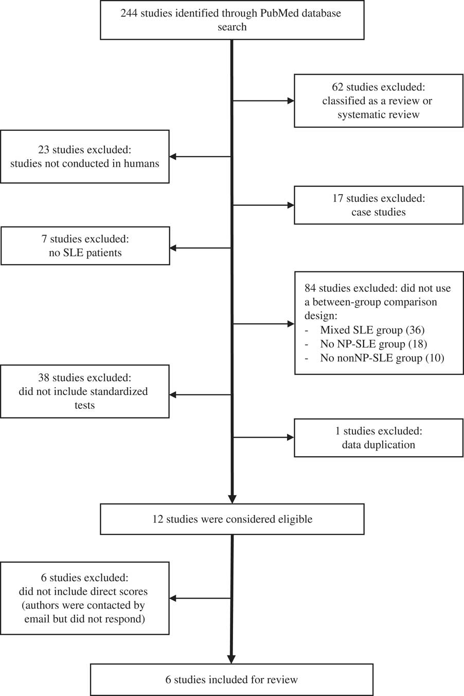
The literature search is shown in Figure 1. We searched the PubMed database for articles written in English that were published from April 1999 (when the ACR criteria for diagnosis for NP-SLE were published) to December 2016. The string used to search for articles was (TITLE-ABS-KEY [“cogn*” OR “neurocogn*”]) AND (TITLE-ABS-KEY [“lupus”]) AND (TITLE-ABS-KEY [“neuropsychiatr*]). A total of 244 documents were retrieved from this initial search.
Flow chart showing the literature search and article selection used by the authors to identify studies on cognitive function in NP-SLE and nonNP-SLE patients.

Two independent and blinded reviewers assessed the eligibility of studies to be included in the meta-analysis. Discrepancies were resolved via arbitration by a third reviewer to reduce selection bias in the literature search. We reviewed the titles and abstracts of all the articles and excluded reviews and meta-analyses, experiments not performed in humans, and single case studies, as well as studies that did not include patients with SLE and those that did not compare subgroups (NP-SLE and nonNP-SLE). We included studies that followed the criteria proposed by the ACR both in the establishment of the diagnoses (NP-SLE vs. nonNP-SLE) and in the selection of the cognitive measures (ACR, 1999). Of the 244 studies retrieved from the search, we excluded 232 and considered 12 eligible for analysis. Since six of these studies did not use a direct score for cognitive measures (data not available), the remaining six were finally included in the meta-analysis.
Neuropsychological Variables
As we found considerable heterogeneity in the selection of tests and batteries for neuropsychological assessment used across the studies, we selected those tests included in the cognitive battery proposed by the ACR for the identification of SLE patients with cognitive impairment (ACR, 1999; Kozora et al., Reference Kozora, Ellison and West2004).
Cognitive tests included
The Revised Wechsler Adult Intelligence Scale (WAIS-R) Digit Symbol Substitution Test (DSST) (Wechsler, Reference Wechsler1981). This task requires psychomotor speed, concentration, and graphomotor abilities and is used to evaluate visuomotor coordination and processing speed. It consists of nine digit-symbol pairs. The subject must draw the symbol corresponding to each number shown in 90 s. The score is the number of correct items completed.
The Trail Making Test (TMT) (Reitan & Wolfson, Reference Reitan and Wolfson1988) includes two tasks. The TMT-A is used to measure simple attention. The subject must use a pencil to join the numbers 1 to 25 in ascending order and as fast as possible. The TMT-B measures psychomotor speed, attention, and cognitive sequencing. The subject must join alternate numbers and letters (sequence altered) in an ascending pattern (1-A-2-B-3, etc.). The score is the time taken to complete each task.
The Stroop Color and Word Test (STROOP) (Golden, Reference Golden1978) measures complex attention, cognitive flexibility and inhibition, information processing, and shifting of set by naming color print for words written in different colors. It comprises three tasks, each of which the subject must complete in 45 s. The three tasks are Stroop-W (reading words), Stroop-C (naming colors), and Stroop-WC (reading words of colors printed in an incongruent color). The combination of these scores makes it possible to calculate an interference score (Stroop-I), which is associated with the individual’s cognitive flexibility. The score used in the studies included in the present meta-analysis is the Stroop-I score.
The California Verbal Learning Test (CVLT) (Delis, Kramer, Kaplan, & Oper, Reference Delis, Kramer, Kaplan and Oper1987) provides information about several components of verbal memory and learning. In this meta-analysis, the learning trial (CVLT-1-5) and short delayed free recall scores were included. Other verbal memory indexes (e.g., long delayed free recall) were not included due to lack of data available in the literature for meta-analytic purposes (minimum requirement: data from two independent studies).
The immediate and 30-min delayed-recall measures of the Rey-Osterrieth Complex Figure test (ROCF) (Corwin & Bylsma, Reference Corwin and Bylsma1993) provide data on visual perception, graphic visuoconstructive ability, visual learning and recall, and organizational and planning ability. The test requires the subject to reproduce a complex geometrical figure, first as a copy of the original model, and then 30 min later from memory.
The Controlled Oral Word Association Test (COWAT) and the Animal Naming Test (Borkowski, Benton, & Spreen, Reference Borkowski, Benton and Spreen1967) were selected as measures of phonetic and semantic verbal fluency. Subjects are asked to generate as many words as possible beginning with a specified letter (F, A, or S) (phonetic fluency), and/or belonging to the same category (e.g., names of animals) (semantic fluency), within 60 s for each task.
Data Processing and Analysis
For each study, we identified and recorded the total number of patients with NP-SLE and nonNP-SLE and the mean (SD) score for the groups in each neuropsychological test. We found that almost all studies provided the raw scores, with the exception of the study by Kozora et al. (Reference Kozora, Ellison and West2004), which provided T-scores. The inclusion of a different metric in our analysis is not a major problem as we used the standardized mean difference (SMD) for the estimation of the effect sizes, assuming that lower raw scores also correspond to lower standardized scores (e.g., T-scores) on almost all the cognitive tests. However, the TMT (A and B) presents an exception, as there is a reversed interpretation for the raw scores (where longer time indicates poorer performance) compared to the standardized scores (where higher T-scores indicate better performance, i.e., finishing the test in less time). For this reason, the TMT (A and B) scores of the study by Kozora et al. (Reference Kozora, Ellison and West2004) have been reversed, so that higher scores indicate poorer performance.
The meta-analysis was performed using R v.i386 3.0.3 (meta library), with the null hypothesis being the absence of differences in cognitive performance between the two subgroups. Both fixed and random effects models were used to combine effect sizes to better ascertain heterogeneity between studies. However, only results for the random effects model were used for interpretation. Beyond the agreement between the fixed and random effects models, heterogeneity was assessed using the I2 statistic. Heterogeneity was assessed using the I2 statistic. Values for I2>60% were considered important for between-study heterogeneity. Significant heterogeneity suggests a non-optimal quality of comparison of data among studies. As mentioned, effect size was calculated using the standardized mean difference (SMD) and 95% confidence intervals (CI) of each test in the meta-analysis. Forest plots were drawn to represent the study results graphically. All the p values were two-tailed, and statistical significance was set at p<.05.
RESULTS
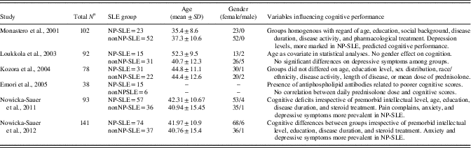
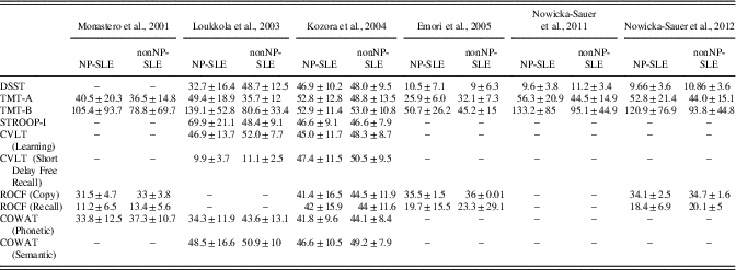
Our search identified 244 articles, of which six met the inclusion criteria and were incorporated in the final analysis. The final cohort comprised 399 SLE patients. Of these, 215 (53.88%) were classified as NP-SLE, and 184 (46.11%) as nonNP-SLE, with an NP-SLE:nonNP-SLE ratio of 1.2:1. The average age range of the participants was 35.4–52.3 years (NP-SLE, 35.4–52.3 years; nonNP-SLE, 37.3–44.4 years). Women were over-represented (n=356 [94%] vs. n=22 [6%]), with a women:men ratio of 16:1 (NP-SLE, n=187 [93.5%] vs. n=13 [6.5%], ratio women:men 14:1; nonNP-SLE, n=169 [95%] vs. n=9 [5%] of men, ratio women:men 19:1) (Table 1). Table 2 shows the cognitive test scores (mean ± SD) obtained by the NP-SLE and nonNP-SLE subgroups in each individual study included in the meta-analysis.
Studies included in the meta-analysis of cognitive impairment in NP-SLE patients in comparison with nonNP-SLE patients

a Some studies provided results from non-SLE subjects, such as healthy controls or another clinical population. These data were not included in the meta-analysis.
NP-SLE=neuropsychiatric systemic lupus erythematosus; nonNP-SLE=non-neuropsychiatric systemic lupus erythematosus; --=No data available.
Cognitive tests scores reported by each study for NP-SLE and nonNP-SLE patients

NP-SLE=neuropsychiatric systemic lupus erythematosus; nonNP-SLE=non-neuropsychiatric systemic lupus erythematosus; DSST=Digit Symbol Substitution Test; TMT=Trail Making Test; STROOP-I=Stroop Color and Word Test-Interference; CVLT=California Verbal Learning Test- Learning (trials 1–5) and Short Delay Free Recall; ROCF=Rey-Osterrieth Complex Figure; COWAT=Controlled Oral Word Association Test; --=no data available.
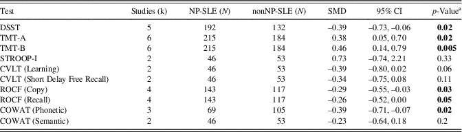
Statistically significant differences were observed in six of the 10 variables included in the meta-analysis (Table 3): DSST, TMT-A, TMT-B, the mean scores of both copy and recall tasks of the ROCF, and the phonetic task of COWAT. In all those variables, NP-SLE patients showed poorer performance and greater cognitive impairment than nonNP-SLE patients.
Results of the meta-analyses according to the cognitive test administered

a Data given in boldface type are significant.
NP-SLE=neuropsychiatric systemic lupus erythematosus; nonNP-SLE=non-neuropsychiatric systemic lupus erythematosus; DSST=Digit Symbol Substitution Test; TMT=Trail Making Test; STROOP-I=Stroop Color and Word Test-Interference; CVLT=California Verbal Learning Test- Learning (trials 1–5) and Short Delay Free Recall; ROCF=Rey-Osterrieth Complex Figure; COWAT=Controlled Oral Word Association Test; SMD=standardized mean difference.
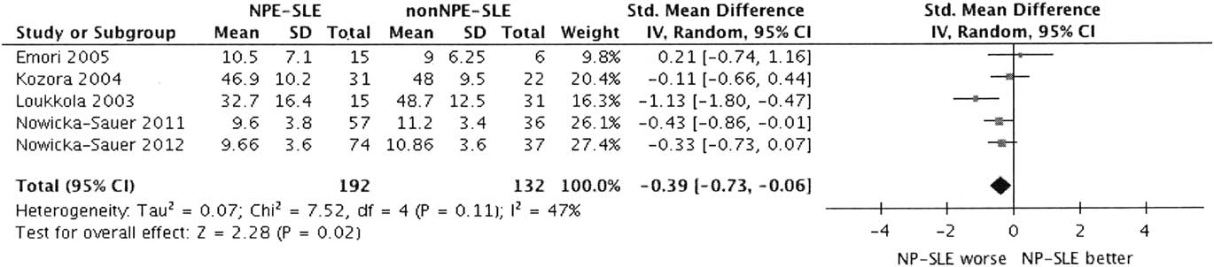
Data from 324 patients were analyzed for the Digit Symbol Substitution Test (DSST) variable. The NP-SLE group had significantly poorer performance than the nonNP-SLE group. The I2 value did not indicate statistically significant heterogeneity (I2=46.8%; p=.1107) (Figure 2).
Forest plot of the Digit Symbol Substitution.

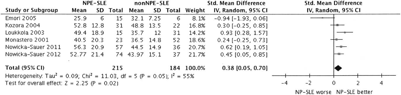
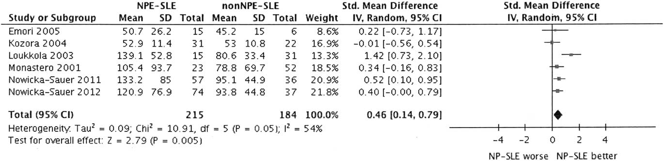
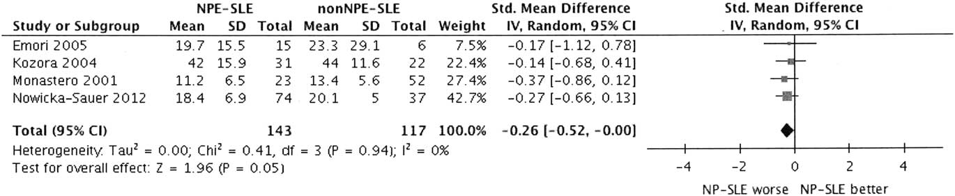
All six studies included in the meta-analysis used the TMT, and, therefore, data from all 399 patients were analyzed for both TMT-A and TMT-B. Statistically significant differences were found between the groups for both TMT-A and TMT-B (Figures 3 and 4). Patients with NP-SLE showed a significant poorer performance than the nonNP-SLE group in both parts of the test. However, data showed statistically significant and moderate heterogeneity for the two parts: TMT-A (I2=55%; p<.05) (Figure 3), TMT-B (I2=54%, p=.05) (Figure 4).
Forest plot of the Trail Making Test-A.

Forest plot of the Trail Making Test-B.

Two studies reported data from the STROOP-I and CVLT and included 99 patients. No statistically significant differences were found between these groups in their performance in the STROOP-I (Figure 5) or in the learning score (Figure 6) or in short-term free recall score (Figure 7) of the CVLT. The I2 value indicated statistically significant and a high heterogeneity for STROOP-I (I2=91%; p<.001) (Figure 5). No heterogeneity was found for the CVLT data in the learning scores (I2=0%; p=.6493) (Figure 6) or in short-term free recall scores (I2=0%; p=.7825) (Figure 7).
Forest plot of the Stroup Color Word Test-Interference.

Forest plot of the California Verbal Learning Test-Learning (trials 155).

Forest plot of the California Verbal Learning Test-Short Delay Free Recall.

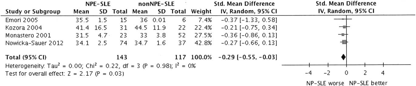
Data from 260 patients were analyzed for the ROCF. Patients in the NP-SLE group had significantly lower scores, indicating poorer performance and greater cognitive impairment, both in copy (Figure 8) and recall (Figure 9). The I2 values did not indicate statistically significant heterogeneity of data.
Forest plot of the Rey-Osterrieth Complex Figure Test- Copy.

Forest plot of the Rey-Osterrieth Complex Figure Test- Recall.

Phonetic task data from COWAT were analyzed for 174 patients. Patients in the NP-SLE group had a significantly lower mean score, suggesting poorer performance for phonetic fluency. The I2 value did not indicate statistically significant heterogeneity (I2=0%; p=.4995) (Figure 10).
Forest plot of the Controlled Oral Word Association Test- Phonetic.

Data from 99 patients who performed the semantic fluency task were analyzed. No statistically significant differences were found for this variable, and data did not show significant heterogeneity (I2=0%; p=.8491) (Figure 11).
Forest plot of the Controlled Oral Word Association Test- Semantic.

DISCUSSION
This is the first systematic review and meta-analysis to compare the cognitive performance of NP-SLE patients with that of nonNP-SLE patients. Cognitive impairment is one of the most prevalent neuropsychiatric presentations in SLE patients (Benedict et al., Reference Benedict, Shucard, Zivadinov and Shucard2008; Huerta et al., Reference Huerta, Gibson, Rey, Huerta and Huerta2015), and although it is well established that cognitive deficits are more severe and extensive in NP-SLE patients than in nonNP-SLE patients (Carlomagno et al., Reference Carlomagno, Migliaresi, Ambrosone, Sannino, Sanges and Iorio2000; Mikdashi et al., Reference Mikdashi, Esdaile, Alarcón, Crofford, Fessler, Shanberg and Schneider2007; Kozora et al., Reference Kozora, Arciniegas, Filley, West, Brown and . . . Zhang2008), a specific profile of cognitive deficits affecting this population has not been defined. The literature on this topic is not extensive, and results are inconclusive owing to important methodological limitations across studies, such as failure to clearly describe the criteria used to define patient groups and a lack of explanation of the specific cognitive measures used in some studies.
In the present systematic review and meta-analysis, we only included studies that based diagnosis of NP-SLE and selection of cognitive tests on the ACR consensus criteria. Although both inclusion criteria are demanding and restrictive and may exclude studies that might provide relevant information on the cognitive profiles of SLE groups, we considered them necessary to overcome the methodological limitations detected in the literature on this topic.
The results of this meta-analysis indicate that the differences between NP-SLE and nonNP-SLE patients were statistically significant for more than half of the cognitive variables used, namely, DSST, TMT-A, TMT-B, ROCF (copy and recall), and COWAT (phonetic). Three of the variables included in meta-analysis, that is, TMT-A, TMT-B, and STROOP-I, revealed statistically significant moderate heterogeneity for the sample.
As expected, and consistent with findings from previous studies (Emori et al., Reference Emori, Matsushima, Aihara, Ohta, Koike, Miyasaka and Kato2005; Kozora et al., Reference Kozora, Ellison and West2004; Loukkola et al., 2003; Monastero et al., Reference Monastero, Bettini, Del Zotto, Cottini, Tincani, Balestrieri and Padovani2001; Nowicka-Sauer, Czuszynska, Smolenska, & Siebert, Reference Nowicka-Sauer, Czuszynska, Smolenska and Siebert2011; Nowicka-Sauer et al., Reference Nowicka-Sauer, Czuszyńska, Majkowicz, Smoleńska, Jarmoszewicz, Olesińska and Siebert2012), NP-SLE patients consistently showed poorer cognitive performance and greater cognitive impairment than nonNP-SLE patients, at least in those variables that showed statistically significant differences, that is, visuomotor coordination, attention, executive function, visual coordination, visual learning and memory, and verbal fluency.
The DSST measures visuomotor coordination and processing speed. NP-SLE patients had a significantly lower mean score than nonNP-SLE patients, and between-group heterogeneity was not significant. Of the five studies in the meta-analysis, three revealed significant differences between the groups (Kozora et al., Reference Kozora, Ellison and West2004; Loukkola et al., 2003; Nowicka-Sauer et al., Reference Nowicka-Sauer, Czuszynska, Smolenska and Siebert2011), and two revealed no statistically significant differences (Emori et al., Reference Emori, Matsushima, Aihara, Ohta, Koike, Miyasaka and Kato2005; Nowicka-Sauer et al., Reference Nowicka-Sauer, Czuszyńska, Majkowicz, Smoleńska, Jarmoszewicz, Olesińska and Siebert2012).
The TMT measures simple attention (TMT-A) and executive function (TMT-B). As expected, NP-SLE patients showed a significantly poorer performance on both TMT-A and B. However, although in the lower limit of the moderate heterogeneity, both parts of the TMT showed a statistically significant heterogeneity (TMT-A I2=55%; p<.05; TMT-B I2=54%; p=.05). It has to be noted that patients from the study of Emori et al. (Reference Emori, Matsushima, Aihara, Ohta, Koike, Miyasaka and Kato2005) appeared to complete both TMT-A and TMT-B much more quickly than the rest of samples, and that this study might be largely responsible for the heterogeneity found. Although socio-demographic data for the individual subsamples are not provided (NP-SLE and nonNP-SLE patients), data for the general sample of the SLE patients do not appear to significantly differ from the other samples as to justify the apparent higher functioning on this test (age 35.1 ± 10.7; female/male=20/1).
NP-SLE patients showed poorer cognitive performance than nonNP-SLE patients on the ROCF test, which assesses visual perception, visuoconstructive ability, visual memory, and planning and organizational ability. NP-SLE patients demonstrated a significantly lower mean score for both copy and recall, and heterogeneity between the groups was not significant. Significant findings were obtained for two of four studies that included ROCF-copy (Monastero et al., Reference Monastero, Bettini, Del Zotto, Cottini, Tincani, Balestrieri and Padovani2001; Nowicka-Sauer et al., Reference Nowicka-Sauer, Czuszyńska, Majkowicz, Smoleńska, Jarmoszewicz, Olesińska and Siebert2012) and for only one of the four studies that included ROCF-recall (Monastero et al., Reference Monastero, Bettini, Del Zotto, Cottini, Tincani, Balestrieri and Padovani2001).
In verbal fluency, NP-SLE patients performed more poorly than nonNP-SLE patients on the phonetic fluency task (COWAT-phonetic). In contrast, no statistically significant group differences were observed on the semantic fluency test (COWAT-semantic), although this result should be interpreted with caution, because only two studies were used in the analysis (Kozora et al., Reference Kozora, Ellison and West2004; Loukkola et al., 2003).
The CVLT makes it possible to obtain values for several components of verbal learning and memory. The analysis did not reveal statistically significant differences between subgroups in verbal learning (CVLT-1-5) or in short-term free recall. Nevertheless, these results should be interpreted with caution, since only two studies were included in the analysis (Loukkola et al., 2003; Kozora et al., Reference Kozora, Ellison and West2004). We were unable to assess long-term verbal memory, as data from at least two studies (minimum requirement for meta-analyses) were not available in the literature.
Finally, the STROOP test is used to measure cognitive flexibility, selective attention, cognitive inhibition, and information processing speed. According to the results, no statistically significant differences were found for this test between NP-SLE and nonNP-SLE patients, possibly because only two studies were used for the analysis (Kozora et al., Reference Kozora, Ellison and West2004; Loukkola et al., 2003) and the heterogeneity of the sample was very high (I2=91%; p<.001).
In general terms and according to the results obtained, it is well established that the profile of cognitive impairment is more pronounced in NP-SLE patients than in nonNP-SLE patients, especially in those cognitive domains that refer to visuomotor coordination, attention, executive function, visual learning and memory, and verbal fluency. Of note, all of the tasks that differentiated the NP-SLE group from the nonNP-SLE group required either a rapid written response (TMT-B, Digit Symbol, ROCF) or a rapid oral motor response (COWAT). Differences between groups may have been mediated by a non-cognitive factor, for example, rudimentary writing speed or oral motor speed. This hypothesis has been suggested in other diseases, such as multiple sclerosis (Arnett, Smith, Barwick, Benedict, & Ahlstrom, Reference Arnett, Smith, Barwick, Benedict and Ahlstrom2008).
On the other hand, although we did not find statistically significant differences between the groups in the cognitive domains of verbal learning and memory, and cognitive flexibility and inhibition, we must interpret our results with caution because of the low number of studies included in the analysis (and, therefore, the low number of participants) and the non-optimal quality of some of them, which was evident in the heterogeneity of some of our analyses. Variability in the management of confounding variables across studies is also evident. Although, as previously reported in the literature, some of the selected studies confirm that the more marked cognitive deficits found in NP-SLE patients are observed irrespective of disease duration (Monastero et al., Reference Monastero, Bettini, Del Zotto, Cottini, Tincani, Balestrieri and Padovani2001; Nowicka-Sauer et al., Reference Nowicka-Sauer, Czuszynska, Smolenska and Siebert2011), disease activity (Monastero et al., Reference Monastero, Bettini, Del Zotto, Cottini, Tincani, Balestrieri and Padovani2001), and pharmacological treatment (Emori et al., Reference Emori, Matsushima, Aihara, Ohta, Koike, Miyasaka and Kato2005; Monastero et al., Reference Monastero, Bettini, Del Zotto, Cottini, Tincani, Balestrieri and Padovani2001; Nowicka-Sauer et al., Reference Nowicka-Sauer, Czuszynska, Smolenska and Siebert2011), only two studies have directly analyzed and described the negative influence of the presence of variables such as depressive symptoms (Monastero et al., Reference Monastero, Bettini, Del Zotto, Cottini, Tincani, Balestrieri and Padovani2001) and antiphospholipid antibodies on cognitive performance.
For this reason, we do not have enough information to analyze the role of these variables in cognitive impairment among SLE patients either by subgroup analysis or meta-regression analysis; consequently, this lack of data is a limitation. Our meta-analysis is subject to additional limitations. For instance, some eligible studies were not included in the meta-analysis because the data on the cognitive variables were not available. We tried to contact the authors by email but received no response, thus preventing us from including their results and obliging us to assume a possible bias in the results obtained. Likewise, the quality of the studies included was not always optimal, and some of the samples were too small to obtain reliable results. However, two relevant issues could enhance the external validity of the results reported: (1) we used a random effects model to interpret the data, thus assuming not an equal effect, but an effect that could vary between studies; and (2) the data presented are from observational studies and are, therefore, more likely to have external validity than randomized clinical trials, which have stricter inclusion criteria.
Regardless of these limitations, the results of this systematic review and meta-analysis accurately define the specific pattern of cognitive deficits in the NP-SLE population, and since it is well established that cognitive impairment has a marked impact on patient functionality, autonomy, and health-related quality of life, our findings may contribute to their early detection and enable us to design and facilitate access to specific and effective interventions for cognitive remediation (Butt et al., Reference Butt, Farman, Khan, Saeed and Ahmad2017). Additionally, this meta-analysis clearly shows the lack of quality data on specific cognitive deficits in NP-SLE patients and associations with other relevant variables such as antiphospholipid antibodies and comorbid psychiatric disorders, mainly depression.
Comparative analysis of the cognitive pattern of NP-SLE and nonNP-SLE patients remains an important issue and can help to explain the etiology and specificity of cognitive dysfunction in SLE. More research is needed to confirm and complete the findings of this meta-analysis. Further studies should include validated criteria for the definition of the clinical SLE patient subgroups and recommended neuropsychological tests for the assessment of cognitive deficits in this population. Neuropsychological assessment is crucial in SLE patients owing to the fact that cognitive decline may be a predictor of brain structural abnormalities and, as defended by some authors, should be a standard element of clinical judgment, care, and monitoring of disease course (Monov & Monova, Reference Monov and Monova2008; Nowicka-Sauer et al., Reference Nowicka-Sauer, Czuszynska, Smolenska and Siebert2011).
In addition, the neuropsychological assessment should be comprehensive and include the assessment of the patient’s emotional status to clarify the role of depression in SLE and its relationship with cognitive functioning. Future studies should also consider the presence of antiphospholipid antibodies as a potential mediator of cognitive performance and shed more light on the nature of cognitive impairment in SLE patients.
ACKNOWLEDGMENTS
We thank the Health Department of the Basque Government (Health Research Fund 2016111021). All the authors of this study certify that they have no affiliations with or involvement in any organization or entity with any financial interest or non-financial interest in the subject matter or materials discussed in this manuscript.