Introduction
Pathologic cyanosis can occur at any time during staged single-ventricle palliation, causing significant morbidity and mortality. Reference Schafstedde, Nordmeyer and Schleiger1 Specifically, following stage 2 (superior cavo-pulmonary connection) and stage 3 (total cavo-pulmonary connection) palliations, cyanosis can occur because of low pulmonary blood flow, baffle leaks, lung disease, veno-venous collaterals, and/or arteriovenous malformations. Reference Mazza, Gribaudo and Agnoletti2
Veno-venous collateral formation leading to cyanosis can be observed any time after stage 2 palliation as the superior vena cava and pulmonary artery pressures are greater than pulmonary vein and inferior vena cava/cardiac pressure by a difference of the transpulmonary gradient. Reference McElhinney, Reddy, Hanley and Moore3 These “pop-off” veno-venous collaterals commonly originate from the jugular, vertebral, subclavian, and innominate veins and course to pulmonary veins and other thoracic and abdominal systemic veins. Reference McElhinney, Reddy, Hanley and Moore3,Reference Lluri, Levi and Aboulhosn4 It is hypothesised that elevated superior vena cava pressure promotes recanalisation of embryologic structures that connected the foetal pulmonary and systemic veins, creating veno-venous collaterals; however, de novo angiogenesis is also still possible. Reference Lluri, Levi and Aboulhosn4 Studies have shown that 31–32% of patients after stage 2 palliation have veno-venous collaterals Reference McElhinney, Reddy, Hanley and Moore3,Reference Magee, McCrindle, Mawson, Benson, Williams and Freedom5 with 47–58% of the veno-venous collaterals ultimately involving the hemiazygos or azygos. Reference McElhinney, Reddy, Hanley and Moore3,Reference Magee, McCrindle, Mawson, Benson, Williams and Freedom5 While the azygos vein is typically ligated during stage 2 palliation, Reference Khajali, Firouzi and Pashapour6 on occasion, patients will have multiple feeder vessels to the para-vertebral veins, which ultimately collect to a reconstituted azygos/accessory hemi-azygos and course below the diaphragm. Reference Schubert, Podrabsky and Berger7 As veno-venous collaterals leading to cyanosis are typically managed with transcatheter occlusion techniques, Reference Masura, Borodacova and Tittel8,Reference Abdel-Aziz, Tanase and Ewert9 situations where all the feeder vessels cannot be accessed are challenging to manage. Veno-venous collaterals following stage 3 palliation also occur in ∼58% of patients, and typically connect with pulmonary veins when presenting with cyanosis. Reference Lluri, Levi and Aboulhosn4
Arteriovenous malformations can form following stage 2 or 3 palliation and cause significant cyanosis-associated morbidity. Reference Dori, Sathanandam and Glatz10 A study showed that 43% of stage 2 and stage 3 patients with heterotaxy and inferior vena cava interruption had arteriovenous malformations, while 8% had portosystemic shunts. Reference McElhinney, Marx and Newburger11 It is postulated that arteriovenous malformations form due to lack of “hepatic factor” being delivered to the lungs, Reference Dori, Sathanandam and Glatz10 putting any anatomy that fails to deliver portal vein to hepatic vein flow to the lungs at risk. This includes patients following stage 2 palliation and polysplenic patients with interrupted inferior vena cava with azygous continuation following stage 3 palliation. Reference Dori, Sathanandam and Glatz10 This azygos-superior vena cava connection is frequently contralateral to the conduit receiving hepatic venous return, thus making it anatomically suboptimal for promoting hepatic venous return to both lungs. Reference Dori, Sathanandam and Glatz10 Heterotaxy patients are also at increased risk of abnormal portal vein connections due to their preponderance of systemic venous abnormalities. Reference McElhinney, Marx and Newburger11
The work-up for cyanosis typically consists of cardiac catheterisation with or without non-invasive axial imaging such as MRI or CT angiography. Reference Stout, Daniels and Aboulhosn12 However, the investigation and subsequent angiographic imaging are usually isolated to the thoracic cavity. In this retrospective case series, we highlight veno-venous collaterals and arteriovenous malformations as causes of cyanosis in patients with single-ventricle physiology after stage 2 or 3 palliation that require diagnosis and management with imaging below the diaphragm.
Materials and method
This study is a retrospective, single-centre case series of six patients approved by the Institutional Review Board of The Children’s Hospital of Philadelphia (Institutional Review Board 24-022836). The Institutional Review Board granted a waiver of consent given the retrospective study design.
Results
Patient 1 – stage 2: left superior vena cava to reconstituted azygos (Figure 1, Movie 1)
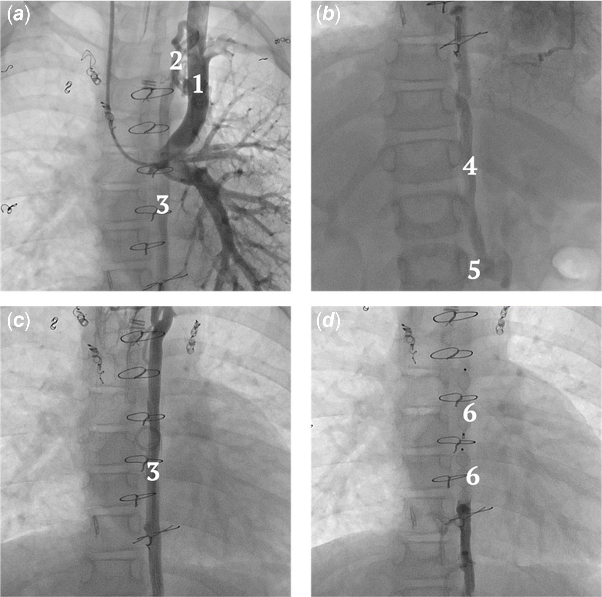
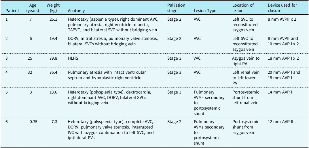
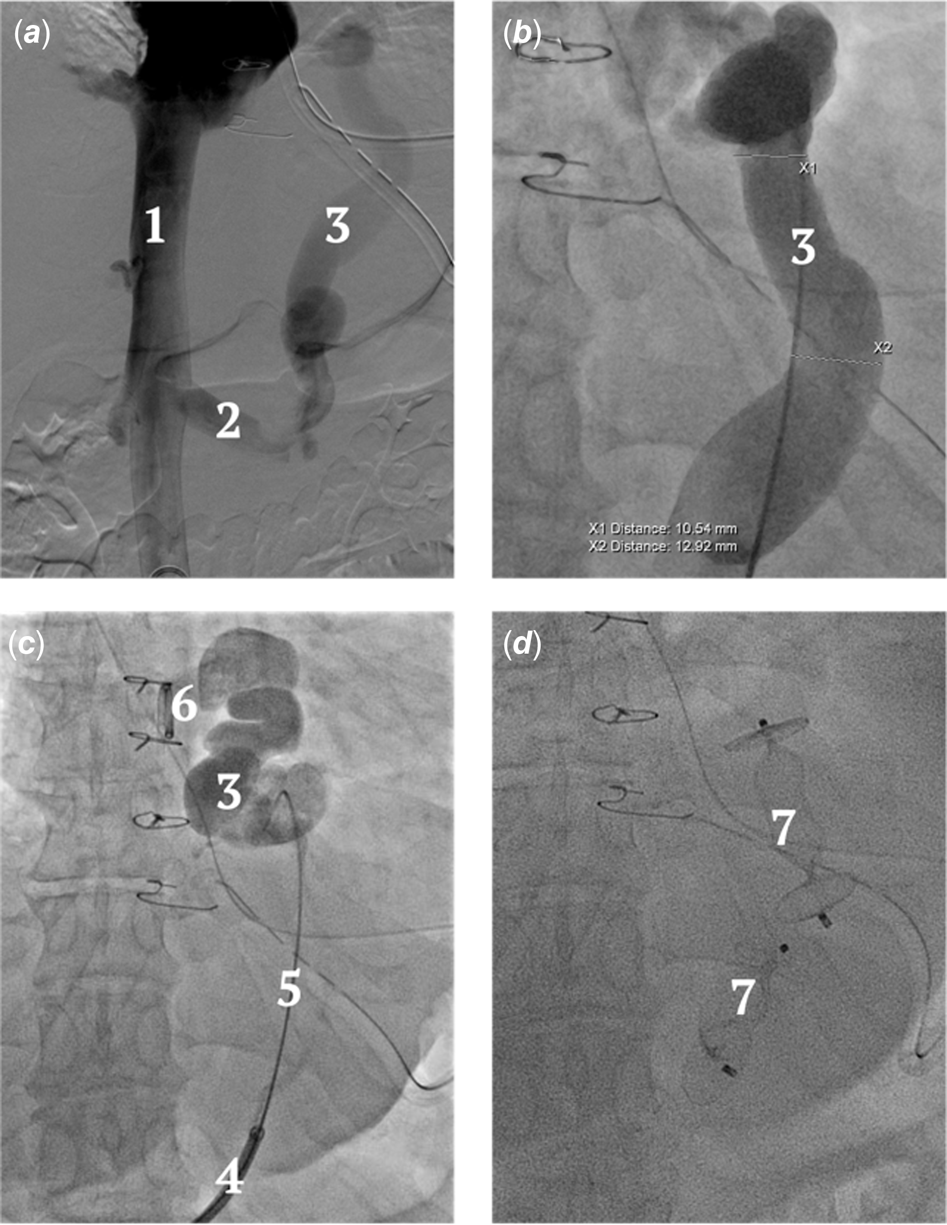
Patient 1 was a seven-year-old girl born with heterotaxy (asplenia type), right dominant atrioventricular canal, pulmonary atresia, right ventricle to aorta, total anomalous pulmonary venous connection, and bilateral superior vena cava without bridging vein who failed stage 3 palliation twice with conversion back to stage 2 (Table 1). Her oxygen saturations were in the mid to high 70s with a haemoglobin of 19.1 g/dL and associated poor appetite and fatigue. She is listed 1b for heart transplantation. Cardiac catheterisation including left superior vena cava angiography with the camera panning to the abdomen demonstrated a network of collaterals arising from the left superior vena cava coursing to a reconstituted azygos with retrograde flow to the inferior vena cava. Given the difficulty in accessing the feeder vessels from the right internal jugular vein venotomy, the azygos vein was accessed from below. A Berenstein glide catheter (Terumo, Elkton, MD, USA) was used to direct a hydrophilic 0.035’’ wire into the azygos vein, over which the catheter was advanced into the thorax and exchanged for a long sheath, through which two 8 mm Amplatzer Vascular Plug II (Abbott Cardiovascular, Green Oaks, IL, USA) were deployed. The patient has been well and at most recent follow-up three months post procedure, her oxygen saturation was 82% on no supplemental oxygen.
Patient 1 – Stage 2: Left SVC to reconstituted azygos. ( a ) Power injection in the left SVC demonstrating multiple VVCs entering a reconstituted azygos. ( b ) Reconstituted azygos draining to left sided IVC. ( c ) Thoracic azygos. ( d ) Status post azygos occlusion with two AVP II devices. 1. Left SVC. 2. VVCs. 3. Reconstituted azygos. 4. Azygos. 5. Left renal vein. 6. Vascular plugs.

Overview of patient and procedural characteristics

Patient 2 – stage 2: left superior vena cava to reconstituted azygos (Figure 2, Movie 2, 3)
Patient 2 was a six-year-old girl born with mitral atresia, double outlet right ventricle, pulmonary valve stenosis, and bilateral superior venae cavae without bridging vein who underwent main pulmonary artery banding and subsequent bilateral bidirectional Glenn. She had an elevated transpulmonary gradient treated with vasodilators and cyanosis, partially due to right-sided veno-venous collaterals for which coil occlusion had been attempted. At age six, she underwent cardiac MRI and cardiac catheterisation to assess candidacy for stage 3. She was cyanotic with an oxygen saturation of 78% and a haemoglobin of 18.4 g/dL. Haemodynamics were unfavourable for a stage 3 palliation. She was found to have a network of numerous small veins arising from the left superior vena cava and coalescing into a reconstituted azygos vein via multiple paravertebral veins. Left superior vena cava angiography with panning to the abdomen demonstrated flow reversal in the azygos vein into the left renal vein. The azygos was accessed with a glide catheter and occluded by placing an 8 mm Amplatzer Vascular Plug II and two 10 mm Amplatzer Vascular Plug II devices through a 5 French long sheath in the thorax. Her oxygen saturation improved to 81% at discharge on no supplemental oxygen.
Patient 2 – Stage 2: Left SVC to reconstituted azygos. ( a ) Power injection in the left SVC demonstrating multiple VVCs entering a reconstituted azygos. ( b ) Glide catheter entering the azygos from the left renal vein. ( c ) Thoracic azygos. ( d ) Status post azygos occlusion with three AVP II devices. 1. Pigtail injecting in left SVC. 2. VVCs. 3. Reconstituted azygos. 4. Glide catheter. 5. Left renal vein. 6. Azygos. 7. Vascular plugs.

Patient 3 - stage 3: right azygos to right upper pulmonary vein (Figure 3, Movie 4)
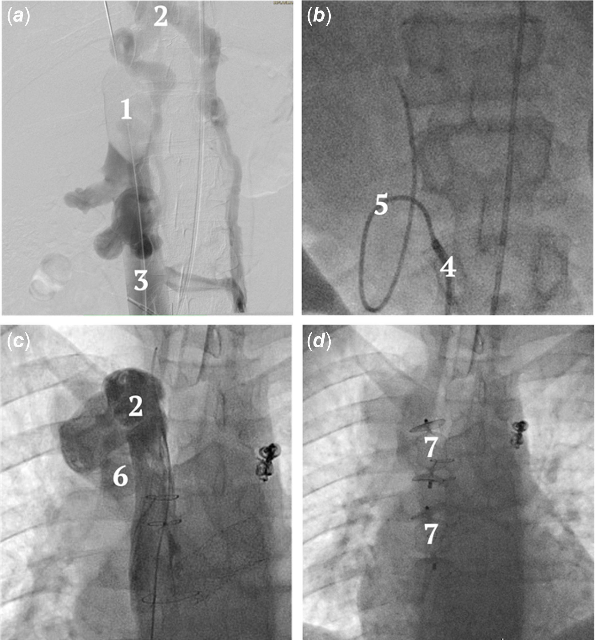
Patient 3 was a 25-year-old man born with hypoplastic left heart syndrome who underwent staged palliation with a Norwood, hemi Fontan, and a lateral tunnel Fontan. At 18 years of age, he underwent coil occlusion of a veno-venous collateral connecting the innominate vein to the left upper pulmonary vein to address cyanosis with modest improvement; oxygen saturations increased from 85 to 87%. Seven years later, he underwent a cardiac MRI for persistent cyanosis (85% by pulse oximetry with a haemoglobin of 18.2 g/dL), which demonstrated a network of collaterals arising from the right internal jugular vein, paravertebral veins, and reconstituted azygos vein to the right pulmonary veins. Cardiac catheterisation including infrarenal inferior vena cava digital subtraction angiography (while occluding the hepatic inferior vena cava with a 25 mm sizing balloon) demonstrated a large veno-venous collateral originating from the azygos vein to the right upper pulmonary vein with additional small feeder vessels from the right superior vena cava. The tortuous azygos vein was accessed from the inferior vena cava using a steerable sheath and glide catheter. An 8 French long sheath was advanced to deploy two 18 mm Amplatzer Vascular Plug II devices. Stent angioplasty for left pulmonary artery stenosis was also performed. Oxygen saturations improved from 82% at admission to 90% at time of discharge with no supplemental oxygen.
Patient 3 – Stage 3: Right azygos to right upper PV. ( a ) Digital subtraction angiography of the infrarenal IVC with balloon occlusion of the hepatic IVC demonstrating collaterals from the left renal vein and IVC connecting to the azygos. ( b ) Glide catheter through a steerable sheath entering the tortuous IVC collateral. ( c ) Large thoracic azygos with connection to the right upper PV. ( d ) Status post azygos occlusion with two AVP II devices. 1. Sizing balloon. 2. Collateral. 3. IVC. 4. Steerable sheath. 5. Glide catheter. 6. Right upper PV. 7. Vascular plugs.

Patient 4 – stage 3: left renal vein collateral to left lower pulmonary vein (Figure 4, Movie 5, 6)
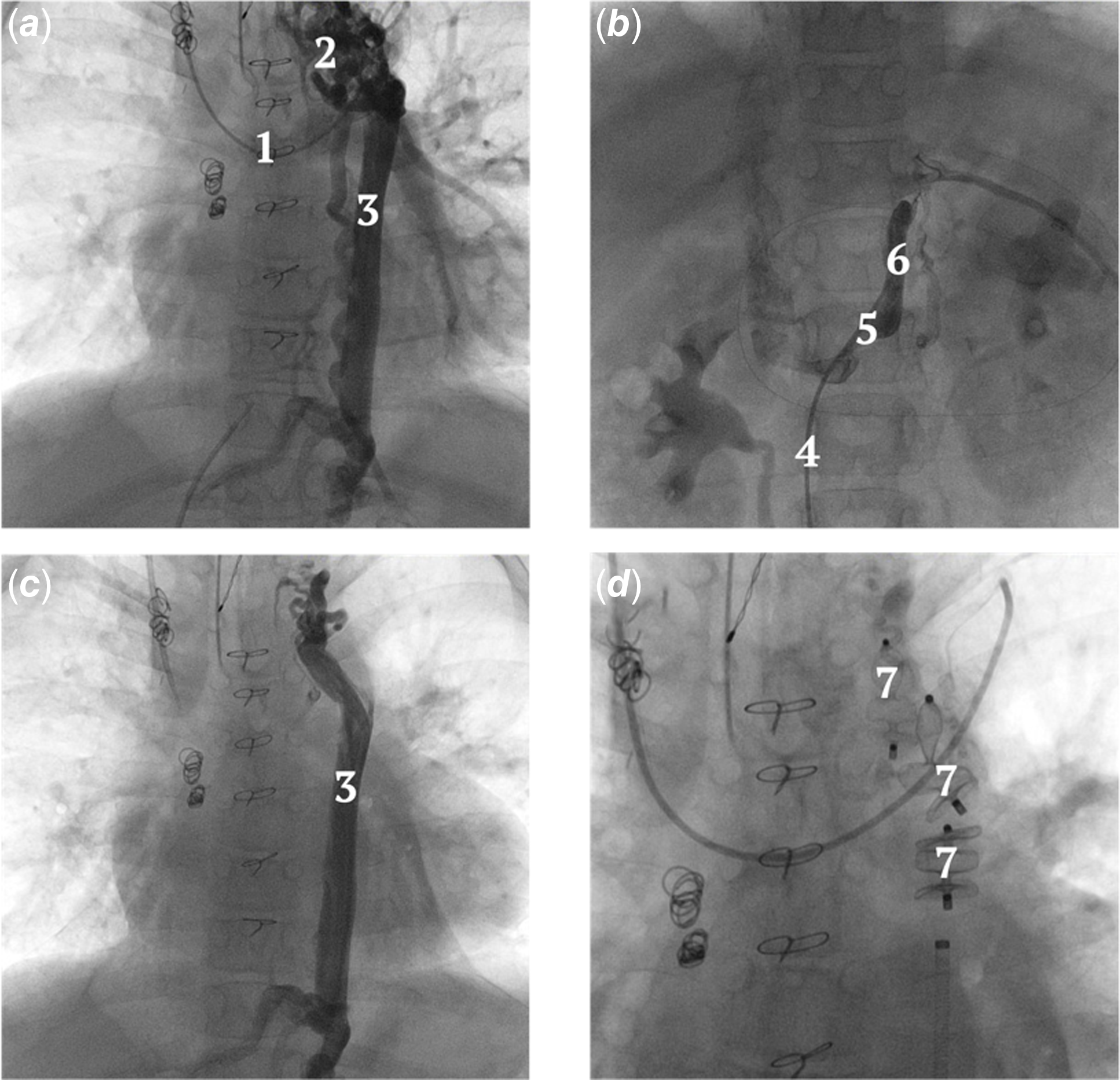
Patient 4 was a 31-year-old woman born with pulmonary atresia with intact ventricular septum and hypoplastic right ventricle who underwent staged palliation culminating in a lateral tunnel Fontan. She underwent a cardiac MRI as part of an initial workup for cyanosis (oxygen saturation of 84%, haemoglobin 14 g/dL) and was found to have a large veno-venous collateral entering the left lower pulmonary vein from a vessel originating below the diaphragm. Cardiac catheterisation demonstrated a dilated Fontan with complex baffle leak and a large tortuous veno-venous collateral arising from the left renal vein and connecting to the left lower pulmonary vein. The veno-venous collateral was profiled using a digital subtraction angiography power injection in the infrarenal inferior vena cava. The veno-venous collateral was accessed with a glide catheter and occluded with a 20 mm Amplatzer Vascular Plug II and an 18 mm Amplatzer Vascular Plug II device in series through a 9 French long sheath. Post-intervention oxygen saturation was 90% at time of discharge with no supplemental oxygen.
Patient 4 – Stage 3: Left renal vein collateral to left lower PV. ( a ) Digital subtraction angiography in the infrarenal IVC demonstrating a VVC arising from the left renal vein to the left lower PV. ( b )/( c ) Large tortuous collateral connecting to the left lower PV. ( d ) Status post collateral occlusion with two AVP II devices. 1. IVC. 2. Left renal vein. 3. VVC. 4. Long sheath. 5. 0.035’’ support wire. 6. Left lower PV. 7. Vascular plugs.

Patient 5 – Stage 3: portosystemic shunt from left renal vein causing bilateral pulmonary arteriovenous malformations (Figure 5, Movie 7)
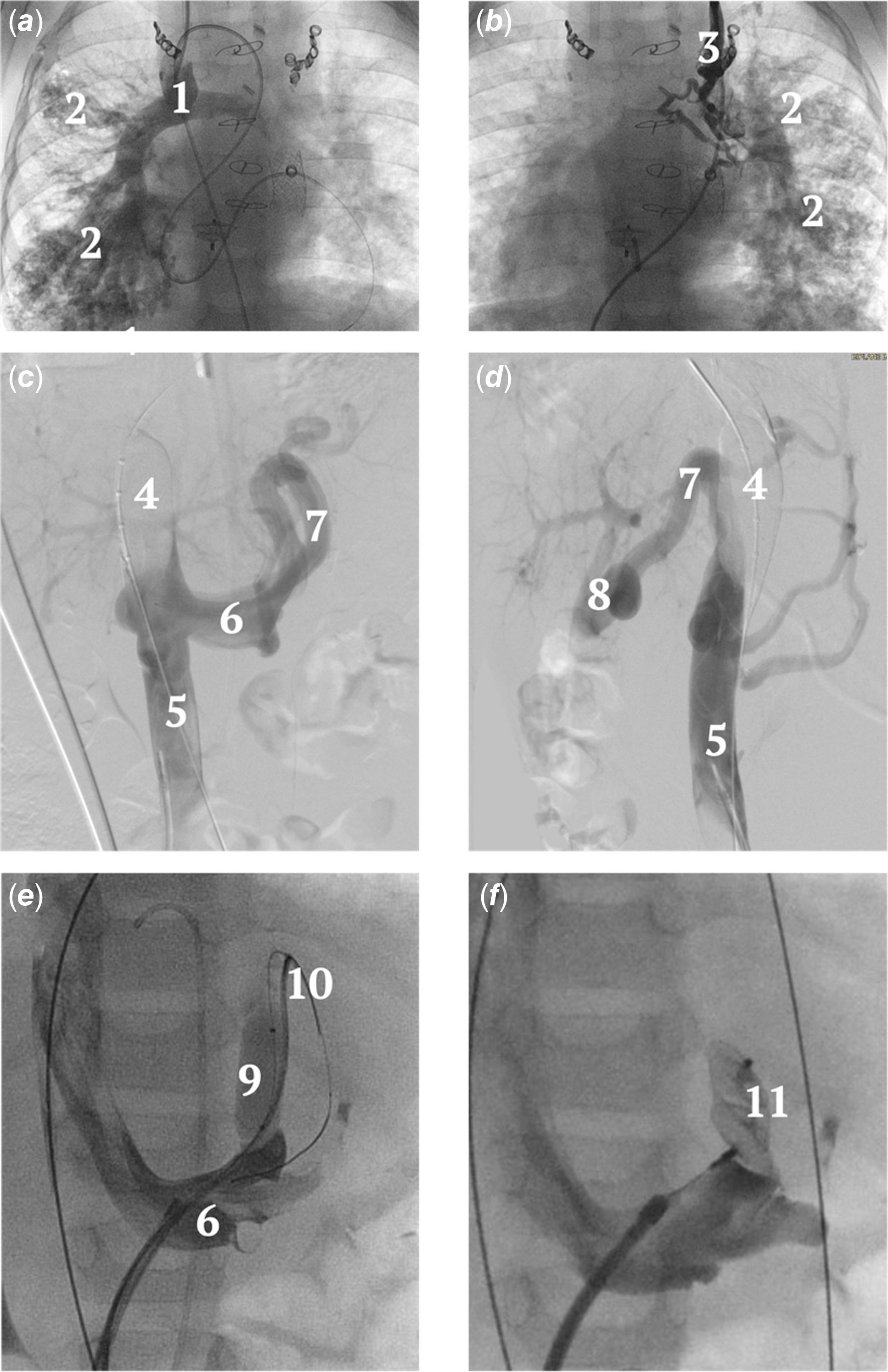
Patient 5 is a three-year-old boy born with heterotaxy polysplenia type, dextrocardia, right dominant atrioventricular canal, double outlet right ventricle, and bilateral superior venae cavae without bridging vein. He underwent staged palliation through bilateral bidirectional Glenn, which was complicated by cyanosis secondary to bilateral pulmonary arteriovenous malformations and was managed with an early stage 3 palliation at 18 months of age. Despite this, he developed progressive cyanosis secondary to persistent arteriovenous malformations and a Fontan baffle leak, which underwent device closure. He continued to have cyanosis with oxygen saturation in the high 70s, desaturations to the 50s with light exercise, and a haemoglobin of 21.4 g/dL. Cardiac catheterisation demonstrated severe microscopic arteriovenous malformation burden in bilateral lungs with a small residual baffle leak, and a small veno-venous collateral from the left superior vena cava to the left upper pulmonary vein. Infrarenal inferior vena cava digital subtraction angiography, while occluding the hepatic inferior vena cava with a 25 mm sizing balloon, demonstrated a large portosystemic shunt arising from the left renal vein. The portal system was well developed without hypoplasia. The shunt was test occluded with a 10 mm x 2 cm compliant balloon while monitoring the portal vein pressure, which increased from 16 to 18 mmHg. The portosystemic shunt was then occluded with a 14 mm Amplatzer Vascular Plug II device through an 8 French long sheath. Next, the left superior vena cava veno-venous collateral was coil occluded, and the residual baffle leak was occluded with a 14 mm covered Cheatham-Platinum stent (NuMED Inc., Hopkinton, NY, USA). Oxygen saturation at discharge was 73% on no supplemental oxygen and has thus far increased to 84% on 3-month follow-up on no supplemental oxygen.
Patient 5 – Stage 3: Portosystemic shunt causing bilateral pulmonary AVMs. ( a ) Power injection in the right SVC demonstrating diffuse AVMs. ( b ) Power injection in the left SVC demonstrating diffuse AVMs. ( c )/( d ) Digital subtraction angiography in the infrarenal IVC with balloon occlusion of the hepatic IVC demonstrating a portosystemic shunt between the portal vein and left renal vein. The portal system appears well developed. ( e ) Test occlusion of the portosystemic shunt with glide catheter distal to the compliant balloon for pressure assessment. ( f ) Unreleased AVP II in the portosystemic shunt at its connection to the left renal vein. 1. Right SVC. 2. AVMs. Left SVC. 4. Sizing balloon. 5. IVC. 6. Renal vein. 7. Portosystemic shunt. 8. Portal vein. 9. Compliant balloon. 10. Glide catheter in portosystemic shunt. 11. Vascular plug.

Patient 6 – Stage 2: portosystemic shunt from azygos causing bilateral pulmonary arteriovenous malformations (Figure 6)
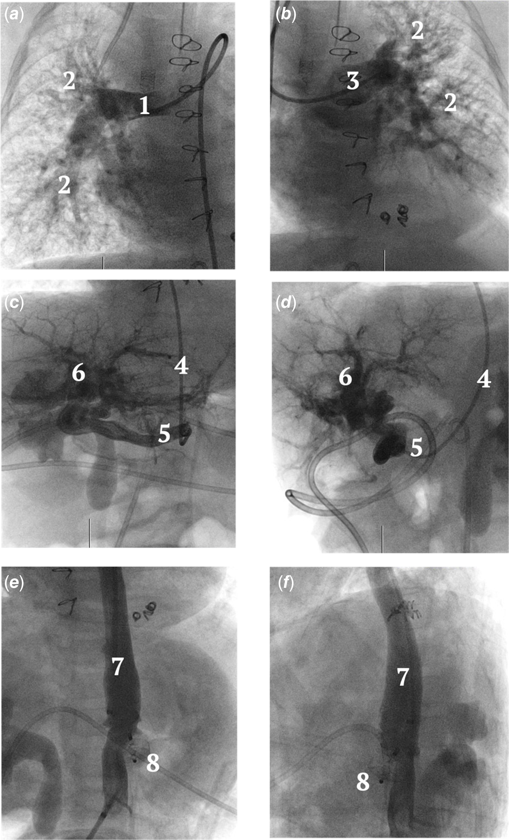
Patient 6 was a nine-month-old female with heterotaxy polysplenia type, complete atrioventricular canal, double outlet right ventricle, pulmonary valve stenosis, interrupted inferior vena cava with azygos continuation to left superior vena cava, and ipsilateral pulmonary veins. She was initially managed with a ductal stent, with a course complicated by persistent hypoxaemia despite adequate pulmonary blood flow calculated on serial cardiac MRI and catheterisations. At 6 months of age, she underwent a right bidirectional Glenn, left Kawashima, ligation and division of main pulmonary artery, ligation of patent ductus arteriosus, and central pulmonary artery plasty. Catheterisation for cyanosis (70’s on 0.50 FiO2) at 9 months of age demonstrated bilateral microscopic pulmonary arteriovenous malformations secondary to a portosystemic shunt between the azygos and portal systemic system. Eight days later she underwent device occlusion of the portosystemic shunt with a 12 mm Amplatzer Vascular Plug II followed by surgical placement of an 8 mm right ventricle to pulmonary artery conduit to secure hepatic factor to the lungs. Over the next two months, saturations improved to 94% on room air. Sadly, the patient was found unresponsive at home and ultimately died at 11 months of age.
Patient 6 – Stage 2: Portosystemic shunt causing bilateral pulmonary AVMs. ( a ) Power injection in the right PA demonstrating diffuse AVMs. ( b ) Power injection in the left PA demonstrating diffuse AVMs. ( c )/( d ) Anterior-posterior and lateral images of a glide catheter in the azygos vein injecting into the portosystemic shunt. The portal system appears well developed. ( e )/( f ) Status post portosystemic shut occlusion with an AVPII device. 1. Right PA. 2. AVMs. 3. Left PA. 4. Glide catheter in azygos vein. 5. Portosystemic shunt. 6. Portal system. 7. Azygos vein. 8. Vascular plug.

Discussion
We present six patients with single-ventricle physiology presenting with cyanosis after stage 2 or 3 palliation who required imaging below the diaphragm for successful diagnosis and management. Traditionally, the workup for cyanosis in this patient population may be limited to the thoracic cavity with non-invasive thoracic axial imaging or cardiac angiography in the superior vena cava and hepatic inferior vena cava only. These cases demonstrate the need for a high index of suspicion for abdominal venous connections that could be contributing to cyanosis. Three groups of patients were identified.
The first group (patients 1 and 2) encompasses stage 2 patients who were ineligible for stage 3 palliation. These patients were found to have multiple feeder vessels connecting to a reconstituted azygos with retrograde flow to below the diaphragm. Accessing numerous small and typically posterior channels for occlusion from above (e.g. internal jugular or subclavian vein access) may be challenging. Even if successful, the efforts may be inadequate as more channels can open over time, particularly if the paravertebral veins are involved. Thus, closure of the reconstituted azygos within the thoracic cavity as close to the entry point of the feeders as possible may be more durable. Accessing the azygos vein requires panning the camera to the abdomen during the superior vena cava injection to identify the origin of the azygos. Balloon occlusion of the hepatic inferior vena cava with concurrent infra-renal contrast injection may assist in identifying the origin of the azygos vein.
The second group of patients (patients 3 and 4) represents stage 3 patients with veno-venous collaterals originating below the diaphragm and connecting to the pulmonary veins. These patients required infrarenal inferior vena cava contrast injection to identify the veno-venous collateral. While there may be feeder vessels from above, the large calibre of the veno-venous collateral from below washes out the contrast injection from above, which can mislead the operator. This was the case in patient 3’s previous catheterisation when the right-sided superior vena cava collaterals were felt to be negligible and there was only a 2% improvement in oxygen saturations (85 to 87%) after occluding a left-sided veno-venous collateral. A preceding cardiac MRI identified a large veno-venous collateral arising from somewhere below the diaphragm in both patients, and thus, dedicated non-invasive abdominal imaging is not mandatory for diagnosing this lesion. Infrarenal cardiac angiography can ultimately delineate the origin of the collateral. However, collaterals under low pressure in an adult-sized patient may be difficult to spontaneously opacify with an infrarenal injection, as contrast may enter the inferior vena cava only with little or no retrograde opacification of renal veins. Thus, simultaneous hepatic inferior vena cava occlusion may be necessary to eliminate inferior vena cava flow and force the entire bolus of contrast retrograde to the renal veins to fully opacify collaterals arising from this area.
The final group (patients 5 and 6) represents heterotaxy patients in either stage 2 or 3 palliation who were found to have bilateral pulmonary arteriovenous malformations secondary to a portosystemic shunt diverting hepatic factor away from the lungs. The first case required balloon occlusion of the hepatic inferior vena cava and infra-renal contrast injection to opacify the shunt as the natural flow was retrograde into the inferior vena cava. The lack of resolution of arteriovenous malformations with stage 3 palliation (patient 5), persistent hypoxaemia despite adequate pulmonary blood flow while a stage 1 (patient 6), history of heterotaxy and the fact that the arteriovenous malformations were bilateral were clues that these patients may have a portosystemic shunt. Lack of hepatic factor to both lungs causing bilateral arteriovenous malformations is suggestive of a portosystemic shunt. This is different from interrupted inferior vena cava with azygos continuation after Kawashima and hepatic vein to pulmonary artery conduit with unilateral arteriovenous malformations due to flow maldistribution of hepatic factor (preference to lung side opposite of azygos with arteriovenous malformations forming in lung receiving azygos). Characterising the maturity of the portal system with angiography and ballon test occlusion prior to occlusion is important to be sure the portal system can accept antegrade flow. Reference Bruckheimer, Dagan and Atar13 If the portal system is significantly hypoplastic or the pressure increases beyond 18 mmHg on test occlusion, a staged approach with transcatheter or surgical restriction prior to occlusion is recommended. Reference Bruckheimer, Dagan and Atar13
The veno-venous collaterals were all initially accessed with a glide catheter with the assistance of various floppy tip wires and in one case a steerable sheath. The higher the tortuosity of the vein, the longer it may take to navigate the equipment to the ideal position. Using two 0.014’’ buddy wires is sometimes helpful to traverse tortuous veins and advance a glide catheter into position. Ultimately, a 0.035’’ stiff support wire was used to advance the appropriately sized long sheath for occlusion device delivery. All lesions were occluded using Amplatzer Vascular Plug II devices, which are the most common devices used in the closure of large venous structures. Reference Masura, Borodacova and Tittel8,Reference Abdel-Aziz, Tanase and Ewert9 The veno-venous collateral’s required multiple devices for adequate occlusion due to the large diameter and/or length of the collaterals. There were no procedural adverse events. The major limitation of the study is that it encompasses short-term follow-up only.
Conclusion
Cardiac catheterisation with imaging below the diaphragm should be considered for the optimal diagnosis and management of stage 2 or stage 3 palliated single-ventricle physiology patients who are presenting with cyanosis.
Supplementary material
The supplementary material for this article can be found at https://doi.org/10.1017/S1047951125100723.
Acknowledgements
None.
Financial support
None.
Competing interests
The author(s) declare none.