Introduction
The Mediterranean is considered a hotspot of sponge diversity, with about 680 species reported including ~265 endemic species (Pansini & Longo, Reference Pansini and Longo2003; Voultsiadou, Reference Voultsiadou2009; Van Soest et al., Reference Van Soest, Boury-Esnault, Vacelet, Dohrmann, Erpenbeck, De Voogd, Santodomingo, Vanhoorne, Kelly and Hooper2012; Xavier & Van Soest, Reference Xavier and Van Soest2012). Moreover, new species and new geographic records are periodically reported in this sea (e.g. Vacelet et al., Reference Vacelet, Bitar, Carteron, Zibrowius and Perez2007; Bertolino et al., Reference Bertolino, Cerrano, Bavestrello, Carella, Pansini and Calcinai2013, Reference Bertolino, Bo, Canese, Bavestrello and Pansini2015; Sitjà & Maldonado, Reference Sitjà and Maldonado2014; Corriero et al., Reference Corriero, Gadaleta and Bavestrello2015; Melis et al., Reference Melis, Riesgo, Taboada and Manconi2016). Knowledge on Mediterranean sponge fauna comes mainly from species living in shallow habitats accessible through scuba diving. Less is known about the circalittoral and bathyal domains (Danovaro et al., Reference Danovaro, Corinaldesi, D'Onghia, Galil, Gambi, Gooday, Lampadariou, Luna, Morigi, Olu, Polymenakou, Ramirez-Llodra, Sabbatini, Sardà, Sibuet and Tselepides2010), although these deep ecosystems may harbour important communities of filter feeding animals, such as corals or sponges, that can also function as habitat engineers (Maldonado et al., Reference Maldonado, Aguilar, Blanco, García, Serrano and Punzón2015). Improving the scientific knowledge of this fauna contributes to the management of these fragile ecosystems and their protection.
The Balearic Promontory, in the Western Mediterranean, is an area of high ecological interest because of the high oligotrophy of its waters, a consequence of the lack of rivers and upwelling zones, the scarcity of rain and the karstic nature of its rocks (Estrada, Reference Estrada1996; Acosta et al., Reference Acosta, Canals, López-Martınez, Munoz, Herranz, Urgeles, Palomo and Casamor2002). Moreover, the oceanographic fronts and currents of the Balearic Archipelago and between the Islands and the Iberian Peninsula may act as genetic barriers to the dispersal of sponges and other benthic organisms, contributing to its isolation (Duran et al., Reference Duran, Pascual, Estoup and Turon2004; Galarza et al., Reference Galarza, Carreras-Carbonell, Macpherson, Pascual, Roques, Turner and Rico2009; Pérez-Portela et al., Reference Pérez-Portela, Noyer and Becerro2015; Pascual et al., Reference Pascual, Palero, García-Merchán, Macpherson, Robainas-Barcia, Mestres, Roda and Abelló2016).
The first taxonomic studies of the sponge fauna in the Balearic Islands date back to the end of the 19th and the beginning of the 20th centuries and focus on samples collected from Maó in Menorca, Palma Bay in Mallorca and Cabrera Island (Lackschewitz, Reference Lackschewitz1886; Ferrer-Hernández, Reference Ferrer-Hernández1916, Reference Ferrer-Hernández1921). Then, there is a gap in the literature until the 1980s, when several authors relaunched sponge research, mainly from shallow waters (Bibiloni & Gili, Reference Bibiloni and Gili1892; Bibiloni et al., Reference Bibiloni, Uriz and Gili1989; Bibiloni, Reference Bibiloni1990; Vacelet & Uriz, Reference Vacelet, Uriz, Reitner and Keupp1991; Martí et al., Reference Martí, Uriz, Ballesteros and Turon2004; Gràcia et al., Reference Gràcia, Clamor, Jaume, Fornós, Uriz, Martín, Gil, Gracia, Febrer and Pons2005, Reference Gràcia, Clamor, Gamundí, Cirer, Fenández, Fornós, Ginés, Ginés, Uriz, Munar, Vicens, Ginard, Betton, Vives, Jaume, Mas, Perelló, Cardona and Timar-Gabor2014; Guzzetti et al., Reference Guzzetti, Salabery, Ferriol, Díaz, Tejada, Faggio and Sureda2019), although with some exceptions from deeper habitats (Uriz & Rosell, Reference Uriz and Rosell1990; Maldonado et al., Reference Maldonado, Aguilar, Blanco, García, Serrano and Punzón2015). Recently, Santín et al. (Reference Santín, Grinyó, Ambroso, Uriz, Gori, Dominguez-Carrió and Gili2018) have analysed the sponges of the Menorca Channel from a community-level perspective.
Within the ecosystem approach to fisheries, the MEDITS surveys programme provides data and samples of benthic and demersal species on the sedimentary bottoms of the continental shelf and upper slopes along the northern Mediterranean (Spedicato et al., Reference Spedicato, Massutí, Mérigot, Tserpes, Jadaud and Relini2019). The aim of this work is to re-describe three poorly known deep-sea sponge species, recorded for the first time from the circalittoral soft bottoms of the Balearic Promontory using an integrative taxonomy approach (combining morphological descriptions and molecular sequences). This is taken as an opportunity to revisit and question the phylogenetic relationships of these species.
Material and methods
Samples
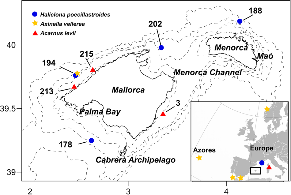
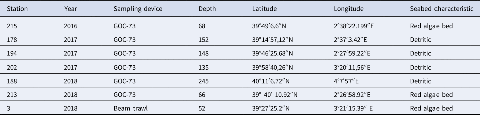
The specimens were collected during the MEDITS surveys developed off the Balearic Islands (western Mediterranean), using the experimental bottom trawl gear GOC-73 (Bertrand et al., Reference Bertrand, de Sola, Papaconstantinou, Relini and Souplet2002) (see Figure 1 and Table 1 for sampling station details). Additional sampling with a Jennings' type beam trawl (Reiss et al., Reference Reiss, Kröncke and Ehrich2006) has also been carried out during these surveys in order to improve the sampling of benthic species. Sponges analysed in this study were collected with both sampling methods during the MEDITS surveys carried out in 2016, 2017 and 2018 around Mallorca and Menorca.
Map of the studied area showing the stations (numbers) where sponges were collected. Coloured symbols represent the distribution of Axinella vellerea, Haliclona poecillastroides and Acarnus levii. The small map shows the previous records of the species in the Mediterranean for A. vellerea (Sitjà & Maldonado, Reference Sitjà and Maldonado2014; Sitjà et al., Reference Sitjà, Maldonado, Farias and Rueda2019) and A. levii (Vacelet, Reference Vacelet1960, Reference Vacelet1961), and previous documented records for H. poecillastroides (Vacelet, Reference Vacelet1969).

Details of the sampling stations

Once on deck, sponges were separated from the rest of the catch and photographed with a Nikon DSLR D300 digital camera on a graph chart. Whole samples were preserved in absolute ethanol (EtOH). Sponge specimens were deposited in the Marine Fauna Collection (http://www.ma.ieo.es/cfm/; CFM-IEOMA) based at the Centro Oceanográfico de Málaga (Instituto Español de Oceanografía) with the following identification reference numbers: CFM-IEOMA-6390–6399.
Morphological descriptions
External morphology, colour and texture were annotated prior to the sample conservation. Spicules preparations and histological sections were made according to the standard methods described by Hooper (Reference Hooper2003).
Spicules were observed with a Nikon S-Ke optical microscope and photographed with a CMOS digital camera. Images were processed with the Fiji software (Schindelin et al., Reference Schindelin, Arganda-Carreras, Frise, Kaynig, Longair, Pietzsch, Preibisch, Rueden, Saalfeld, Schmid, Tinevez, White, Hartenstein, Eliceiri, Tomancak and Cardona2012). For each sample, 30 spicules per spicule class or category were counted. Spicular sizes are provided as minimum-mean-maximum in chord length and minimum-mean-maximum in width and expressed in microns (μm). Thick sections of both tangential-surface and transverse-surface sections were made with a scalpel and, when necessary, dehydrated with alcohol and cleared with xylene. Cleared sections were re-hydrated with water, included in mounting media and observed with the microscope or a Leica M165C stereomicroscope. Aliquots of suspended spicules were transferred onto aluminium foil, air dried, sputter coated with gold and observed under a HITACHI S-3400N scanning electron microscope (SEM).
The terminology applied for the morphological description follows Boury-Esnault & Rützler (Reference Boury-Esnault and Rützler1997). Identification was according to Topsent (Reference Topsent1904), Sitjà & Maldonado (Reference Sitjà and Maldonado2014) and Vacelet (Reference Vacelet1960, Reference Vacelet1969).
Molecular analyses
DNA was extracted from a piece of choanosomal tissue (~2 cm3) using the DNeasy Blood and Tissue Extraction kit (QIAGEN). Polymerase chain reaction (PCR) was used to amplify the Folmer fragment of the cytochrome oxidase subunit I (COI; DNA barcoding), with the universal primers LCO1490 and HCO2198 (Folmer et al., Reference Folmer, Black, Hoeh, Lutz and Vrijenhoek1994). PCR was performed in 25 μl volume reaction (17.2 μl ddH2O, 2.5 μl Mangobuffer, 1 μl DNTPs, 1.75 MgCl2 0.5 μl of each primer, 0.5 μl BSA, 0.05 μl TAQ and 1 μl DNA). The PCR thermal profile applied was: initial stage of 94°C for 5 min, then 37 cycles at 94°C for 15 s, 46°C for 15 s and 72°C for 15 s, followed by a final extension at 72°C for 7 min. PCR amplification of A. vellerea did not work, including another set of primers (LCO and Tetract-minibarR1) designed to amplify the COI mini-barcode (the first 130 bp of the Folmer fragment) (Cárdenas & Moore, Reference Cárdenas and Moore2019). PCR products were purified using the QIAquickR PCR Purification Kit (QIAGEN). Both heavy and light strands were sequenced on an ABI 3130 sequencer using the ABI Prism Terminator BigDyeR Terminator Cycle Sequencing Reaction Kit (Applied Biosystems).
Sequences were imported into BioEdit 7.0.5.2. (Hall, Reference Hall1999) and checked for quality and accuracy with nucleotide base assignment. Multiple sequence alignments (MSA) were obtained with ClustalW (Thompson et al., Reference Thompson, Higgins and Gibson1994). The DNA sequences obtained were deposited in the GenBank database (http://www.ncbi.nlm.nih.gov/genbank/) under the following accession numbers: MN508968, MN508969 and MN508967. Sequences were validated using the BLAST function from the GenBank database (Altschul et al., Reference Altschul, Gish, Miller, Myers and Lipman1990). The sequences were also used to reconstruct the phylogenetic relationships between species, through a phylogenetic tree based on Bayesian inference (BI) and Maximum likelihood (ML). To build our alignments, we made a selection of the related sequences obtained after the BLAST search (Supplementary Table S1). BI and ML trees containing H. poecillastroides (specimen CFM-IEOMA-6392) and proximal sequences of the order Haplosclerida (25 of which belong to Petrosiidae, 11 to Chalinidae, 5 to Callyspongiidae, 5 to Phloeodictyidae and 6 to Niphatidae) were run. An additional sequence identified as Haplosclerida sp. was also used (GenBank ID MK833931). Sequences of Axinella polyploides, Geodia barretti and Baikalospongia intermedia were used as outgroups. For A. levii (specimens CFM-IEOMA-6397 and CFM-IEOMA-6398), BI and ML trees with sequences of the order Poecilosclerida (38 belong to the family Microcionidae, 9 Acarnidae, 7 Iotrochotidae, 4 Hymedesmiidae, 4 Myxillidae, 2 Coelosphaeridae and 1 Podospongiidae), and also one sequence of Axinellida (Acantheurypon pilosella), were run. Sequences of Axinella polyploides and Xestospongia muta were used as outgroups.
The Hasegawa–Kishino–Yano (Hasegawa et al., Reference Hasegawa, Kishino and Yano1985) plus gamma distribution and proportion of invariable sites model (HKY + G + I) was assigned as the optimal substitution model of molecular evolution. This model was selected following Bayesian Criteria (BIC) and the Akaike Information Criterion corrected (AICc) using MEGA v.10.0.5 (Kumar et al., Reference Kumar, Stecher, Li, Knyaz and Tamura2018). The codon positions that were included were 1st + 2nd + 3rd. BI and ML analyses were performed with the CIPRES science gateway (http://www.phylo.org) (Miller et al., Reference Miller, Pfeiffer and Schwartz2010), with Mr Bayes v 3.6.2. (Ronquist et al., Reference Ronquist, Teslenko, Van Der Mark, Ayres, Darling, Hohna and Huelsenbeck2012) conducting four independent MCMC runs (with four chains each) for 5 million generations, sampling every 1000 generations and discarding the first 25% of samples as burn-in. Convergence was assessed by effective sample size (ESS) calculation and was visualized using TRACER v.1.5. RAXML (Stamatakis, Reference Stamatakis2014), performed with 1000 bootstrap iterations. Genetic distance (p-distance) and number of base differences between pairs of DNA sequences were estimated with MEGA v10.0.5 software (Kumar et al., Reference Kumar, Stecher, Li, Knyaz and Tamura2018).
Results
SYSTEMATICS
Class DEMOSPONGIAE Sollas, 1885
Subclass HETEROSCLEROMORPHA Cárdenas, Pérez and
Boury-Esnault, 2012
Order AXINELLIDA Lévi, 1953
Family AXINELLIDAE Carter, 1875
Genus Axinella Schmidt, 1862
Axinella vellerea Topsent, Reference Topsent1904
Material examined
Samples CFM-IEOMA-6390 and CFM-IEOMA-6391, station 194, 148 m (Table 1), collector P Ferriol and JA Díaz.
Macroscopic description
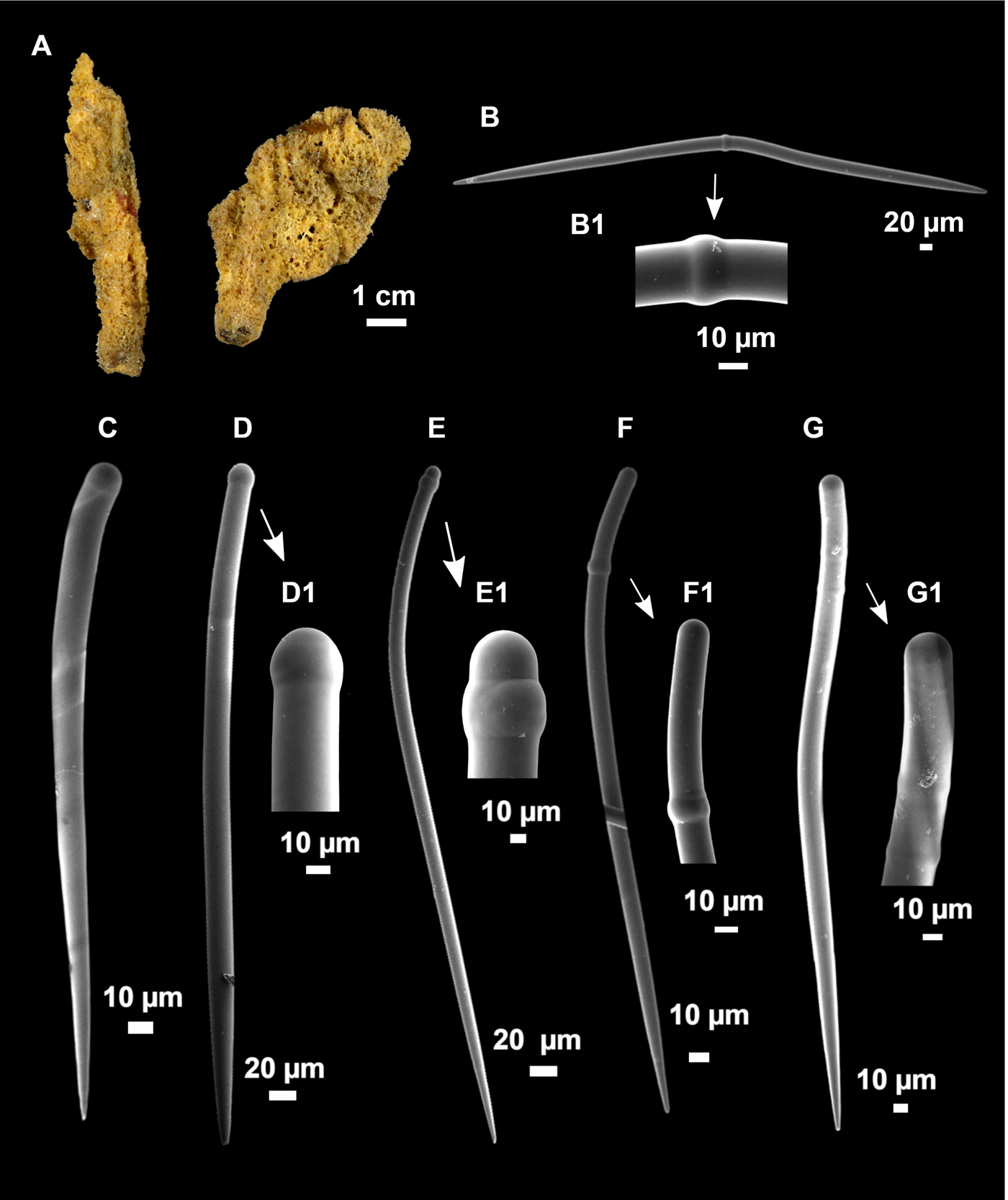
Two individuals (Figure 2A), 7 × 3 cm in 6390 and 5.5 × 1.5 cm in 6391, both supported by a tough peduncle of 1 cm in diameter. Colour in life orange-yellow and ochre to light brown after preserved in EtOH. Consistency hard but slightly compressible, rough to the touch. Localized hispidation visible to the naked eye. Surface irregular and pores scattered through the body, up to 1 mm in diameter. There was a translucid dermal membrane, patent in some parts of the ectosome and inconspicuous in other zones.
Axinella vellerea Topsent, Reference Topsent1904. (A) habitus of CFM-IEOMA-6390 (right) and CFM-IEOMA-6391 (left); B–G1, spicules of CFM-IEOMA-6390; (B) centrotylote oxea, with central swelling (B1); (C–G) tylostyles occurring in a varying size and shape, with diversity of heads and terminal swellings (D1, E1, F1 and G1).

Spicules
Oxeas very rare and angulate (Figure 2B), occasional marked swelling near the centre of the shaft (Figure 2B1), measuring 748–1065/21–26 μm (N = 4) in 6390 and 623–1091/16–20 μm (N = 4) in 6391.
Styles in a wide range of shapes and sizes (Figure 2C–G), with terminal, subterminal, single or multiple swellings (Figure 2D1–G1). They were slightly sinuous or curved in different grades, often resembling rhabdostyles. There was a large variation in size range, but no defined size categories. Measurements were 442–763–1345/13–22–31 μm in 6390 and 462–726–1309/13–19–25 μm in 6391.
Strongyles very rare and slightly curved, with symmetrical and roundish ends, same size range as styles.
Ecology notes
The species was found in north-western Mallorca, an area under the seasonal influence of the so-called Balearic Current (Garcia-Ladona et al., Reference Garcia-Ladona, Castellon, Font and Tintore1996). This current appears in spring, flowing along the northern continental shelf edge off Mallorca and may have some influence on the communities of filter-feeding animals. The station was rich in filter-feeding invertebrates such as Gryphus vitreus (Born, 1778) and Funiculina quadrangularis (Pallas, 1766). There was also a high abundance of echinoderms and sponges (see Supplementary Table S2 for the rest of accompanying fauna).
Taxonomic remarks
The specimens matched the descriptions provided by Topsent (Reference Topsent1904) and Sitjà & Maldonado (Reference Sitjà and Maldonado2014) in both spicular complement and abundance. Styles were the most abundant spicules, while diactines (mostly oxeas, some with strongylote modification) were rare, and its presence subjected to individual variation. The only difference is that in our specimens, styles are shorter (1345 μm) than specimens from the Atlantic (1800 μm) and Alboran Sea (1725 μm). The external morphology similarity with Axinella centrotylota Pansini, Reference Pansini1982 should be noted. However, they differ in spicular complement and spicular sizes. In A. centrotylota oxeas are very abundant and measure 400–490/10–15 μm, while in A. vellerea they are extremely rare and much larger (623–1091/16–26 μm). Moreover, styles of A. centrotylota are smaller (220–1270/8–15 μm) and can be divided in two categories, instead of a single category in A. vellerea.
Order HAPLOSCLERIDA
Family CHALINIDAE Gray, 1867
Genus Haliclona Grant, 1841
Haliclona poecillastroides (Vacelet, Reference Vacelet1969)
Reniera poecillastroides Vacelet, Reference Vacelet1969
Material examined
Samples CFM-IEOMA-6392, station 178, 152 m, collectors P Ferriol and JA Díaz; CFM-IEOMA-6393, station 194, 148 m, collectors P Ferriol and JA Díaz; CFM-IEOMA-6394, station 202, 135 m, collectors P Ferriol and JA Díaz; CFM-IEOMA-6395 and CFM-IEOMA-6396, station 188, 245 m, collector JA Díaz (Table 1).
Comparative material examined
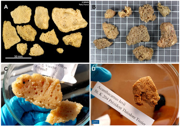
Haliclona poecillastroides, paratype (Station Marine d'Endoume collection), Cassidaigne Canyon, off Marseille, France, Gulf of Lion, 150 m (Figure 3C).
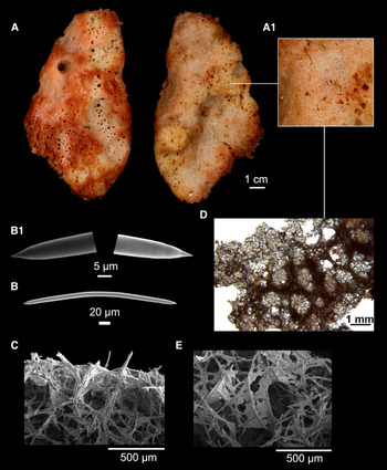
Haliclona poecillastroides (Vacelet, Reference Vacelet1969). (A) habitus of CFM-IEOMA-6392 with detail of the ectosome (A1); B–E, spicules and skeletal arrangement of CFM-IEOMA-6392; (B) oxea, with detail of heads (B1); (C) transversal view of the choanosome, with the subectosomal condensation of spicule tracks; (D) tangential view of the ectosome and the supporting subdermal tracks; (E) tangential view of the dermal membrane.

Macroscopic description
Massive to lamellar, somehow irregular shape, up to 7 cm in diameter and 2 cm thick (Figures 3A and 4A–C). Colour on deck variable, 6394 and 6396 were whitish, while 6392 was dirty beige with pink shades (Figure 3A) and 6395 and 6393 were dark grey. Once the specimens were fixed in EtOH, the colour remained similar to the fresh state. It is remarkable that pinkish specimens coloured the EtOH dark lilac, while the other specimens did not. Copious amounts of mucus were expelled when specimens were examined fresh on deck.
(A) Holotype of Haliclona poecillastroides (https://science.mnhn.fr/institution/mnhn/collection/ip/item/2015-576). (B) Fresh image of the habitus of CFM-IEOMA-6393 (scale grid 1 cm). (C) Paratype of Haliclona poecillastroides (Station Marine d'Endoume collection). (D) Paratype of Acanthacarnus levii (Station Marine d'Endoume collection).

Consistency slightly hard to the touch and very friable. Surface slightly rough, but no hispidation visible to the naked eye. Inhalant and exhalant faces present, the former with a detachable crust, with small pores (Figure 3A right, and Figure 3A1 for close-up), while the latter without crust and with abundant rounded oscules of 1–5 mm diameter (Figure 3A, left).
Spicules
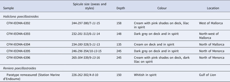
Spicules were oxeas (Figure 3B, B1), with some occasional stylote modifications. They were gently curved or straight, rarely sinuous, with tapered tips. Heterogeneous tips were found in some cases, with one tip more roundish, being probably intermedium forms of the stylote modifications. The oxeas sizes of each studied specimen are presented in Table 2, overall measuring 232–295–380/6–12–16 μm, but spicules >350 μm were very rare. Some thinner juvenile oxeas of 150/4 μm were also present.
Comparative table of the Haliclona poecillastroides individuals analysed in this work

ID are the reference numbers of the CFM-IEOMA. Measures are given as minimum-mean-maximum for total length/minimum-mean-maximum for total width (as they appear in the cited texts). All measurements are expressed in μm.
Skeletal structure
Choanosome composed by an isotropic and irregular net of spicule tracks, uni- to pauci-spicular, drawing heterogeneous meshes. These meshes were overlaid by oxeas in confusion, being only visible in some areas. The tracks condensed towards the surface and then ran in parallel to the surface, supporting the ectosome and generating rounded meshes (Figure 3C, D).
Ectosome made of an isodictyal layer of single or occasionally double spicule mesh (Figure 3E), forming a detachable crust that only persisted in some individuals. Under this crust, there were broad and characteristic subdermal spaces.
Ecology notes
The species was found at several stations in western, northern and southern Mallorca and northern Menorca, from 135–245 m depth (see Supplementary Table S2 for the rest of accompanying fauna), at gravel and bathyal mud bottoms.
Remarks
Our material matched the description of H. poecillastroides in the external morphology, the shape of the spicules, the presence of styles of the same length as the oxeas and immature, smaller oxeas. However, the colour appeared to be variable, with some individuals with pinkish shades. The spicules of our specimens are larger than those from the Gulf of Lion (Vacelet, Reference Vacelet1969; Griessinger, Reference Griessinger1971, Table 2), showing that the species may have a regional/seasonal variability in the spicule sizes, as indicated by these authors.
Order POECILOSCLERIDA Topsent 1928
Family ACARNIDAE Dendy, 1922
Genus Acarnus Gray, 1867
Acarnus levii (Vacelet, Reference Vacelet1960)
Acanthacarnus levii Vacelet, Reference Vacelet1960
Material examined
CFM-IEOMA-6397, station 215, 68 m, collector P Ferriol; CFM-IEOMA-6398, station 3, 52 m, collector JA Díaz; and CFM-IEOMA-6399, station 213, 66 m, collector JA Díaz.
Comparative material examined
Acarnus levii (Figure 4D), paratype (Station Marine d'Endoume collection), R/V ‘President Theodore Tissier’, Corsica Channel, station K. 204 (exogenous), 335–367 m.
Macroscopic description
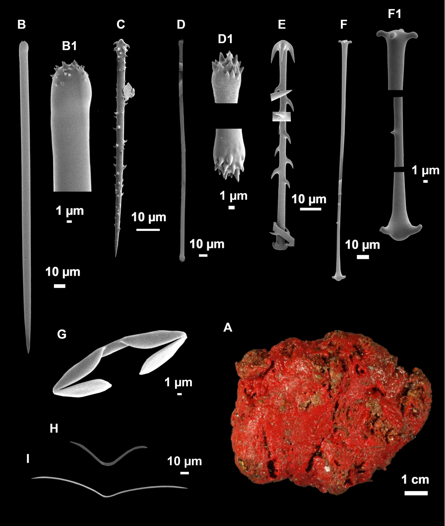
Individuals were massive, up to 5 cm diameter (Figure 5A), with a tendency to become roundish. Some specimens were fleshy (6397 and 6399), while others incorporated large amounts of detritus in the choanosome (6398). Colour in life bright red, brownish in EtOH, which became orange immediately after fixation of the sponge. Consistency compressible, a mark remains after pressed with the tweezers. Surface slightly rough to the touch. Ectosome consistent, not easily detachable. Openings were not visible.
Acarnus levii (Vacelet, Reference Vacelet1960). (A) habitus of CFM-IEOMA-6397; B–I, spicules of CFM-IEOMA-6397; (B) styles, with detail of the microspined head (B1); (C) acanthostyle; (D) tylote, with detail of the microspined heads (D1); (E) cladotylote II; (F) inmature cladotylote, with details of the cladomes and the spination of the shaft (F1); (G) palmate isochela; (H) oxhorn toxas; (I) accolada toxa.

Spicules
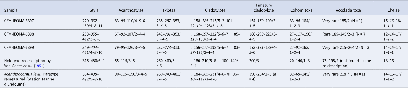
The spicule sizes of all studied specimens are detailed in Table 3, overall mean measurements and description are as follows:
-
Megascleres:
• Styles straight or slightly curved (Figure 5B), with microspined ends (Figure 5B1), 279–373–481/3–7–11 μm.
• Acanthostyles straight, with a marked head, finely and homogeneously spined (Figure 5C), except just beneath the head, where spines were rare, 67–97–200/2–4–6 μm, although sizes >130 μm were very rare.
• Tylotes straight or slightly contoured, rarely tortuous (Figure 5D), with spined heads (Figure 5D1), 232–284–353/3–4–5 μm.
• Cladotylote I, normal-shaped, abundant, stout and strongly hooked, with one cladome more developed than the other, measuring 156–191–231/4–6–10 μm.
• Cladotylote II, (Figure 5E) same morphology as cladotylote I, measuring 83–105–140/3–4–7 μm.
• Immature cladotylote, slightly spined (Figure 5F), with similar sized cladomes (Figure 5F1), 154–186–222/3–4–6 μm. These spicules, already described by Vacelet (Reference Vacelet1960), were considered immature stages of cladotylotes I–II by Van Soest et al. (Reference Van Soest, Hooper and Hiemstra1991).
Table 3.Comparative table of the spicular set of the Acarnus levii individuals analysed in this work (including a paratype from Corsica) and the Holotype

Numbers are the reference numbers of the CFM-IEOMA. Measures are given as minimum – mean – maximum for total length / minimum – mean – maximum for total width (or as they appear in the cited texts). All measurements are expressed in μm.
-
Microscleres:
• Palmate isochela (Figure 5G), very common, 12–15–18/1–1–2 μm.
• Oxhorn toxas (Figure 5H), in a large size variation, but without distinguishable categories. The smaller ones tended to have a more pronounced curvature, 27–103–196/1–2–4 μm.
• Accolada toxas (Figure 5I), very rare, thin and straight, with a short and shallow curvature at the centre, 185–195–264/2–2–3 μm.

Skeletal structure
A tangential ectosome composed of bundles of tylotes. Choanosomal structure composed by plumose spicule tracks, regularly connected by single large styles.
Ecology notes
The specimens were collected from red algae beds at 52–68 m depth, dominated by red algae, both calcareous and ‘soft’ species, the green algae Codium bursa (Olivi) C. Agardh, 1817, the brown algae Laminaria rodriguezii Bornet, 1888 and fauna such as echinoderms and ascidians, including a high diversity and abundance of mesophotic sponges (see Supplementary Table S2 for the rest of accompanying flora and fauna).
Taxonomic remarks
There are currently 26 species of Acarnus in the world. In the Mediterranean and the North-east Atlantic, three species of this genus have been reported: A. souriei (Levii, 1952), A. tortilis (Topsent, 1892) and A. levii. Acarnus levii differs from A. sourei in the habitus (massive vs incrusting) and the possession of a single category of acanthostyles and three categories of cladotyles. It differs from A. tortilis by the presence of acanthostyles. Other similar species are the Pacific A. peruanus, the Indo-Pacific A. bicladotylota, the southern Atlantic A. nicoleae and the Mexican A. michoacanensis (Van Soest et al., Reference Van Soest, Hooper and Hiemstra1991; Aguilar-Camacho et al., Reference Aguilar-Camacho, Carballo and Cruz-Barraza2013). The differences between A. levii and A. peruanus are minimal and reduced to spicular dimensions of the megascleres and toxas. However, due to the vast geographic distances between both species (western Mediterranean and eastern Central Pacific) they are unlikely to be the same species. Acarnus levii differs with A. bicladotylotus in the reduced spination of cladotylote I, with A. nicoleae in the dimensions of the megascleres (Van Soest et al., Reference Van Soest, Hooper and Hiemstra1991) and with A. michoacanensis in the possession of a single cladotylote category (vs two in A. levii) (Aguilar-Camacho et al., Reference Aguilar-Camacho, Carballo and Cruz-Barraza2013).
The accolada toxas were reported in the original description of this species, but not found in the re-description by Van Soest et al. (Reference Van Soest, Hooper and Hiemstra1991). We found them in all the studied specimens and in a paratype from the Corsica Channel, albeit in very low numbers. It is probable that they were overlooked by Van Soest et al. (Reference Van Soest, Hooper and Hiemstra1991), who thought that Vacelet reported ‘thin growth stages of the choanosomal styles’ instead of true toxas.
Genetics
Haliclona poecillastroides (specimen 6392) gave 613 bp COI Folmer fragment. For A. levii, sequences of two specimens (6397 and 6398) were obtained, resulting in edited fragments of 458 bp (POR246) and 617 bp, (POR615) respectively.
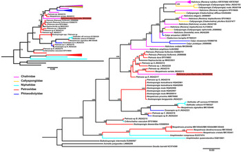
We generated, for each species (H. poecillastroides and A. levii) a BI and a ML tree. For H. poecillastroides (Figure 6, large and small trees, respectively), both trees show similar topologies with all haplosclerid families being polyphyletic. The Bayesian reconstruction suggested that H. poecillastroides groups within a well-supported Petrosia/Neopetrosia clade, and not with any Haliclona species (Figure 6, large tree). The ML tree placed H. poecillastroides in a basal branch of a cluster that included both Chalinidae and Petrosiidae sequences, however, this allocation was poorly supported.
Phylogenetic tree topology constructed with Maximum likelihood (small simplified tree) and Bayesian inference (large tree) for the COI sequences of Haliclona poecillastroides described in the present study and other related Haplosclerida. Branch colours represent different families. A sequence of Axinella polypoides, Geodia barretti and Baikalospongia intermedia are used as outgroups.

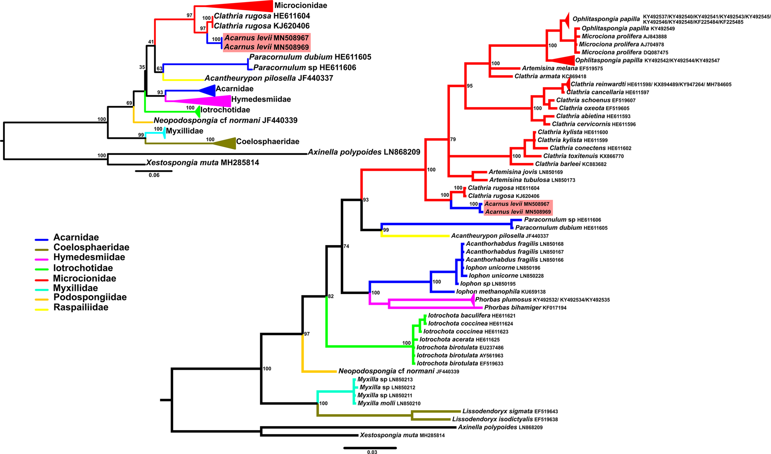
The BI and ML trees including A. levii were congruent (Figure 7, large and small trees, respectively) and showed the family Acarnidae as polyphyletic. The Acarnidae are distributed in three well-supported clades: (1) a clade contained Iophon spp. and Acanthorhabdus fragilis, (2) a clade contained Paracornulum spp. and finally, (3) a clade containing A. levii, nested in the Microcionidae and sister to Clathria rugosa.
Phylogenetic tree topology constructed with a Bayesian inference for the COI sequences of Acarnus levii described in the present study and other related sequences (large tree) and simplified tree topology obtained with RAXML (small tree). Branch colours represent different families. A sequence of Xestospongia muta and Axinella polypoides are used as outgroups.

For H. poecillastroides, lower P-values and bp differences were obtained when compared with Petrosia sp., Petrosia sp. E and N. seriata (7–8% p-distance and 41–43 bp differences). Otherwise, for A. levii, the lowest genetic distances were found when comparing A. levii with C. rugosa, with a p-distance of 2% and 11 bp differences.
Discussion
Biogeography
The results obtained improve the knowledge of the sponge diversity in the western Mediterranean. We studied the sedimentary bottoms of the continental shelf and slope off Balearic Islands and reported the presence of three poorly known species. For Axinella vellerea the present work represents its second record in the Mediterranean, as it was recently reported from Alboran Island (Sitjà & Maldonado, Reference Sitjà and Maldonado2014). Moreover, this species had only been reported from three other localities: the type locality in the Azores Archipelago (Topsent, Reference Topsent1904), the Gulf of Cadiz (Sitjà et al., Reference Sitjà, Maldonado, Farias and Rueda2019) and the Folden fiord, in Norway (Burton, Reference Burton1931). In their work, Sitjà & Maldonado (Reference Sitjà and Maldonado2014) considered this species as Atlantic, explaining its presence in Alboran Island as a result of a penetration towards the Mediterranean. If the Atlantic origin is correct, a Balearic presence must mean that A. vellerea populations expanded into the Mediterranean despite important oceanographic barriers, like the Almeria-Oran front and the Ibiza Channel, which are efficient in retaining both larval and adult stages from many phyla (Patarnello et al., Reference Patarnello, Volckaert and Castilho2007; Mokhtar-Jamaï et al., Reference Mokhtar-Jamaï, Pascual, Ledoux, Coma, Féral, Garrabou and Aurelle2011; García-Merchán et al., Reference García-Merchán, Robainas-Barcia, Abelló, Macpherson, Palero, García-Rodríguez, Gil de Sola and Pascual2012). Although current knowledge on the genetic dynamics of sponge populations is scarce, in general terms, dispersive potential of the phylum is considered to be low (Duran et al., Reference Duran, Pascual, Estoup and Turon2004; Guardiola et al., Reference Guardiola, Frotscher and Uriz2012; Pérez-Portela et al., Reference Pérez-Portela, Noyer and Becerro2015). Moreover, it is well known that the number of recorded individuals in the Mediterranean and the Atlantic is very similar. Considering the limited dispersive potential of the sponges, it is plausible to consider this species as Atlanto-Mediterranean instead of Atlantic.
The species Haliclona poecillastroides is documented for the first time in the Balearic Islands. Previously, this species was only known from two records in the Gulf of Lion and undocumented reports from the Menorca Channel (Santín et al., Reference Santín, Grinyó, Ambroso, Uriz, Gori, Dominguez-Carrió and Gili2018). Finally, Acarnus levii is reported for the first time elsewhere than Corsica, representing the third record of the species and expanding its geographic range westwards (Figure 1).
The present records highlight the singularity of the Balearic Islands ecosystems, whose oceanographic characteristics lead to conditions in which singular and poorly known sponge assemblages develop (Bibiloni, Reference Bibiloni1990). Moreover, bottom trawl fishing pressure around the archipelago is not as high as in adjacent waters off the Iberian Peninsula, thus sedimentary insular bottoms present a better conservation state (Quetglas et al., Reference Quetglas, Guijarro, Ordines and Massutí2012). To this fact it must be added that in soft circalittoral bottoms around the islands two widely distributed red algae facies (Peyssonnelia and maërl beds) provide substrate and increment the structural complexity, creating habitats with high biodiversity (Ballesteros, Reference Ballesteros1992, Reference Ballesteros1994; Barberá et al., Reference Barberá, Moranta, Ordines, Ramón, De Mesa, Díaz-Valdés, Grau A and Massutí2012; Ordines et al., Reference Ordines, Ramón, Rivera, Rodriguez-Prieto, Farriols, Guijarro, Pascual and Massutí2017).
All the sponge species here included were collected in the mesophotic zone, between 40 and 300 m depth, a transitional zone between shallow and deep communities that includes the circalittoral and the offshore circalittoral habitats (Evans et al., Reference Evans, Aish, Boon, Condé, Connor, Gelabert, Michez, Parry, Richard, Salvati and Tunesi2016). Here, the ecological factors (e.g. light, nutrients or temperature) are under a marked gradient that generates a biological mixture of shallow, deep and strictly mesophotic communities (Soares et al., Reference Soares, Tavares and Carneiro2019). In the Mediterranean and regarding sponges, many of the works published recently are focused on these transient communities (Bertolino et al., Reference Bertolino, Bo, Canese, Bavestrello and Pansini2015; Idan et al., Reference Idan, Shefer, Feldstein, Yahel, Huchon and Ilan2018; Longo et al., Reference Longo, Cardone, Pierri, Mercurio, Mucciolo, Marzano and Corriero2018; Santin et al., Reference Santín, Grinyó, Ambroso, Uriz, Gori, Dominguez-Carrió and Gili2018). However, sampling on these depths is problematic and prone to logistical limitations. Currently, the objectives of MEDITS surveys include not only the assessment of the demersal resources but the entire benthic ecosystem exploited in the Mediterranean bottom trawl fishery. In this regard, the potential of MEDITS surveys to study deep-sea sponges is clear since the experimental bottom trawl used in the sampling is not only effective for the capture of fishes, decapod crustaceans and cephalopods, the target species for which it was designed (Dremière et al., Reference Dremière, Fiorentini, Cosimi, Leonori, Sala and Spagnolo1999; Fiorentini et al., Reference Fiorentini, Dremière, Leonori, Sala and Palumbo1999), but other mega-benthic organisms. In fact, MEDITS data is being used to model the distribution of sessile species (e.g. Lauria et al., Reference Lauria, Garofalo, Fiorentino, Massi, Milisenda, Piraino, Russo and Gristina2017) and in the Balearic Islands it has also been used to study essential and sensitive habitats and the influence of benthic communities on demersal resources (Ordines & Massutí, Reference Ordines and Massutí2009; Ordines et al., Reference Ordines, Quetglas, Massutí and Moranta2009, Reference Ordines, Jordà, Quetglas, Flexas, Moranta and Massutí2011). Moreover, clear advances in biodiversity knowledge are being made from samples obtained during the MEDITS surveys, particularly in the case of small benthic fishes, such as the Callionymidae and Gobidae fishes, but also other fishes and echinoderms (Farias et al., Reference Farias, Ordines, García-Ruiz and Fricke2016; Kovačić et al., Reference Kovačić, Ordines and Schliewen2017, Reference Kovačić, Ordines and Schliewen2018, Reference Kovačić, Ordines, Ramirez-Amaro and Schliewen2019; Fricke & Ordines, Reference Fricke and Ordines2017a, Reference Fricke and Ordines2017b; Ordines et al., Reference Ordines, Fricke, Williston, Guijarro and Massuti2018, Reference Ordines, Ferriol, Moya, Farias, Rueda and Garcia-Ruiz2019a, Reference Ordines, Kovačić, Vivas, García-Ruiz and Guijarro2019b).
Taxonomic incongruences
Our results show that Acarnus levii is, by both molecular analysis and morphology, more related to the family Microcionidae (genus Clathria) than to Acarnidae. Specifically, the species has a genetic proximity with Clathria rugosa (Hooper & Levi, Reference Hooper and Levi1993), a New Caledonian microcionid species with singular ‘quasidiactinal subtylostyles’ in the ectosome. These spicules resemble diactinal forms and are also spined in their base and tips, a well-known fact since the presence of diactines in the ectosome is considered a diagnostic trait to separate Acarnidae and Microcionidae (Hooper & Van Soest, Reference Hooper and Van Soest2002). In fact, the literature has other examples of Clathria spp. with dubious ‘quasidiactinal’ spicules in the ectosome and other mixed features between Clathria and Acarnus: e.g. (1) Clathria (Microciona) acarnoides from the Caribbean has ‘cladotylote acanthostyles’, which are very similar to the cladotylotes of Acarnus; and (2) Clathria (Clathria) nicoleae has diactines in the ectosome, consisting of ‘tylostrongiles’ strongly spined in the head and resembling the tylotes of some Acarnus spp. (Aguilar-Camacho et al., Reference Aguilar-Camacho, Carballo and Cruz-Barraza2013; Vieira de Barros et al., Reference Vieira de Barros, Santos and Pinheiro2013; Van Soest et al., Reference Van Soest, Meesters and Becking2014). Moreover, other works have revealed the genetic proximity of the type species of the genus Acarnus, A. innominatus Gray 1867, to the family Microcionidae, through 16S, 18S and 28S markers (Hajdu et al., Reference Hajdu, de Paula, Redmond, Cosme, Collins and Lôbo-Hajdu2013; Redmond et al., Reference Redmond, Morrow, Thacker, Diaz, Boury-Esnault, Cárdenas, Hajdu, Lobo-Hajdu, Picton, Pomponi and Kayal2013).
The genus Acarnus is characterized by the presence of Cladotylotes, a strong synapomorphy that unites the group. For this reason, the relatedness of A. levii and A. innominatus with Microcionidae is potentially shared with all the Acarnids. If that is the case, the reallocation of the whole Acarnus genus to Microcionidae seems the right choice. In fact, Lévi (Reference Lévi and Grassé1973) already placed Acarnus in Clathriidae, a decision that was later refused by Van Soest (Reference Van Soest1984), who considered the genus to be more related to Myxillidae and Coelosphaeridae. However, since Acarnus is the type genus of Acarnidae, reassigning this genus to Microcionidae would require assigning a new family to the rest of the 12 remaining genera now in Acarnidae, and to expand the current definition of Microcionidae to include species with diactinal spicules in the ectosome, two actions that are far beyond the scope of the present work. Therefore, we refrain from moving either the species A. levii or the genus Acarnus until a revision of both families is made, including sequences of other Acarnus spp. and Clathria spp. with Acarnus-like features.
On the other hand, and according to our COI phylogenetic analyses, H. poecillastroides is more related to the family Petrosiidae than to the Chalinidae. Both molecular and morphological results support a Petrosiidae relationship: (1) choanosomal skeleton isotropic and confused, instead of composed by primary ascending tracks regularly interconnected by secondary tracks of Chalinidae; (2) presence of a isodictyal ectosomal crust and a large subdermal space beyond; (3) condensation of the choanosomal tracks near the surface to support the ectosome, from which extend parallel and circular meshes; and (4) stony but friable consistency. Indeed, all these features are considered typical of Petrosiidae (Van Soest, Reference Van Soest1980; Desqueyroux-Faúndez & Valentine, Reference Desqueyroux-Faúndez, Valentine, Hooper and van Soest2002). It should be noted that the species H. poecillastroides was originally assigned to the family Renieridae Schmidt, 1870 (now a synonym of Chalinidae) and the genus Reniera Schmidt, 1862 (now the subgenus Haliclona (Reniera)). That classification was very broad and emphasized the organization of the skeleton and the amount of spongin/spicules as a character with systematic value. Thus, sponges with a disorganized skeleton and mostly composed by spicules were included in the family Renieridae, while those with a more organized, spongin-reinforced skeleton were considered as Haliclonidae (de Laubenfels, Reference de Laubenfels1932; Griessinger, Reference Griessinger1971). Later, Wiedenmayer (Reference Wiedenmayer1977) used Nepheliospongiidae Clarke, 1900 to group species ‘characterized by strong development of megascleres in relation to fleshy parts and spongin, hence by a stiff and hard, occasionally friable consistency. The spicules may be packed into stout, crowded fibres, or tracts, agglutinated by spongin, or they may be packed in confusion around canals and alveoles. The basic architecture is that of ascending tracts, diverging and becoming radial towards the surface, connected by tangential tracts arranged in layers parallel to the surface, i.e. accretive.’ This definition overlapped with that previously given to Renieridae. Nepheliospongiidae was later split in two families by Van Soest (Reference Van Soest1980), who erected the family Petrosiidae for those species ‘with an ectosomal skeleton consisting of an isotropic reticulation of single spicules or spicule tracts, and choanosomal skeleton verging towards an isotropic reticulation of spicule tracts, in which primary and secondary tracts are indistinct’, and Oceanapiidae for species with an ‘ectosomal skeleton consisting of an often mutilayered isotropic reticulation of single spicules, […] and an irregular system of tangential thick spicule tracks’. The H. poecillastroides skeleton matches the definition of Petrosiidae, a family with four accepted genera: Petrosia, Neopetrosia, Xestospongia and Acanthostrongylophora (Hooper, 1984). It differs from both Petrosia and Acanthostrongylophora in not having a hispid ectosome, and with Petrosia also in lacking diverse spicule categories. It is, however, very similar to both Xestospongia and Neopetrosia, two genera with unclear relationship and overlapping diagnostic characters. These species are divided on the basis of the size of the spicules and skeletal arrangement: those having spicules >200 μm belong to Xestospongia and if they are <200 μm to Neopetrosia (Desqueyroux-Faúndez & Valentine, Reference Desqueyroux-Faúndez, Valentine, Hooper and van Soest2002). However, this dichotomy is not always met, such as in Neopetrosia carbonaria (Lamarck, 1815), Neopetrosia dominicana (Pulitzer-Finali, 1986), Neopetrosia dutchi (van Soest et al., Reference Van Soest, Meesters and Becking2014), Neopetrosia ovata (Meesters & Becking, 2014) and Neopetrosia sigmatifera (Vicente et al., Reference Vicente, Ríos, Zea and Toonen2019), all presenting oxeas >200 μm (see Santos et al., Reference Santos, Sandes, Cabral and Pinheiro2016; Vicente et al., Reference Vicente, Ríos, Zea and Toonen2019). Conversely, in species like Xestospongia dubia (Ristau, 1978), Xestospongia emphasis (de Laubenfels, 1954), Xestospongia mammillata (Pulitzer-Finali, 1982), Xestospongia menzeli (Little, 1963), Xestospongia tuberosa (Pulitzer-Finali, 1993) and Xestospongia vansoesti (Bakus & Nishiyama, 2000) oxeas may be <200 μm (revised in Carvalho et al., Reference Carvalho, Lopes, Cosme and Hajdu2016). A possible cause that might explain differences in spicule size between the members of Xestospongia and Neopetrosia is the depth at which they live, the availability of nutrients and the temperature (Valisano et al., Reference Valisano, Pozzolini, Giovine, Cerrano, Maldonado, Becerro, Turon and Uriz2012). In other demosponges, individuals inhabiting deeper habitats develop longer spicules than those inhabiting shallower ones (Bavestrello et al., Reference Bavestrello, Bonito and Sarà1993; Uriz et al., Reference Uriz, Turon, Becerro and Agell2003). However, to date this has not been tested in any Neopetrosia nor Xestospongia species. Another way to discern between both genera is the comparison of the skeletal arrangement, because the ectosome of Neopetrosia is ‘a simple tangential unispicular isodictyal network of small spicules’, while in Xestospongia it consists of a ‘dense ectosomal brushes of large spicules’. In addition, the choanosome of Neopetrosia is ‘more compact’ than those of Xestospongia (Desqueyroux-Faúndez & Valentine, Reference Desqueyroux-Faúndez, Valentine, Hooper and van Soest2002). In H. poecillastroides, spicules are >200 μm, but both ectosomal and choanosomal architectures are intermediate between Neopetrosia and Xestospongia. Moreover, the genetic results are not conclusive, and H. poecillastroides shows genetics similarities with both N. seriata, an Indo-Pacific shallow water species, and two Petrosia sp. A possible explanation for the somewhat ambiguous position of H. poecillastroides inside Petrosiidae is the lack of genetic studies specifically dealing with this family. We foresee that H. poecillastroides may represent a potential new Petrosiidae genus with mixed characters between Neopetrosia and Xestospongia, gathering several chalinid species with Petrosiidae characters, such as: Haliclona (Halichoclona) magna (Vacelet, Reference Vacelet1969), H. (Halichoclona) fistulosa (Bowerbank, 1866) or some Haliclona (flagellia) species (Fourt et al., Reference Fourt, Goujard, Pérez and Chevaldonné2017; Van Soest, Reference Van Soest2017; Dinn, Reference Dinn2020). Those species are large, massive, massive-encrusting or tubular, with low spongin content, having a characteristic layer in the ectosome, large subectosomal spaces, and a low-organized choanosome with some ascending track of spicules (de Weerdt, Reference de Weerdt2000). Like the cases of Xestospongia plana and Xestospongi friabilis, two temperate species first included in Haliclona but later transferred to the genus Xestospongia, we propose to transfer H. poecillastroides to Xestospongia based on skeletal architecture morphology (Costello et al., Reference Costello, Emblow and White2001).
The phylogenetic tree shows the fairly well-known polyphyly of the order Haplosclerida (Redmond et al., Reference Redmond, Raleigh, van Soest, Kelly, Travers, Bradshaw, Vartia, Stephens and McCormack2011). The group is specially challenging because it has a very limited set of morphological traits, low number of synapomorphies and an elevated number of species (Griessinger, Reference Griessinger1971). In this sense, the combined use of morphological characters and molecular markers has the potential to solve the relationship of its members because it allows detection of morphological homoplasies and phylogenetic relatedness. Contrary to Haplosclerida, the poecilosclerids sponges emerge as a different case because they possess a very large and heterogeneous set of skeletal elements.
Supplementary material
The supplementary material for this article can be found at https://doi.org/10.1017/S0025315420001071.
Acknowledgements
The authors wish to thank the captain and crew of R/V ‘Miguel Oliver’ as well as the participants in MEDITS surveys developed around the Balearic Islands in 2016, 2017 and 2018. Special thanks to Dr Jean Vacelet (Station Marine d'Endoume, Marseille) for kindly lending the loan of type material of H. poecillastroides and Acarnus levii, to Dr Pierre Chevaldonné (Station Marine d'Endoume, Marseille) for discussions on the H. poecillastroides sequence and Mr Ferran Hierro (University of the Balearic Islands) for the technical assistance at the scanning electron microscope.
Financial support
The MEDITS surveys are co-funded by the European Union through the European Maritime and Fisheries Fund (EMFF) within the National Program of collection, management and use of data in the fisheries sector and support for scientific advice regarding the Common Fisheries Policy. Julio A. Diaz is supported by FPI contract co-funded by the Regional Government of the Balearic Islands and the European Social Fund. Paco Cárdenas received support from the European Union's Horizon 2020 research and innovation programme through the SponGES project (grant agreement No. 679849). This document reflects only the authors' view and the Executive Agency for Small and Medium-sized Enterprises (EASME) is not responsible for any use that may be made of the information it contains.