INTRODUCTION
Trypanosomes are ubiquitous blood parasites that infect all classes of vertebrates and are the aetiological agents of severe diseases in both animals and humans. Little is known about the prevalence and pathogenesis of trypanosomes in Australian marsupials. To date 10 trypanosome species/genotypes have been identified in marsupials; Trypanosoma thylacis in the northern brown bandicoot (Isoodon macrourus) (Mackerras, Reference Mackerras1959; Mackerras and Mackerras, Reference Mackerras and Mackerras1960), T. binneyi from the platypus (Ornithorhynchus anatinus) (McMillan and Bancroft, Reference McMillan and Bancroft1974), and more recently novel Trypanosoma spp. have been identified from the eastern barred bandicoot (Perameles gunnii) (Bettiol et al. Reference Bettiol, Goldsmid, Le and Driessen1996), the southern brown bandicoot (Isoodon obesulus) (Bettiol et al. Reference Bettiol, Jakes, Le, Goldsmid and Hocking1998), the woylie (Bettongia penicillata) (Smith et al. Reference Smith, Clark, Averis, Lymbery, Wayne, Morris and Thompson2008), the chuditch (Dasyrus geoffroiii) (Smith et al. Reference Smith, Clark, Averis, Lymbery, Wayne, Morris and Thompson2008), the eastern grey kangaroo (Macropus giganteus), the common wombat (Vombatus ursinus) (Noyes et al. Reference Noyes, Stevens, Teixeira, Phelan and Holz1999) and the swamp wallaby (Wallabia bicolor) (Hamilton et al. Reference Hamilton, Stevens, Gidley, Holz and Gibson2005). A new species of trypanosome (T. copemani n. sp.) has also recently been described from Gilbert's potoroo (Potorous gilbertii) and quokka (Setonix brachyurus) (Austen et al. Reference Austen, Jefferies, Friend, Adams, Ryan and Reid2009).
The koala (Phascolarctos cinereus) has a fragmented distribution in the eastern states of Australia ranging from north-east Queensland (Qld) to the Eyre Peninsula in South Australia. A variety of factors have contributed to dramatic population declines over much of the koala's geographical range, including habitat loss and fragmentation, urbanization pressures such as increased incidents of motor vehicle strike and dog attacks and a high prevalence of disease. The koala is currently classified as vulnerable to extinction in N. S. W. and Qld under the N. S. W. Threatened Species Conservation Act 1995 and the Qld Nature Conservation (Wildlife) Regulation Act 2006.
Over the past 2 years a number of koalas have presented to the Australian Wildlife Hospital in Beerwah, Qld with serious illness associated with regenerative anaemia and trypanosome infections. The present study describes the morphological, pathological and genetic characterization of a trypanosome identified in the blood of one of these sick koalas as well as a PCR survey of other koalas presented to the hospital for unrelated conditions. We consider that the trypanosome infecting koalas is a new species and propose the name Trypanosoma irwini n. sp.
MATERIALS AND METHODS
Sources of isolates and sample collection
The Australian Wildlife Hospital at Beerwah, Qld provides veterinary care for sick and injured wildlife, including approximately 600 koalas per annum. These koalas are generally presented to the hospital because they have been injured or are suffering from overt disease. Most koalas presented to the hospital originated from south-east Qld or northern N.S.W.
A total of 68 blood samples were collected during routine veterinary examinations of koalas admitted to the Australian Wildlife Hospital between October 2006 and September 2008. Details of the geographical origin of each of the koalas as well as sex, age and reason for admission were recorded. For clinical examination and blood collection, the koalas were anaesthetized with an intramuscular administration of alfaxalone (Alfaxan® CD RTU, Jurox Australia) at an approximate dose rate of 3 mg/kg. Anaesthesia was maintained with a combination of oxygen and isoflurane delivered by either mask or endotracheal intubation. Blood samples of approximately 2 ml were collected by venepuncture of the cephalic vein. The blood was mixed with EDTA in a Vacutainer® tube (Becton-Dickinson, New Jersey, USA) and stored at 4°C. The sentinel koala, a seriously ill adult male koala from Lismore N. S. W., was euthanased and a post-mortem examination was conducted. Tissues were fixed in 10% neutral buffered formalin, stained with haematoxylin and eosin and processed for histology using standard methods. All veterinary procedures were performed by registered wildlife veterinarians in accordance with standard veterinary practice.
Morphological measurements
Thin-blood smears were prepared using blood from the sentinel koala treated at the Australian Wildlife Hospital, Beerwah, Qld, stained with Diff-Quick stain (Dade Behring Diagnostics Australia Pty Ltd, Lane Cove, N. S. W.) and examined microscopically. Each slide was immersed in methanol for 30 sec, followed by immersion in Diff-Quick Solution I for 30 sec before counterstaining with Diff-Quick Solution II for 30 sec. The slides were then rinsed in tap water, air-dried and a cover-slip mounted using DePeX mounting medium (Merck Pty. Limited, Kilsyth, Vic., Australia).
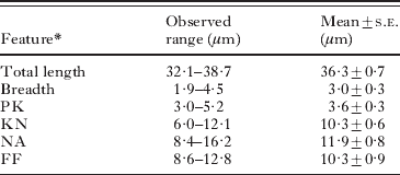
Digital light micrograph images of trypanosomes in blood films were taken at ×1000 magnification and measurements of key morphological features (see Table 2) were performed using Image Pro Express version 5.1 (Media Cybernetics Inc., USA).
In vitro culture
Attempts were made to culture trypanosomes from koala blood by adding 20 μl of fresh blood into 1·8 ml cryopreservation vials containing 1 ml of Modified Sloppy Evans Medium (MSEM) (Noyes et al. Reference Noyes, Stevens, Teixeira, Phelan and Holz1999). Cultures were incubated in the dark at room temperature for approximately 10–14 days. Microscopic examination of wet-smear preparations of the medium from each culture was performed weekly after the initial 10 to 14-day incubation to detect motile trypanosomes at 200× and 400× magnification.
DNA extraction
Blood samples were preserved in EDTA vials and whole genomic DNA extracted using a MasterPure™ DNA Purification Kit (EPICENTRE® Biotechnologies, Madison, Wisconsin, USA). DNA was eluted in 50 μl and stored at −20°C until use. Additional DNA samples were kindly provided by a number of researchers for sequencing and phylogenetic analysis. They included: T. cyclops (LV492), T. dionsii (P3), T. bennetti (KT-2), T. corvi (LSHTM), T. cruzi marinkellei (B3), T. sp. (KG1), T. avium (sp30), T. avium (sp40), T. avium (SIM3), T. avium (Buzzard) and T. avium (A1073).
18S rDNA amplification and sequencing
Two approximately 900 bp fragments of the 18S rRNA gene were amplified using 2 nested PCRs. The fragments partially overlapped and covered an approximately 1·5 kb fragment of the 18S rRNA gene. Both nested PCRs utilized the same external primers, SLF (5′ gcttgtttcaaggacttagc 3′) and S762 (5′ gacttttgcttcctctaatg 3′). One nested PCR used internal primers S825F (5′ accgtttcggcttttgttgg 3′) with S662 (5′ gactacaatggtctctaatc 3′) (~904 bp product) and the other nested PCR used primers S825 (5′ accgtttcggcttttgttgg 3′) and SLIR (5′ acattgtagtgcgcgtgtc 3′) (~959 bp product). Primers S662, S762R, S825 were sourced from Maslov et al. (Reference Maslov, Lukes, Jirku and Simpson1996) and SLF and SLIR were designed during the present project. The PCR reactions were performed using 1 μl of DNA in a 25 μl reaction containing 1×PCR buffer, 2 mm MgCl2, 0·4 mm dNTPs, 0·8 μm of each primer and 0·04 U/μl Tth+ Taq DNA polymerase (Fisher Biotec Australia, Wembley, W. A., Australia). The PCR conditions consisted of a pre-PCR step with 95°C for 5 min, 50°C for 2 min and an extension of 72°C for 4 min followed by 35 cycles of 94°C for 30 sec, 52°C for 30 sec and an extension of 72°C for 2 min and 20 sec after which a final extension of 72°C for 7 min. All positive PCR products were purified using a MO BIO UltraClean™ 15 DNA Purification Kit (MO BIO Laboratories Inc. West Carlsbad, California, USA) and sequenced using an ABI Prism™ Terminator Cycle Sequencing kit (Applied Biosystems, Foster City, California, USA) on an Applied Biosystem 3730 DNA Analyzer.
gGAPDH amplification and sequencing
An approximately 880 bp fragment of the glycosomal glyceraldehyde phosphate dehydrogenase (gGAPDH) gene was PCR amplified using a hemi-nested PCR. The primary PCR used primers GAPDHF (5′ ctymtcggnamkgagatygayg 3′) and GAPDHR (5′ grtksgartadccccactcg 3′) (designed during this study), followed by a secondary reaction using forward primer GAPDHF (5′ ctymtcggnamkgagatygayg 3′) and reverse primer G4a (5′ gttytgcagsgtcgccttgg 3″) sourced from Hamilton et al. (Reference Hamilton, Stevens, Gaunt, Gidley and Gibson2004). Clustal W alignments were used to design the GAPDHF and GAPDHR primers to be able to amplify Leishmania, Trypanosoma brucei, Trypanosoma vivax and Trypanoplasma borrelli gGAPDH sequences but not bovine and mouse gGAPDH sequences. The PCR reactions were performed using 1 μl of DNA in a 25 μl reaction containing 1×PCR buffer, 2 mm MgCl2, 0·4 mm dNTPs, 0·8 μm of each primer, and 0·04 U/μl Tth+ Taq DNA polymerase (Fisher Biotec Australia). The PCR conditions consisted of a pre-PCR step with 95°C for 5 min, 50°C for 2 min and an extension of 72°C for 4 min followed by 35 cycles of 94°C for 30 sec, 50°C for 30 sec (primary PCR) or 52°C for 30 sec (secondary PCR) with an extension of 72°C for 2 min and 20 sec and finishing with an extension of 72°C for 7 min. DNA sequencing was conducted as described above.
Phylogenetic analysis
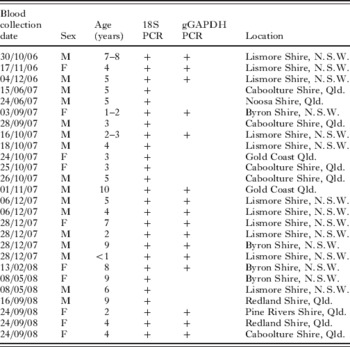
The details of the 18S PCR-positive koalas are detailed in Table 1. Nucleotide sequences generated for koala-derived trypanosomes for the 18S rDNA and gGAPDH loci were combined with sequences from a number of related trypanosomatids from GenBank and phylogeny inferred from Maximum Likelihood analysis by the program PhyML (Dereeper et al. Reference Dereeper, Guignon, Blanc, Audic, Buffet, Chevenet, Dufayard, Guindon, Lefort, Lescot, Claverie and Gascuel2008). The reliability of the inferred trees was assessed by the approximate likelihood ratio test (aLRT) (Anisimova and Gascuel, Reference Anisimova and Gascuel2006), a statistical test of branch support and an alternative to non-parametric bootstrap branch support estimation. Novel nucleotide sequence for T. irwini at both the 18S rDNA and gGAPDH loci were submitted to GenBank under the following Accession numbers (FJ649479 and FJ649485). In addition, novel gGAPDH sequences for T. bennetti (KT-2) (FJ649486), T. cyclops (LV492) (FJ649493), T. dionsii (P3) (FJ649494), T. corvi (LSHTM) (FJ649496), T. avium (sp30) (FJ649490), T. avium (sp40) (FJ649489), T. avium (Buzzard) (FJ649491), T. avium (A1073) (FJ649488), T. avium (SIM3) (FJ649487), T. cruzi marinkellei (B3) (FJ649495) and T. sp. (KG1) (FJ649492) and novel 18S rDNA sequences for T. avium (sp30) (FJ649482), T. avium (sp40) (FJ649483), T. avium (Buzzard) (FJ649481), T. avium (A1073) (FJ649480) and T. cruzi marinkellei (B3) (FJ649484) generated during the present study for the phylogenetic analysis have also been submitted to GenBank.
Koalas examined and found positive for trypanosome infections in this study

Parsimony analysis and measurement of genetic distance were conducted using Mega version 4 (Mega4: Molecular Evolutionary Genetics Analysis software, Arizona State University, Tempe, Arizona, USA) (Tamura et al. Reference Tamura, Dudley, Nei and Kumar2007).
RESULTS
Species description
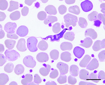
Trypanosoma irwini n. sp. (Fig. 1)
Bloodstream trypomastigote in a Giemsa-stained blood smear from koala (Phascolarctos cinereus) from Lismore, N. S. W. (sentinel koala). Scale bar represents 10 μm.

Diagnosis: Parasites observed in blood smears were long, thin organisms with a single flagellum, an undulating membrane, a kinetoplast and a nucleus, characteristics consistent with the trypomastigote stage of a trypanosomatid.
Taxonomic summary
Vertebrate host: Koala (Phascolarctos cinereus)
Invertebrate host: Unknown
Type location: Lismore, N.S.W., Australia
Additional locations: A range of locations in Qld, Australia
Site of infection: Blood
Pre-patent and patent periods: Unknown
Etymology: This species is named Trypanosoma irwini in honour of the wildlife conservationist Steve Irwin.
Parasite measurements
The morphology of the trypanosomes detected in blood smears from the sentinel koala was consistent with trypomastigote life-cycle stages of other described Trypanosoma spp. according to Hoare (Reference Hoare1972). The trypomastigote-like stages had a prominent kinetoplast, undulating membrane, a pointed posterior end and a long free flagellum (Fig. 1). A summary of the morphological features of the bloodstream forms of T. irwini observed in 9 trypomastigotes is presented in Table 2.
The mean dimensions and standard errors (s.e.) of morphological features of novel Trypanosoma sp. isolated from bloodstream forms from the koala (Phascolarctos cinereus) determined from the sentinel koala case
(Measurements determined from 9 trypomastigotes.)
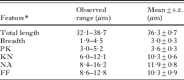
* Total length: body length measured along mid-line including free flagellum (total length).
- Breadth:
maximum breadth measured at nucleus level (including undulating membrane).
- PK:
distance between the posterior end and the kinetoplast.
- KN:
distance between the kinetoplast and posterior edge of the nucleus.
- NA:
distance between the anterior edge of the nucleus and the anterior end of the body.
- FF:
length of the free flagellum.
In vitro culture
Numerous attempts to culture the parasite were unsuccessful.
Clinical and pathological findings in the sentinel koala
Of the 26 T. irwini PCR positives identified, 9 koalas sampled appeared to be free of any disease at the time of sampling. Clinical and pathological findings of the sentinel koala are outlined below. On initial presentation to the hospital, the sentinel koala displayed depression, pale mucous membranes, unilateral chronic keratoconjunctivitis and generalized weakness. Significant clinical pathological findings included profound and regenerative anaemia (packed cell volume (PCV) of 9% when normal PCV is 30–40% and haemoglobin 32 g/l, where the normal range is 88–140 g/l). Examination of a blood film revealed a marked regenerative response to the anaemia and low numbers of parasites morphologically consistent with the trypomastigote stages of a trypanosomatid organism (Fig. 1). Cytological examination of bone marrow revealed erythroid hyperplasia. The koala was euthanized after 1 month due to hepatic and renal failure. A necropsy examination revealed osteochondromas affecting a number of ribs, haemosiderosis and extramedullary haematopoiesis of the spleen, atrophy of lymphoid tissues, bone marrow erythroid and myeloid hyperplasia, severe extensive periacinar necrosis of the liver, interstitial nephritis and oxalate nephrosis of the kidneys and interstitial pneumonia.
Genetic characterization
Of a total of 68 koalas screened for the presence of trypanosomes, using a PCR to amplify the 18S rDNA locus, 26 were determined to be infected with a Trypanosoma sp. (38·2%) (Table 1). Eleven of the trypanosome PCR-positive koalas were from Lismore Shire, N. S. W., 4 were from Byron Shire in N.S.W., 11 from various locations (over 5 shires) in Qld. Clustal W alignment of the partial 18S rDNA sequences amplified from all 26 koalas showed the DNA sequences to be 100% identical. For the purpose of phylogenetics analysis, some of the 26 18S rDNA-positive samples were also PCR analysed at the gGAPDH locus and 16 were successfully amplified at the gGAPDH locus and the resultant DNA sequences were found to be 100% identical. Representative DNA sequences for the 18S rDNA and gGAPDH loci of the novel trypanosome were deposited in the GenBank database as described above.
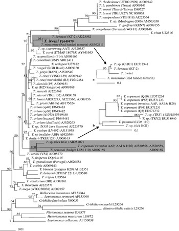
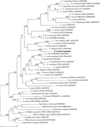
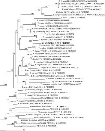
Maximum likelihood and parsimony analysis produced similar trees (data not shown). Phylogenetic analysis at both the 18S rDNA and gGAPDH loci revealed that T. irwini was genetically distinct and grouped with an avian trypanosome Trypanosoma bennetti (Figs. 2 and 3, PhyML trees shown). At the 18S rDNA locus, for which more sequence information was available from GenBank, T. irwini also grouped with Trypanosoma minasense, a trypanosome of neotropical primates and an isolate from an Australian marsupial, the chuditch (T. sp. CHU1). A composite tree of 18S rDNA and gGAPDH sequences also grouped T. irwini most closely with T. bennetti (Fig. 4. PhyML tree shown).
Phylogenetic analysis of the relationships of Trypanosoma spp. and novel trypanosomatid Trypanosoma irwini isolates (26 in total) from the koala using Maximum Likelihood analysis performed by PhyML 3.0 based on partial 18S rRNA gene sequences (~1500 bp). Branch support values (>60%) determined by aLRT. a) Demonstrates the phylogenetic position of T. sp. isolate Chuditch (CHU1) (~600 bp sequences). b) Phylogenetic position of T. copemani isolates (Q10, Q3, P94 and P63) and T. sp. TRY1 and TRY2 (~600 bp sequences). Scale bar represents the number of substitutions per site.

Phylogenetic analysis of the relationships of Trypanosoma spp. and the novel trypanosomatid Trypanosoma irwini isolates (16 in total) from the koala using Maximum Likelihood analysis performed by PhyML 3.0 based on partial gGAPDH sequences (~900 bp). Branch support values (>60%) determined by aLRT. Scale bar represents the number of substitutions per site.

Phylogenetic analysis of the relationships of Trypanosoma spp. and the novel trypanosomatid Trypanosoma irwini isolates (16 in total) from the koala based on composite gGAPDH and 18S rDNA partial sequences (~900 and ~1500 bp respectively) using Maximum Likelihood analysis performed by PhyML 3.0. Branch support values (>60%) determined by aLRT. Scale bar represents the number of substitutions per site.

At the 18S rDNA locus, T. irwini had a 0·9% and 1·7% genetic distance from T. bennetti and T. minasense respectively. At the gGAPDH gene the percentage genetic distance between T. irwini and T. bennetti was 10·7% (gGAPDH sequence for T. minasense was unavailable).
DISCUSSION
This is the first description of a Trypanosoma sp. isolated from koalas in Australia. The observation that the 26 trypanosome-positive koalas were geographically dispersed suggests that T. irwini appears to have a wide distribution in N.S.W. and Qld. The pathogenicity of T. irwini in koalas is still unclear. Some of the clinical and pathological findings in the sentinel koala, particularly those suggestive of extravascular haemolysis and generalized immune reaction, are entirely consistent with trypanosomiasis. However, there were other lesions in this koala that are unlikely to be aetiologically related to the trypanosome infection: the presence of various and diverse pathology precludes a definitive diagnosis of trypanosomiasis as the primary cause of illness in this case. Interestingly, approximately one third (9/26) of the T. irwini-positive koalas sampled for this study did not exhibit any clinical disease referable to that infection at the time they were sampled (data not shown). These koalas were admitted to the hospital as the result of traumatic injuries (by car or dog).
Immunosuppression and immunodeficiency syndromes are commonly associated with pathogenic retrovirus infection and immunosuppression has also been shown to be associated with trypanosomiasis as noted in Trypanosoma evansi infections (Dargantes et al. Reference Dargantes, Campbell, Copeman and Reid2005). Infection with the koala retrovirus (KoRV) is widespread in both captive and wild koalas and associated disease incidence is high (Hanger et al. Reference Hanger, Bromham, McKee, O'brien and Robinson2000, Reference Hanger, Mckee, Tarlington and Yates2003; Tarlinton et al. Reference Tarlinton, Meers, Hanger and Young2005). It is possible that a trypanosome infection in a healthy koala remains innocuous but when the immune status of the koala changes due to infection such as with KoRV, clinical signs of trypanosomiasis manifest. The level of retroviral RNA in serum was not measured in the euthanased koala, and therefore it is not possible to comment on the likely involvement of the KoRV in the disease observed in that individual. However, the fact that Trypanosoma irwini n. sp. was also identified in koalas without any disease referable to this infection suggests that it may be non-pathogenic or have low pathogenicity in many individuals, presumably those with intact immune systems.
Although it is possible that T. irwini n. sp. co-evolved with its host and may have limited pathogenicity in many individuals, its pathogenic potential (and therefore consequences for conservation) should not be ignored given the currently incomplete understanding of KoRV-associated disease, particularly the immunodeficiency syndrome. Further work is required to investigate the prevalence and pathogenic potential of T. irwini in koalas, particularly in those co-infected with KoRV.
Current phylogenetic evidence suggests that T. irwini is genetically most closely related to the avian trypanosome T. bennetti, which was isolated from an American kestrel (Falco sparverius) (Kirkpatrick and Terway-Thompson, Reference Kirkpatrick and Terway-Thompson1986) and T. minasense, which infects over 32 species of neotropical primates (Ziccardi et al. Reference Ziccardi, Lourenco De Oliveira and Nogueira1996). Most of the morphological measurements of the bloodstream form of T. irwini are within the range of those published for cultured T. bennetti and T. minasense with the exception of the measurement from the posterior end to the kinetoplast (PK). Trypanosoma minasense is usually reported to have a posterior to kinetoplast measurement of 5·7–15 μm (Hoare, Reference Hoare1972; Sato et al. Reference Sato, Leo, Katakai, Takano, Akari, Nakamura and Une2008) and T. bennetti 6·7–12·3 μm (Kirkpatrick and Terway-Thompson, Reference Kirkpatrick and Terway-Thompson1986). In contrast, the PK for T. irwini is considerably shorter at 3–5·2 μm. The utility of delineating trypanosome species based on morphometrics is however questionable as pleomorphism of bloodstream trypomatigotes of numerous T. spp. has been documented. Pleomorphism of bloodstream forms of T. minasense has been reported in different experimentally infected hosts, namely squirrel monkeys and marmosets, where the level of variation did not conform to previously measured T. minasense blood forms (Ziccardi and Lourenco-de-Oliveira, Reference Ziccardi and Lourenco-De-Oliveira1999). Pleomorphism of bloodstream forms of T. bennetti has also been reported with an increase in in vitro culturing temperature producing a posterior migration of the kinetoplast, affecting the PK values (Kirkpatrick and Terway-Thompson, Reference Kirkpatrick and Terway-Thompson1986).
There are no established criteria for delimiting species within the genus Trypanosoma. However, it has been proposed that the genetic distance at the 18S rDNA locus of 0·7% and 3·4% is sufficient to classify distinct species for Theileria sp. and Babesia sp. respectively (Schnittger et al. Reference Schnittger, Yin, Gubbels, Beyer, Niemann, Jongejan and Ahmed2003). In the present study, molecular and phylogenetic analysis at 2 unlinked loci indicated that T. irwini warrants separate species status. The genetic distance between T. irwini and its closest relative T. bennetti at the 18S rDNA locus is 0·9%, whereas the genetic difference between 2 established species, T. avium isolates and T. grayi is 0·6–0·8%. At the gGAPDH locus, established species T. conorhini, T. vespertilionis, T. mega and T. rotarium have between 4·4% and 5·3% genetic distances, whilst intraspecies genetic distance within T. avium is 0·9–2%. The genetic distance between T. bennetti and T. irwini at the gGAPDH locus is 10·7%, which is equal to or greater than genetic distances between established species.
The 3 phylogenetic trees produced in order to determine the evolutionary position of T. irwini amongst the trypanosomes largely produced the same topology of other researchers with a few exceptions. The commonly reported aquatic, avian, T. lewisi, T. cruzi, T. brucei and T. theileri clades reported by Hamilton et al. (Reference Hamilton, Gibson and Stevens2007) were generated in the 18S rDNA, gGAPDH and concatenated analyses with the exception of a separation of the avian clade in the 18S rDNA tree. This split in the avian clade has also been produced in 18S rDNA ML analysis by other researchers (Hamilton et al. Reference Hamilton, Stevens, Gaunt, Gidley and Gibson2004, Reference Hamilton, Stevens, Gidley, Holz and Gibson2005; Votypka et al. Reference Votypka, Lukes and Obornik2004). It has been suggested, however, that the 18S rDNA locus is an unreliable marker for inferring deep level trypanosome phylogenetic analysis (Hamilton et al. Reference Hamilton, Stevens, Gaunt, Gidley and Gibson2004) due to the fact that the 18S rRNA genes of taxa in the Trypanosoma genus are evolving at least 4 significantly different rates (Stevens and Rambaut, Reference Stevens and Rambaut2001). This is primarily why the gGAPDH locus was also analysed as it is increasingly used in Trypanosoma phylogeny and is under different evolutionary constraints to the 18S rRNA gene (Hamilton et al. Reference Hamilton, Stevens, Gaunt, Gidley and Gibson2004). The topology of the gGAPDH and concatenated 18S rDNA and gGAPDH trees were nearly identical and very similar to equivalent trees presented by Hamilton et al. (Reference Hamilton, Gibson and Stevens2007) and Viola et al. (Reference Viola, Almeida, Ferreira, Campaner, Takata, Rodrigues, Paiva, Camargo and Teixeira2009). The variation between the gGAPDH and concatenated tree was primarily due to lack of sequence data for the gGAPDH locus as the ‘T. irwini’ clade only comprised T. irwini and T. bennetti due to T. minasense gGAPDH sequence being unavailable. In addition, the gGAPDH analysis of the ‘T. irwini’ clade showed low aLRT branch support (37) for association with the ‘T. theileri’ clade, whilst in the concatenated analysis the ‘T. irwini’ clade is related to the ‘lizard’ clade with moderate branch support (64). The addition of the T. minasense gGAPDH sequence will perhaps help to resolve the potential relationship with the ‘lizard’ clade in the future.
The phylogenetic relationships of T. irwini may also be further clarified once more marsupial-derived trypanosome species are characterized. The relationship between T. irwini and T. thylacis from the northern brown bandicoot (Isoodon macrourus) (Mackerras, Reference Mackerras1959; Mackerras and Mackerras, Reference Mackerras and Mackerras1960), the Trypanosoma spp. from the southern brown bandicoots (Isoodon obesulus) (Bettiol et al. Reference Bettiol, Jakes, Le, Goldsmid and Hocking1998) and eastern barred bandicoots (Perameles gunnii) (Bettiol et al. Reference Bettiol, Goldsmid, Le and Driessen1996) cannot be evaluated because there are no genetic sequences available for these trypanosomes. Despite the wombat being the closest living relative to the koala (Osborne et al. Reference Osborne, Christidis and Norman2002), the T. copemani genotypes (AAI, AAP and H26) isolated from wombats are genetically distinct (genetic distance 5·6% at 18S rDNA and 20·4% at gGAPDH) from T. irwini. This is also true for quokka and Gilbert's potoroo isolates of T. copemani (genetic distance 5·3% at 18S rDNA) and woylie-derived Trypanosoma isolates (TRY1 and TRY2) (genetic distance 11·4% and 10·6% at 18S rDNA respectively) which are phylogenetically associated with T. copemani (Austen et al. Reference Austen, Jefferies, Friend, Adams, Ryan and Reid2009). Of all of the marsupial trypanosome genotypes identified thus far, the chuditch isolate (CHU1) was the closest marsupial-derived genotype to T. irwini with a genetic distance of 2·8% at the 18S rDNA locus, creating a clade with T. minasense, T. bennetti and T. irwini. The relationship of the chuditch isolate could not be examined in detail, as the available 18S rDNA sequence was short (only 563 bp) and to date no gGAPDH sequence has been generated.
The ‘T. irwini’ clade formed is a perplexing mix of isolates with vast differences in geography and hosts, with T. bennetti isolated from a bird in North America (also found in a PCR survey in African songbirds (Sehgal et al. Reference Sehgal, Jones and Smith2001), T. minasense from neotropical primates in South America, the chuditch (CHU1) or western quoll, a carnivorous Australian marsupial and T. irwini from the koala. The clade formed in the analysis could be evidence of host switching as has been suggested for T. binneyi, the platypus trypanosome and T. therezieni, a trypanosome of Madagascan chameleons that both fall in the ‘aquatic’ clade of trypanosomes from fish and amphibians. The phylogenetic placement of these trypanosomes indicates that host ecology may be more influential than host phylogeny in the evolution of some trypanosomes (Stevens et al. Reference Stevens, Noyes, Schofield and Gibson2001). Host switching could also be evidence of trypanosome and vector co-evolution rather than host co-evolution. It should be noted that the ‘T. irwini’ clade presents the first evidence of possible mammalian-avian host switching in the trypanosomes. The vast differences in host phylogeny, host ecology and lack of knowledge about vectors of the trypanosomes of the ‘T. irwini’ clade do, however, prevent formation of a plausible hypothesis regarding what factors other than genetics unite the trypanosomes of this clade.
Despite numerous attempts with fresh blood samples from trypanosome-infected koalas, establishment of in vitro cultures in Modified Sloppy Evans Media (MSEM) was not successful. With the benefit of the phylogenetic analysis, culturing attempts in the future could focus on testing media used in the in vitro culturing of the genetically most similar trypanosomes T. bennetti (Kirkpatrick and Terway-Thompson, Reference Kirkpatrick and Terway-Thompson1986) and T. minasense (Ziccardi et al. Reference Ziccardi, Lourenco De Oliveira and Nogueira1996). A feeder cell line system (possibly even host derived) may also need to be developed as used for the successful isolation and propagation of numerous trypanosomes including Trypanosoma isolate KG1 from a tick (Thekisoe et al. Reference Thekisoe, Honda, Fujita, Battsetseg, Hatta, Fujisaki, Sugimoto and Inoue2007) and Trypanosoma isolate TSD1 isolated from a Sika deer (Hatama et al. Reference Hatama, Shibahara, Suzuki, Kadota, Uchida and Kanno2007).
Determining the vector of T. irwini is important to understanding the transmission and phylogenetic relationships of this parasite. However, the vectors of T. bennetti and T. minasense are currently unknown and can therefore not offer any suggestions for potential vectors of T. irwini. Trypanosome vectors are commonly haematophagous arthropods such as flies, fleas and bugs. Ectoparasites, which have been identified on koalas, include ticks (Ixodes spp. and Haemophalus spp.), fleas (e.g. Ctenophalus felis), mites (Austrochirus perkinsi, Sarcoptes scabiei, Demodex spp. and Notoedres cati) (Jackson et al. Reference Jackson, Reid, Spittal, Romer and Jackson2003) and mosquitoes. Mosquitoes and flies are often observed worrying the noses and the eartips of koalas (Gillett, A., personal communication). The wombat flea (Lycopsylla nova) has been suggested as a possible vector for the wombat isolate H26 (Noyes et al. Reference Noyes, Stevens, Teixeira, Phelan and Holz1999). Ticks are also a possible vector for T. irwini because they have been shown to be naturally infected with trypanosomes in a number of studies (el Kady, Reference El Kady1998; Latif et al. Reference Latif, Bakheit, Mohamed and Zweygarth2004; Thekisoe et al. Reference Thekisoe, Honda, Fujita, Battsetseg, Hatta, Fujisaki, Sugimoto and Inoue2007).
In conclusion, the genetic analysis of 2 unlinked loci confirm that the trypanosome identified to be infecting and potentially causing disease in the koala is a new species; Trypanosoma irwini and the first to be described in the koala.
The authors thank the staff and carers at, and connected with, the Australia Wildlife Hospital, Beerwah, Qld. Additional DNA samples were gratefully received for 18S rDNA and gGAPDH phylogenetic tree construction: T. cyclops (LV492) and T. dionsii (P3) from Wendy Gibson, T. bennetti (KT-2; ATCC 50102), T. corvi (LSHTM), T. avium (Buzzard), T. avium (sp30), T. avium (sp40), and T. avium (A1073) from Julius Lukes and Jan Votypka, T. cruzi marinkellei (B3) from Christian Barnabe and T. sp. (KG1) was obtained from Noburu Inoue and Oriel Thekisoe. The authors would also like to thank Patrick Hamilton for invaluable phylogenetic advice.