Introduction
The adult mammalian ovary is a complex organ composed of various cell types including oocytes, granulosa, theca, stroma and surface epithelial cells. These cell types are further divided into various subtypes. For example, the granulosa cells can be further differentiated into mural, cumulus, corona radiata or luteal cells, while theca cells develop into internal, external and luteal cells. The coordinated control of proliferation, differentiation and apoptosis of these cell types forms the underlying basis for menstrual or estrous cycles in mammals. The mechanism by which each cell type obtains its state of proliferation and/or differentiation is the subject of intense study and it has been shown that, as well as endocrine compounds, locally produced factors can regulate or modulate these developmental processes (Eppig, Reference Eppig2001).
Kit ligand (KL), encoded by the Steel (Sl) gene, is a locally produced factor that is thought to have many roles in ovarian function (Yoshida et al., Reference Yoshida, Takakura, Kataoka, Kunisada, Okamura and Nishikawa1997). KL mRNA expression in follicles is, however, localized to granulosa cells in all species studied so far (Manova et al., Reference Manova, Nocka, Besmer and Bachvarova1990; Motro & Bernstein, Reference Huang, Manova, Packer, Sanchez, Bachvarova and Besmer1993; Laitinen et al., Reference Laitinen, Rutanen and Ritvos1995; Ismail et al., Reference Ismail, Okawara, Fryer and Vanderhyden1996; Tisdall et al., Reference Tisdall, Quirke, Smith and McNatty1997, Reference Tisdall, Fidler, Smith, Quirke, Stent, Heath and McNatty1999), and can be expressed as either a membrane-bound or a soluble protein, depending on how the mRNA is spliced (Huang et al., Reference Huang, Nocka, Buck and Besmer1992). Both transcripts, when translated, yield membrane-associated products, but KL-1 is efficiently cleaved and released as a soluble product due to a proteolytic cleavage site encoded by an 84-base pair exon. The other form, KL-2, lacks this cleavage site and therefore remains membrane-bound (Huang et al., Reference Huang, Nocka, Buck and Besmer1992). The membrane-bound KL is the more potent of the two forms with regard to its ability to induce the proliferation of primordial germ cells (Dolci et al., Reference Dolci, Williams, Ernst, Resnick, Brannan, Lock, Lyman, Boswell and Donovan1991; Allard et al., Reference Allard, Blanchard and Boekelheide1996). Both membrane-bound and soluble forms of KL are present in the mouse ovary (Manova et al., Reference Manova, Nocka, Besmer and Bachvarova1990).
The receptor for KL is c-Kit, a member of the tyrosine kinase receptor family encoded by a proto-oncogene at the W locus. During postnatal ovarian development, both c-Kit mRNA and protein are found in oocytes at all stages of follicle development, at least in mice (Manova et al., Reference Manova, Nocka, Besmer and Bachvarova1990; Horie et al., Reference Horie, Takakura, Taii, Narimoto, Noda, Nishikawa, Nakayama, Fujita and Mori1991; Motro & Bernstein, Reference Motro and Bernstein1993) and sheep (Clark et al., Reference Clark, Tisdall, Fidler and McNatty1996). In addition, c-Kit expression is found in interstitial and theca cells of antral follicles in mice and sheep (Manova et al., Reference Manova, Nocka, Besmer and Bachvarova1990; Motro & Bernstein, Reference Motro and Bernstein1993; Clark et al., Reference Clark, Tisdall, Fidler and McNatty1996). In sheep, c-Kit mRNA has also been found in granulosa cells (Clark et al., Reference Clark, Tisdall, Fidler and McNatty1996; Juengel et al., Reference Juengel, Quirke, Tisdall, Smith, Hudson and McNatty2000), suggesting that its full range of functions may differ between species. In vitro studies with rodents and sheep showed that the KL/c-Kit system has been implicated in proliferation of primordial germ cells, activation of primordial follicles, oocyte growth, proliferation of granulosa cells and recruitment of theca cells (reviewed by Driancourt et al., Reference Driancourt, Reynaud, Cortvrindt and Smitz2000, and van den Hurk & Zhao, Reference van den Hurk and Zhao2005).
Although there is evidence for an intraovarian KL/c-Kit system that is important for ovarian function, information on its localization and function has mainly been obtained from murine, ovine and primate models. The distribution of KL and c-Kit in the goat ovary has not been described. Knowledge of the factors that control folliculogenesis in goats is important for improving the effectiveness of in vitro techniques such as culture of early follicles, maturation and fertilization of oocytes, which facilitate the production of large numbers of embryos from genetically valuable animals.
The aim of the present study was to examine the expression of KL and c-Kit mRNA and protein in the ovaries of goats, to obtain evidence for the presence of a KL/c-Kit system that may play an important role during folliculogenesis. To this end, mRNA expression was detected by reverse transcriptase polymerase chain reaction (RT-PCR) and protein distribution was evaluated using immunohistochemistry.
Materials and methods
Ovaries
During the breeding season, ovaries (n = 60) were recovered from slaughtered adult mixed-breed goats and transported to the laboratory in a thermos flask, within 1 h. Twenty ovaries from 10 randomly chosen goats were fixed overnight at room temperature in 4% (w/v) paraformaldehyde in phosphate-buffered saline (PBS, pH 7.4), and subsequently dehydrated and embedded in paraffin wax (Histoplast, Shandon Scientific) in preparation for immunohistochemical studies. The remaining 40 ovaries were used to recover cells and tissues for RT-PCR.
Immunohistochemical localization of KL and c-Kit
Immunohistochemical study for KL and c-Kit was performed on serial 5 µm sections cut from ovaries of 10 different goats. These sections were mounted on poly-l-lysine coated slides, dried overnight at 37 °C, deparaffinized in xylene and rehydrated in a graded ethanol series. Endogenous peroxidase was blocked by incubating the deparaffinized sections in 3% hydrogen peroxide in methanol for 10 min. The sections were then washed with PBS (pH 7.4) and the epitopes were activated by microwaving the sections for 7 min at 900 W in 0.01 M citrate buffer (pH 6.0). Following microwave treatment, the sections were washed in PBS/0.05% Tween (Merck) before being incubated for 30 min with 5% of either normal goat or horse serum in PBS to minimize non-specific binding. The primary antibodies used were: (1) rabbit polyclonal anti-c-Kit antibody (C-19, Santa Cruz Biotechnology) diluted 1:50 (4 µg/ml), and (2) mouse monoclonal anti-KL antibody (G-3, Santa Cruz Biotechnology) diluted 1:20 (10 µg/ml) in PBS containing 5% normal goat or horse serum. The sections were incubated overnight at 4 °C in appropriate dilutions of the antibodies. All other incubations and washes were performed at room temperature. After incubation with an antibody, sections were washed three times with PBS/0.05% Tween and incubated for 45 min with an appropriate biotinylated secondary antibody, i.e. goat anti-rabbit IgG for c-Kit and either goat or horse anti-mouse IgG for the KL antibody (both from Vector Laboratories), diluted 1:200 in PBS containing 5% normal goat or horse serum. Next, the sections were washed three times in PBS/0.05% Tween before being incubated for 45 min with an avidin–biotin complex (1:600, Vectastain Elite ABC kits; Vector Laboratories). The sections were then washed three times in PBS and stained with diaminobenzidine (DAB; 0.05% DAB in Tris/HCl pH 7.6, 0.03% H2O2 – Sigma tablets) for 10 min. The stained sections were rinsed in PBS and water, and counterstained for 10 s in Mayer's haematoxylin. Finally, the sections were washed for 10 min in running tap water, dehydrated in a graded ethanol series and then xylene, and mounted in Depex. The staining intensity for both KL and c-Kit immunoreactive protein expression was scored as follows: absent (−), weak (+), moderate (+ +) or strong (+++). Five randomly chosen sections from each ovary (n = 8) from eight different goats were analysed in this way by two independent researchers.
Controls for non-specific staining were performed by: (1) replacing the primary antibody with IgGs from the same species in which the specific antibody was raised, at the same concentration; (2) incubation with DAB reagent alone to exclude the possibility of non-suppressed endogenous peroxidase activity; (3) preabsorbing the c-Kit antibody overnight at 4 °C with its blocking peptide at 20-fold excess (Santa Cruz Biotechnology); and (4) western blotting analysis to confirm the specificity of KL antibody, since a respective blocking peptide is not available. For western blotting, goat (n = 3) and mouse (n = 2, positive control; specific reaction according to manufacturer) ovaries were homogenized in lysis buffer (20 mm Tris (pH 7.4), 150 mM NaCl, 1% Triton X-100, 10% glycerol and 1% protease inhibitor cocktail). After centrifugation at 13 000 g for 15 min, the supernatant was removed and used for analysis. For each sample, 20 µl was boiled in the presence of 2ß-mercaptoethanol and electrophoresed in a 12% polyacrylamide gel. Following electrophoresis, gels were electrotransferred for 1 h to nitrocellulose membranes (Amersham Pharmacia). Membranes were then blocked in Tris-buffered saline (50 mM Tris (pH 7.4) and 150 mM NaCl) with 5% non-fat dried milk, incubated with primary antibody (the same one used for immunohistochemistry) diluted 1:200, washed twice with blocking buffer, and incubated with secondary antibody (goat anti-mouse conjugated with horseradish peroxidase, Santa Cruz Biotechnology), at 1:2000 dilution. In the negative control, the primary antibody was replaced with IgGs from the same species in which the specific antibody was raised. After washing three times, detection was performed using DAB (0.05% DAB in Tris/HCl pH 7.6, 0.03% H2O2; Sigma).
Classification of follicles and statistical analysis
Ovarian follicles were classified as: (1) primordial (one layer of flattened granulosa cells, or a mixture of flattened and cuboidal granulosa cells around the oocyte); (2) primary (a single layer of cuboidal granulosa cells); (3) secondary (two or more layers of cuboidal granulosa cells); (4) small antral follicles (<3 mm in diameter; with multiple granulosa cells enclosing an antrum); and (5) large antral follicles (3–6 mm). The diameter of follicles was calculated according to the method described by van den Hurk et al. (Reference van den Hurk, Dijkstra, Hulshof and Vos1994).
One-way ANOVA and Duncan's test were used to compare the number of follicles of different categories with oocyte, granulosa or theca cells positive for either KL or c-Kit among four different ovaries. The differences were considered significant when p < 0.05.
Collection of cells and tissues for RT-PCR
The recovered ovaries were rinsed in saline (0.9% NaCl) containing antibiotics (100 IU/ml penicillin and 100 µg/ml streptomycin). Ten ovaries were dedicated to the isolation of preantral follicles and the others were used to provide antral follicles, oocytes, cumulus cells, mural granulosa cells and samples of corpora lutea, medulla and ovarian surface.
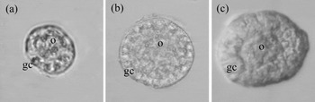
Early-stage follicles, i.e. primordial, primary and secondary, were isolated using the mechanical procedure described previously (Lucci et al., Reference Lucci, Amorim, Bao, Figueiredo, Rodrigues, Silva and Goncalves1999). Briefly, ovaries were cut individually into small fragments using a tissue chopper (Mickle Laboratory Engineering) adjusted to 75 µm. The fragments were then placed in PBS containing 5% bovine serum albumin (Sigma) and aspirated 40 times using a large Pasteur pipette (diameter ∼1600 µm) and 40 times with a smaller pipette (diameter ∼600 µm). The suspension was then filtered successively through 500 and 100 µm nylon mesh filters. After repeated washing to completely remove the stromal cells, 15 primordial (Fig. 1a), primary (Fig. 1b) or secondary (Fig. 1c) follicles were carefully selected based on the morphological shape and number of granulosa cell layers around the oocyte and placed in separate Eppendorf tubes. All samples were stored at −80 °C until the RNA was extracted.
Isolated goat ovarian follicles. (a) Primordial follicle, an oocyte surrounded by single layer of flattened/cuboidal granulosa cells, (b) primary follicle with one layer of cuboidal granulosa cells and (c) secondary follicle with more than two layers of granulosa cells. gc, granulosa cells; o, oocyte.

From a second group of ovaries (n = 20), cumulus–oocyte complexes (COCs) were aspirated from small (1–3 mm) and large (3–6 mm) antral follicles using an 18-gauge needle attached to a tube in line with a vacuum pump. From the follicle content thus collected, compact COCs were selected as described by van Tol & Bevers (Reference van Tol and Bevers1998). Thereafter, the cumulus was separated from the oocyte by a combination of vortexing and aspiration via a narrow-bore Pasteur pipette. Mural granulosa cells were scraped off from follicular walls recovered from dissected antral follicles in which the COCs had been removed to avoid contamination by cumulus cells. Denuded oocytes, cumulus and mural granulosa cells were washed four times in PBS and packed in tubes in groups of either 10 denuded oocytes, cumulus cells from 10 COCs, or samples of mural granulosa, and then stored at −80 °C until RNA extraction.
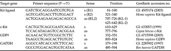
To collect theca cells, small (n = 10) and large antral follicles (n = 10) were isolated from goat ovaries (n = 5) and dissected free of stromal tissue using forceps, as described previously for bovine ovaries (van Tol & Bevers, 1998). The follicles were then bisected and the granulosa cells scraped off using a scalpel blade. Next, the theca cell layers were vortexed for 1 min in 1 ml HEPES-buffered M199 (Gibco BRL, Paisley, UK) supplemented with penicillin/streptomycin, trans-ferred to a fresh 1 ml of buffer, vortexed for another minute, washed twice in 2 ml HEPES-buffered M199, collected and stored at −80 °C. To investigate the possibility of theca cell contamination by adhering mural granulosa cells we used specific primers (Table 1) to detect growth differentiation factor 9 (GDF9) that is expressed in goat mural granulosa cells but not in the theca (Silva et al., Reference Silva, van den Hurk, van Tol, Roelen and Figueiredo2004). From another group of ovaries (n = 5), small pieces of corpus luteum, medulla and surface epithelium were collected and stored at −80 °C until RNA extraction. Three samples of each tissue sample were recovered and analysed.
Oligonucleotide primers used for PCR analysis of goat cells and tissues

s, sense; as, antisense; R1, round 1; R2, round 2 or heminesting.
Extraction of total RNA and reverse transcription
Isolation of total RNA combined with on-column DNase digestion was performed using the RNeasy mini-kit and the RNase-free DNAse set (Qiagen). According to the manufacturer's instructions, 350 µl lysis buffer was added to each frozen samples and the lysate aspirated through a 20-gauge needle before being centrifuged at 10 000 g for 3 min at room temperature. The lysates of theca cells, corpus luteum, medulla and ovarian surface samples were then subjected to a proteinase K treatment (6.7 mAU/ml, Qiagen) at 55 °C for 10 min. Thereafter, all lysates were diluted 1:1 with 70% ethanol and introduced onto a mini-column. After binding of the RNA to the column, DNA digestion was performed using RNase-free DNase (340 Kunitz units/ml) for 15 min at room temperature. After washing the column three times, the RNA was eluted with 30 µl RNAse-free water.
Prior to the reverse transcription reaction, the eluted RNA samples were incubated for 5 min at 70 °C, and chilled on ice. Reverse transcription was then performed in a total volume of 20 µl made up of 10 µl of sample RNA, 4 µl 5× reverse transcriptase buffer (Gibco BRL), 8 units RNasin, 150 units Superscript II reverse transcriptase (BRL), 0.036 U random primers (Life Technologies) and containing 10 mM dithiothreitol (DTT) and 0.5 mM of each dNTP. The mixture was incubated for 1 h at 42 °C, for 5 min at 80 °C and then stored at −20 °C. Minus RT blanks were prepared under the same conditions, but without inclusion of reverse transcriptase.
Amplification of KL and c-Kit cDNA by PCR
PCR reactions were carried out in 200 µl tubes (Biozym), using 1 µl cDNA as template in 25 µl of a mixture containing 2 mM MgCl2, 200 µM of each dNTP, and 0.5 µM each of primers and 0.625 units Taq DNA polymerase (HotStarTaq, Qiagen) in 1× PCR buffer. The primers used for amplification of KL, c-Kit, GDF9 and glyceraldehyde-3-phosphate dehydrogenase (GAPDH) are presented in Table 1. GAPDH was used as an internal control, since transcripts for this gene are expected to be present at the same level in all cell types. For KL, the primers spanned the alternatively spliced exon, thus enabling simultaneous detection of mRNA for both the soluble form (KL-1) and the membrane-bound form (KL-2). Since the position of the missing exon (KL-2) in the goat sequence of KL is not known, the primers for KL were designed based on the human sequence.
The thermal cycling profile for the first round of PCR was: initial denaturation and activation of the polymerase for 15 min at 94 °C, followed by 40 cycles of 15 s at 94 °C, 30 s at 55 °C and 45 s at 72 °C. Final extension was for 10 min at 72 °C. During the amplification of KL cDNA, heminesting was used to increase the specificity and sensitivity, using a different sense primer. For heminesting, 1 µl of the first round product was transferred to another 200 µl tube containing 24 µl amplification mixture, and amplified for 30 cycles using the same thermal cycling profile. All reactions were performed in a 24-well thermocycler (Perkin-Elmer). Finally, 10 µl of the product was resolved by electrophoresis in 1% agarose gels containing ethidium bromide. A 100 base pair (bp) DNA ladder (Gibco BRL) was included as a reference for fragment size.
A standard sequencing procedure (ABI PRISM 310 Genetic Analyzer, Applied Biosystems) was used to verify the specificity of the PCR products.
Results
Immunohistochemistry
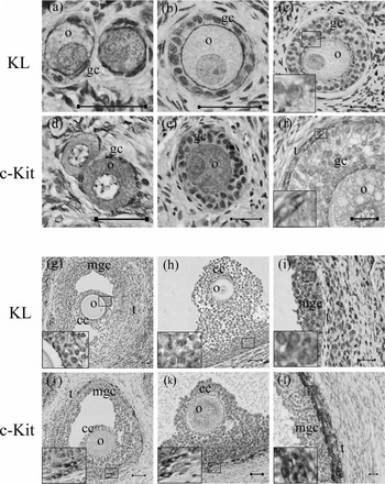
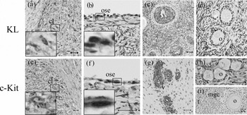
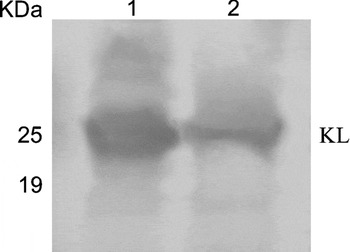
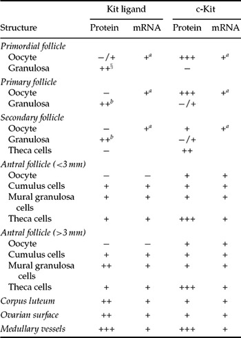
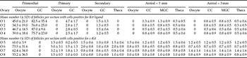
All stages of follicle development (primordial, primary, secondary and antral follicles) and corpora lutea were identified within the ovarian sections. KL was detected in granulosa cells of follicles from the primordial stage onwards (Fig. 2, Table 2) and, in primordial, primary and secondary follicles the most intensive reaction was observed at the apical side of the granulosa cells where they border the oocyte (Fig. 2a–c). Independent of the animal, approximately half of the oocytes from primordial follicles also stained positively for KL (Fig. 2a, Table 3). In small antral follicles, KL staining intensity was weaker than in preantral follicles and distributed equally across the cumulus and mural granulosa cells (Fig. 2g, Table 2). In large antral follicles, both cumulus and theca cells showed a weaker immunoreaction than the corresponding mural granulosa cells (Fig. 2h, i). In addition, strong KL immunoreactivity is observed in corpora lutea (Fig. 3a), ovarian surface epithelium (Fig. 3b) and vascular smooth muscle (Fig. 3c). In the control sections for KL protein (Fig. 3d), in which the specific antibody was replaced with normal IgG, no positive staining was observed. Additionally, western blot analysis showed, in goat and mouse ovaries, a band of molecular size approximately 25 kDa (Fig. 4), which is consistent with the size of KL protein.
Kit ligand (KL) and c-Kit immunoreactivity in goat ovarian follicles. (a, d) Primordial follicle, (b, e) primary follicle, (c, f) secondary follicle, (g, j) small antral follicle, (h, k) COC of a large antral follicle, (i, l) mural granulosa and theca cells from a large antral follicles. Inserts: higher magnification showing immunoreaction in the cell cytoplasm. cc, cumulus cells; gc, granulosa cells; mgc, mural granulosa cells; o, oocyte; t, theca cells. Scale bars represent 25 µm.

Kit ligand (KL) and c-Kit immunoreactivity in goat: (a, e) corpus luteum, (b, f) ovarian surface epithelium, (c, g) blood vessels and (d, h, i) negative control. Inserts: higher magnification showing immunoreaction in the cell cytoplasm. cl, corpus luteum; cc, cumulus cells; gc, granulosa cells; mgc, mural granulosa cells; o, oocyte; ose, ovarian surface epithelium; t, theca cells; v, vessels. Scale bars represent 25 µm.

Western blot analysis of Kit ligand (KL) expression in mouse (1) and goat ovaries (2). The band of approximately 25 kDa is consistent with the size of KL protein.

Localization of mRNA and relative intensity of immunohistochemical staining for KL and c-Kit in the ovaries of goats
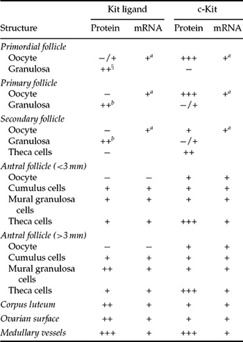
Immunoreaction: −/+, occasionally found; −, absent; +, weak; ++, moderate; +++, strong.
a Whole follicles.
b Reaction mainly observed at the junction of granulosa cells and oocyte.
Mean number of follicles (± SD) per section with oocytes, granulosa or theca cells positive for Kit ligand or c-Kit in eight different ovaries
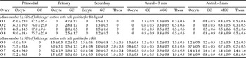
Data are from 20 sections from four different ovaries.
GC, granulosa cells; MGC, mural granulosa cells; O, ovary.
c-Kit was immunohistochemically demonstrable in the oocytes of follicles at all stages, i.e. primordial, primary, secondary, small and large antral follicles (Fig. 2, Table 2). The staining intensity in oocytes of primordial and primary follicles was stronger than in late-staged follicles. In addition, moderate to strong c-Kit staining was observed in theca cells of late secondary, small and large antral follicles (details in Fig. 2f, j, k, l). Occasionally, weak c-Kit immunoreaction was observed in the granulosa cell of early or late-staged follicles (Fig. 2e, f, j, k). Finally, immunoreactivity for c-Kit was clearly visible in corpora lutea (Fig. 3e), ovarian surface epithelium and vascular smooth muscle and endothelial cells (Fig. 3f, g). Table 3 shows the number of follicles from different categories analysed in eight different ovaries. No significant inter-ovary variation (p > 0.05) in the immunohistochemical staining for either KL or c-Kit was observed (Table 3). Control sections for c-Kit, in which the specific antibody was replaced with normal IgG, showed absence of staining (Fig. 3h). When the antibody was preabsorbed with its blocking peptide, only a weak background staining was observed (Fig. 3i).
Expression of mRNA for KL and c-Kit in goat ovaries
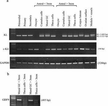
The first round of amplification using primers for KL yielded specific products for both KL-1 and KL-2 only in samples of cDNA prepared from mural granulosa cells of antral follicles and corpora lutea. After heminesting, however, amplification of cDNA from primordial, primary and secondary follicles resulted in specific products for soluble KL-1 mRNA, but not for KL-2 mRNA. When cDNA from either cumulus, mural granulosa or theca cells collected from small or large antral follicles was amplified, products for both KL-1 and KL-2 were observed after heminest-ing. KL mRNA expression was not detected in oocytes from either small or large antral follicles (Fig. 5a), which confirms the absence of contaminating cumulus cells. In addition, we detected GDF9 mRNA in mural granulosa cells but not in the theca (Fig. 5b), confirming the purity of theca samples. KL-1 and KL-2 mRNA expression were also detected in corpus luteum, ovarian medulla and ovarian surface epithelium (Fig. 5a). Amplification of – RT blanks or water controls yielded no specific products in any of the reactions (results not shown).
Expression of (A) KL, c-Kit and GAPDH mRNA in different follicle and cell types in goat ovaries and (B) GDF9 in mural granulosa and theca cells. Follicle and cell types are indicated at the top. One-hundred base pair ladders are included as markers for fragment size.

Amplification of cDNA from primordial, primary and secondary follicles and from oocytes, cumulus, mural granulosa and theca cells from small or large antral follicles using specific primers for c-Kit, resulted in abundant product after one round of amplifica-tion in all cases. c-Kit expression was also detected in corpus luteum, ovarian surface epithelium and medullary tissue (Fig. 5a). The expression of the housekeeping gene (GAPDH) is also illustrated in Fig. 5a. Amplification of – RTblanks or water controls yielded no specific products in any of the reactions (results not shown).
Sequence analysis of the amplified c-Kit and KL products confirmed their specificity when compared with the published c-Kit (GI: 633053) and KL-1 (GI: 16580734) mRNA from goats. Sequencing of KL-2 showed that, compared with the sequence of KL-1 (GI: 16580734), an 84 base pair exon is absent between nucleotides 680 and 764.
Discussion
Over the last decade, several papers have demonstrated the presence of a functional KL/c-Kit system in mammalian ovaries, in particular those of the mouse and the sheep (for reviews see Driancourt et al., Reference Driancourt, Reynaud, Cortvrindt and Smitz2000, and van den Hurk & Zhao, Reference van den Hurk and Zhao2005). The present study examined the distribution of KL and c-Kit mRNA and protein in goat ovaries, to explore possible differences among species. With regard to KL, we demonstrated the presence of protein in granulosa cells of primordial, primary and secondary follicles, particularly where the granulosa borders the oocyte and a zona pellucida has not yet been formed. A similar distribution of KL was described previously for monkey primordial follicles (Gougeon & Busso, Reference Gougeon and Busso2000). On some occasions we detected KL protein in the oocyte of caprine primordial follicles, a finding that confirmed the previous descriptions of KL in murine and human oogonia and oocytes from primordial follicles (Kang et al., Reference Kang, Lee, Lee, Rha, Song and Park2003; Hoyer et al., Reference Hoyer, Byskov and Mollgard2005). Using RT-PCR, the current study demonstrated the expression of mRNA for the soluble KL-1 in caprine primordial, primary and secondary follicles. The detection of KL mRNA in granulosa cells of early-stage follicles was previously described in sheep (Tisdall et al., Reference Tisdall, Quirke, Smith and McNatty1997; McNatty et al., Reference McNatty, Heath, Lundy, Fidler, Quirke, O'Connell, Smith, Groome and Tisdall1999), mouse (Motro & Bernstein, Reference Motro and Bernstein1993) and human (Laitinen et al., Reference Laitinen, Rutanen and Ritvos1995).
c-Kit mRNA and protein were both detected in the oocyte cytoplasm of both early- and later-staged goat follicles. This suggests that, in the goat, the oocyte is a target for granulosa cell-derived KL, as has been proposed in the sheep (Clark et al., Reference Clark, Tisdall, Fidler and McNatty1996; Tisdall et al., Reference Tisdall, Fidler, Smith, Quirke, Stent, Heath and McNatty1999), mouse (Motro & Bernstein, Reference Motro and Bernstein1993) and monkey (Gougeon & Busso, Reference Gougeon and Busso2000). In vitro, KL has been shown to be essential for mouse primordial follicle activation (Parrott & Skinner, Reference Parrott and Skinner1999). Similarly, injection of a KL antibody into the ovaries of mice severely retarded early folliculogenesis (Yoshida et al., Reference Yoshida, Takakura, Kataoka, Kunisada, Okamura and Nishikawa1997), while ovaries of mice carrying a mutation in the Steel gene (Kuroda et al., Reference Kuroda, Terada, Nakayama, Matsumoto and Kitamura1988; Huang et al., Reference Huang, Manova, Packer, Sanchez, Bachvarova and Besmer1993; Bedell et al., Reference Bedell, Brannan, Evans, Copeland, Jenkins and Donovan1995) contained only follicles arrested at early stages of development. Granulosa cell-derived KL also appeared to promote the formation of theca cell layers around mouse primary and secondary follicles (Parrott & Skinner, Reference Parrott and Skinner1997, Reference Parrott and Skinner2000), which suggests that KL may act as a theca cell organizer.
In caprine antral follicles, KL protein was present in cumulus and mural granulosa cells, and occasionally in theca cells. At these sites, mRNA for both the soluble and membrane-bound KL subtypes were also detectable. With regard to its presence in theca cells it is unclear whether this mRNA is derived from theca cells per se or from blood vessel tissue (endothelium or smooth muscle) present within the thecal layer. The expression of c-Kit by oocytes, cumulus, mural granulosa and thecal cells of both small and large antral follicles argues for autocrine and paracrine roles for KL in directing the development of goat antral follicles. Patterns of c-Kit expression were similar in sheep (Clark et al., Reference Clark, Tisdall, Fidler and McNatty1996; Tisdall et al., Reference Tisdall, Fidler, Smith, Quirke, Stent, Heath and McNatty1999; Juengel et al., Reference Juengel, Quirke, Tisdall, Smith, Hudson and McNatty2000), but different in mice (Motro & Bernstein, Reference Motro and Bernstein1993) and brushtail possums (Eckery et al., Reference Eckery, Lawrence, Juengel, Greenwood, McNatty and Fidler2002), in which c-Kit was not expressed by granulosa cells. In vitro studies with antral follicles have demonstrated that KL can promote mouse oocyte growth (Eppig, Reference Eppig2001; Nilsson & Skinner, Reference Nilsson and Skinner2001) and inhibits expression of bone morphogenetic protein-15 mRNA in mice (Otsuka & Shimasaki, Reference Otsuka and Shimasaki2002) and meiotic maturation of rat oocytes (Ismail et al., Reference Ismail, Okawara, Fryer and Vanderhyden1996). In addition, KL promoted granulosa cell proliferation and steroidogenesis in mice (Reynaud et al., Reference Reynaud, Cortvrindt, Smitz and Driancourt2000) and sows (Brankin et al., Reference Brankin, Mitchell, Webb and Hunter2003) as well as growth and differentiation of theca cells in cattle (Parrott & Skinner, Reference Parrott and Skinner1997, Reference Parrott and Skinner2000) and rats (Huang et al., Reference Huang, Weitsman, Dykes and Magoffin2001). These data imply that, depending on the species, different folli-cular compartments may contain the receptor for KL.
In addition to follicles, c-Kit and KL (-1 and -2) mRNA and protein were both also detected in goat corpora lutea, suggesting a possible role of the KL/c-Kit system in luteal activity. Similar immunolocalization of KL and c-Kit has been described previously in ovine luteal cells (Gentry et al., Reference Gentry, Smith, Anthony, Zhang, Long and Smith1996, Reference Gentry, Smith, Leighr, Bao and Smith1998). KL was, however, undetectable in murine corpora lutea (Manova et al., Reference Manova, Huang, Angeles, De Leon, Sanchez, Pronovost, Besmer and Bachvarova1993). During their development, maintenance and regression, luteal cells change in composition, size and function. In tissues other than corpora lutea, such as hematopoietic and muscular tissue, KL has been shown to play a critical role in the regulation of such processes (Broudy, Reference Broudy1997; Miyamoto et al., Reference Miyamoto, Sasaguri, Sasaguri, Azakami, Yasukawa, Kato, Arima, Sugama and Morimatsum1997). In sheep, the level of KL mRNA expression within corpora lutea does not change during the luteal phase (Gentry et al., Reference Gentry, Smith, Anthony, Zhang, Long and Smith1996), which could mean that KL has a continuous role during development, maintenance and regression of a corpus luteum.
The detectable expression of both mRNA (KL-1 and KL-2) and proteins for KL and c-Kit in goat ovarian surface epithelium also suggests a potential role for KL/c-Kit at this site. Expression of KL and its receptor c-Kit in ovarian surface epithelium has been described previously in human, cattle and sheep (Tisdall et al., Reference Tisdall, Quirke, Smith and McNatty1997; Parrott et al., Reference Parrott, Mosher, Kim and Skinner2000). In the rat, ovarian surface epithelium cells expressed predominantly KL-1 mRNA (Ismail et al., Reference Ismail, Cada and Vanderhyden1999). In vitro, KL has been shown to stimulate growth of ovarian surface epithelium in mice (Parrott et al., Reference Parrott, Mosher, Kim and Skinner2000).
In the present study with goats, KL-1, KL-2 and c-Kit mRNA were also expressed in ovarian medullary tissue. These mRNAs could be derived from blood vessel walls, since the corresponding KL and c-Kit proteins were demonstrated at these sites. Such demonstration is supported by similar findings that were obtained with sheep ovaries, using in situ hybridization (Tisdall et al., Reference Tisdall, Quirke, Smith and McNatty1997, Reference Tisdall, Fidler, Smith, Quirke, Stent, Heath and McNatty1999). The presence of KL and c-Kit in blood vessel walls probably reflects a local function within the circulatory system (Miyamoto et al., Reference Miyamoto, Sasaguri, Sasaguri, Azakami, Yasukawa, Kato, Arima, Sugama and Morimatsum1997), although a paracrine influence on folliculogenesis and/or luteogenesis cannot be excluded.
In summary, the present study demonstrates a KL/c-Kit system in goat ovarian follicles at all stages of follicle development, corpora lutea, ovarian surface epithelium and ovarian medulla. This widespread distribution of the KL/c-Kit system shows that, in goat ovaries, it may play an important role in various processes, including folliculogenesis and luteal activity.
Acknowledgements
This work was supported by Coordenação de Aperfeiçoamento de Pessoal de Nível Superior – CAPES. The authors thank Mr Fritz Kindt (Faculty of Biology, Utrecht, The Netherlands) for helping with photography and Dr Tom Stout (University of Utrecht) for suggestions and correcting the English in the manuscript. We would also like to acknowledge Karianne Peterson (University of Utrecht, The Netherlands) for providing some of the goat ovaries, and Susana M. Chuva de Sousa Lopes (Hubrecht Laboratory, Utrecht, The Netherlands) for providing mouse ovaries.