Introduction
Schistosoma is a blood fluke causing a complex chronic disease in humans. According to recent estimates, schistosomiasis afflicts 252 million people, including 40 million women at reproductive age (Olveda et al., Reference Olveda, McManus and Ross2016; Friedman et al., Reference Friedman, Olveda and Mirochnick2018). The World Health Organization reported that in 2014, at least 258 million people worldwide required regular and frequent preventive treatment for schistosomiasis. However, the disease has recently appeared in Europe on the French island of Corsica (Merrifield et al., Reference Merrifield, Hotez and Beaumier2016). Among human schistosomes, Schistosoma mansoni is the main cause of intestinal schistosomiasis, a disease characterized by weakness, weight loss, headaches, nausea, vomiting, dysentery, hepatosplenomegaly and eosinophilia.
Praziquantel (PZQ) is the main drug used for treatment of all forms of human schistosomiasis, being effective and well-tolerated. However, the development of resistance in some strains necessitates searching for new schistosomicidal agents (El Bialy et al., Reference El Bialy, Taman and El-Beshbishi2013; Aleixo de Carvalho et al., Reference Aleixo de Carvalho, Geraldo and de Moraes2015; Taman et al., Reference Taman, El-Beshbishi and Bardicy2016). This drug research is mainly performed by testing the anti-parasitic activity of natural compounds (Aleixo de Carvalho et al., Reference Aleixo de Carvalho, Geraldo and de Moraes2015; de Moraes, Reference de Moraes2015; El-Beshbishi et al., Reference El-Beshbishi, Saleh and Abd El-Mageed2019), drug re-purposing (El-Beshbishi et al., Reference El-Beshbishi, Taman and El-Malky2013a, Reference El-Beshbishi, Taman and El-Malkyb; Taman et al., Reference Taman, El-Beshbishi, El-Tantawy, El-Hawary and Azab2014) and by the synthesis of new series of compounds with anthelmintic activity (Moreira et al., Reference Moreira, Piló-Veloso and de Mello2007; El Bialy et al., Reference El Bialy, Taman and El-Beshbishi2013; Spivak et al., Reference Spivak, Keiser and Vargas2014).
Schistosomes are known to feed on blood, which is important for development and sexual maturation. Several proteolytic enzymes can degrade haemoglobin into peptides, amino acids and heme (Oliveira et al., Reference Oliveira, d'Avila and Torres2000), but free heme has deleterious effects on blood-feeders; thus, the crystallization of heme into hemozoin (HZ) is the detoxification mechanism by which schistosomes escape the harmful effects of heme (Oliveira et al., Reference Oliveira, d'Avila and Tempone2004).
Quinoline pharmacophore has a diverse variety of pharmacological activities, which include anti-tubercular (Fan et al., Reference Fan, Wu, Cheng, Zhang and Feng2018a), anti-bacterial (Zhang et al., Reference Zhang, Zhang and Pan2018), anti-protozoan (Fan et al., Reference Fan, Cheng, Wu, Liu, Zhang, Xu and Feng2018b), anti-fungal (Musiol et al., Reference Musiol, Jampilek and Buchta2006), anti-oxidant (Wilhelm et al., Reference Wilhelm, Ferreira and Pinz2017), anti-proliferative (Zhang et al., Reference Zhang, Xiao and Su2017), anti-tumour (Sharma et al., Reference Sharma, Monika and Archana2013), anti-inflammatory (Upadhyay et al., Reference Upadhyay, Dodia, Khunt, Chaniara and Shah2018), anti-hypertensive (Muruganantham et al., Reference Muruganantham, Sivakumar, Anbalagan, Gunasekaran and Leonard2004) and anti-HIV effects (Luo et al., Reference Luo, Tan and Zeng2010). Moreover, quinolines can be used for treatment of lupus (Bengtsson et al., Reference Bengtsson, Sturfelt and Lood2012) and neurodegenerative diseases (Wang et al., Reference Wang, Hu, Yang, Feng, Li, Huang and Chan2018).
Quinoline compounds have been used for many years as a potent treatment of malaria through hindering the crystallization of heme into HZ, subsequently killing the blood stages of Plasmodium through oxidative stress (Foley & Tilley, Reference Foley and Tilley1998). The same mechanism has been proven to be employed in schistosomes. Consequently, interference with HZ formation represents an attractive anti-schistosomal pathway. Besides, this pathway is specific for blood parasites and is not present in the host, which makes it an interesting drug target (Chen et al., Reference Chen, Shi and Sullivan2001).
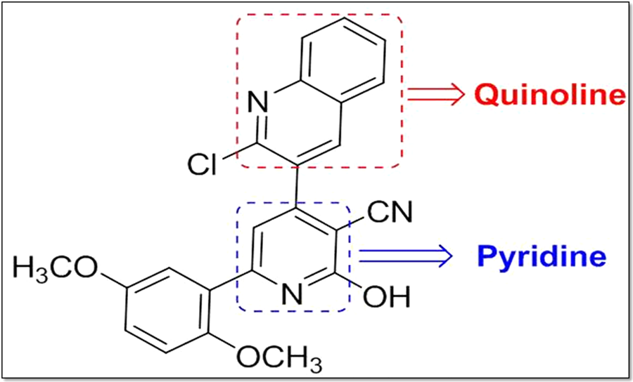
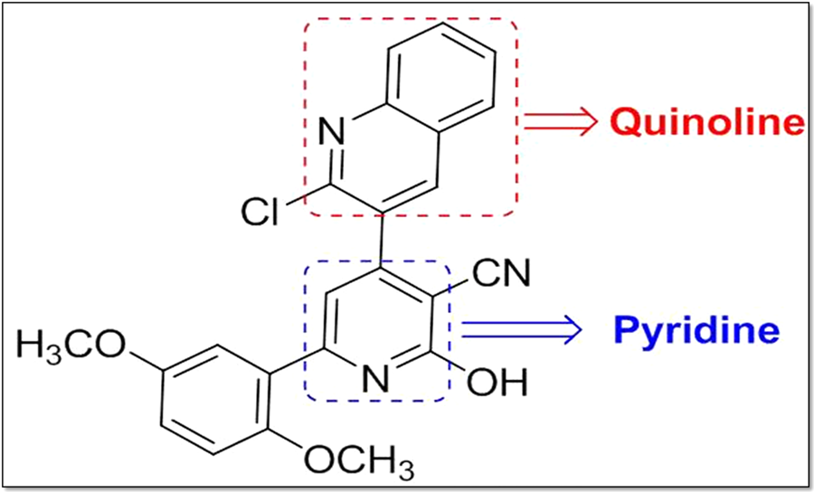
The compound PPQ-8 (4-[2-chloroquinolin-3-yl]-6-[2,5-dimethoxyphenyl]-2-oxo-1,2-dihydropyridine-3-carbonitrile) is a novel biological potent heterocyclic agent. It was designed and synthesized via a combination of both therapeutically active moieties: 2-chloroquinoline as lipophilic and the diverse heterocyclic ring structure (having a known anti-protozoan activity) directly attached to C3 of the quinoline ring.
PPQ-8 was previously reported as an anti-Toxoplasma agent (Abd Elgawad et al., Reference Abd Elgawad, Alhusseiny and Taman2019). Herein, we investigated the effects of PPQ-8 on S. mansoni-infected mice by evaluating some parasitological, histopathological, general and biochemical parameters.
Materials and methods
Tested compound
Compound PPQ-8 (fig. 1) was previously described (Abd Elgawad et al., Reference Abd Elgawad, Alhusseiny and Taman2019).
Chemical structure of compound PPQ-8 (4-[2-chloroquinolin-3-yl]-6-[2,5-dimethoxyphenyl]-2-oxo-1,2-dihydropyridine-3-carbonitrile).
Animals, parasite and PPQ-8 treatment in vivo
Female Balb/c mice (6–7 weeks, 25–30 gm) were infected with the Egyptian strain of S. mansoni by subcutaneous injection of 80 ± 5 cercariae, freshly shed from Biomphalaria alexandrina snails.
Snails were purchased from the Schistosome Biological Supply Centre, Theodor Bilharz Research Institute, Imbaba, Giza, Egypt. Mice were kept at the animal care facility at Mansoura Experimental Research Center in controlled environmental conditions (22–25°C, 12/12 h dark/light cycle), and fed on a balanced dry standard commercial pellet diet and sterile water ad libitum.
Infected mice were randomly allocated into four groups, each containing ten mice:
– Group I: infected untreated;
– Group II: infected and treated with dimethyl sulfoxide (DMSO) as vehicle control;
– Group III: infected and treated with PPQ-8 (20 mg/kg); and
– Group IV: infected and treated with PPQ-8 (40 mg/kg).
Treatment protocol was a single dose administered at day 49 post infection (PI).
PPQ-8 was dissolved in DMSO, and then given by oral gavage using a ball-tipped feeding needle in a volume of 200 µl/mouse. Mice were deprived of food 2 h before administration, and then allowed to eat 1 h after treatment. Ten weeks PI, mice were weighed and then euthanized by intraperitoneal injection of 100 mg/kg of sodium thiopental.
Evaluation of PPQ-8 compound efficacy
Study of parasitological criteria
After being euthanized, worms were recovered by mesenteric perfusion (Smithers & Terry, Reference Smithers and Terry1965), separated by sex, counted and examined by bright field microscopy. Liver and small intestine of each mouse were collected, washed with phosphate-buffered saline (PBS), sliced into small pieces and subsequently digested overnight in 10 ml of 4% potassium hydroxide at 37°C, as previously described (Cheever, Reference Cheever1968). Aliquots of digested tissues (10 µl) were placed onto a glass coverslip and the number of eggs was counted using light microscopy then multiplied by 103 to determine egg/gm (Kamel et al., Reference Kamel, Cheever and Elwi1977). The percentages of the different egg developmental stages (oogram pattern) were examined according to Pellegrino et al. (Reference Pellegrino, Oliveira, Faria and Cunha1962).
Histopathological studies
Livers collected after mesenteric perfusion from controls and PPQ-8-treated mice were washed with PBS, sliced into small pieces, fixed in 5 ml of 2.5% glutaraldehyde and 4% formalin. Tissue slices were embedded in paraffin, stained with haematoxylin and eosin, and observed under the microscope. Granuloma count and diameter were determined in three successive microscopic fields of serial tissue sections > 250 µm apart. The measurement of granuloma diameter was carried out using an ocular micrometre.
Liver and spleen indices
The weights of liver and spleen were recorded to calculate the ratio between their weights to the total body weight (liver and spleen indices).
Nitric oxide determination
Level of nitric oxide (NO) in liver tissue homogenate was measured using a nitric oxide assay kit (Biodiagnostic Company, Giza, Egypt, catalogue number 2533), according to the manufacturer's recommendations.
Data analysis
Data were analysed using Statistical Package for Social Sciences (SPSS) software (SPSS Inc., Chicago, USA), version 16.0. The normality of data was first tested with the one-sample Kolmogorov–Smirnov test. Continuous variables were presented as means ± standard deviation. Analysis of variance followed by post-hoc testing using Fisher's least significant difference were used to compare the means of more than two groups. The results were considered significant when P ≤ 0.05.
Comparison was made between the infected non-treated group and different treatment groups (II–IV) to detect any significant differences in measured variables, and between different treatment groups to evaluate variable responses obtained when using the compound PPQ-8 at two different doses. Superscripts ‘*’ were used to refer to statistically significant difference between PPQ-8-treated group(s) and both control groups, while the letter superscripts were used to describe statistically significant differences between PPQ-8-treated group(s).
The percentage of reduction between the treated group and the untreated control group was assessed using the following formula: (mean value of the untreated group − mean value of the treated group) × 100 / mean value of the untreated group.
Results
Parasitological studies
The oral treatment of S. mansoni-infected mice with 20 mg/kg PPQ-8 resulted in significant reductions in total worm count, male and female worms burden, with percentage reductions of 32.2%, 23.3% and 39.7%, respectively, while PPQ-8 given as 40 mg/kg resulted in a higher reduction in total worm count (55.2%), male worms (48.4%) and female worms (61.2%), in comparison with untreated mice, with statistically significant difference between the two treated groups (table 1).
Effect of compound PPQ-8 on adult worm burden in Schistosoma mansoni-infected mice.

N, number of mice dead; DMSO, dimethyl sulfoxide.
Values are expressed as means ± standard deviation. Values between parentheses refer to the percentage of reduction compared with infected non-treated group.
a Analysis of variance test.
* Significant difference from infected non-treated and DMSO-treated groups at P < 0.05.
** Significant difference from infected non-treated and DMSO-treated groups at P < 0.001.
*** Significant difference from PPQ-8 (20 mg/kg)-treated group at P < 0.05.
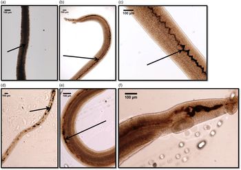
Examination by bright field microscopy revealed control worms with well-developed gut and HZ content filling the gut (fig. 2a–c), while in worms recovered after treatment with PPQ-8, a reduction in HZ content was clearly visible in both male and female worms (fig. 2d–f), with no visible destructive effect on the tegument.
Bright field microscopy showing (a) female, (b, c) male adult control worms with well-developed gut and HZ content filling the gut. With 20 mg/kg PPQ-8, a reduction in HZ content was clearly visible in both female (d) and male (e) worms, with no visible destructive effect on the tegument. The same observations were reported with male worms treated with 40 mg/kg PPQ-8 (f). Arrows point to HZ content in the intestinal caeca.

The treatment of S. mansoni-infected mice with 20 mg/kg PPQ-8 induced a significant reduction in hepatic and intestinal egg load by 63% and 57.4%, respectively, while doubling the dose resulted in a more significant reduction in tissue egg load (by 83.6% and 73.5%, respectively), and the difference between the PPQ-8-treated groups was significant (table 2).
Effect of compound PPQ-8 on hepatic and intestinal egg load in Schistosoma mansoni-infected mice.

N, number of mice dead; DMSO, dimethyl sulfoxide.
Values are expressed as means ± standard deviation. Values between parentheses refer to the percentage of reduction compared with infected non-treated group.
a Analysis of variance test.
* Significant difference from infected non-treated and DMSO-treated groups at P < 0.001.
** Significant difference from PPQ-8 (20 mg/kg)-treated group at P < 0.001.
*** Significant difference from PPQ-8 (20 mg/kg)-treated group at P < 0.05.
On studying the effects of PPQ-8 on the percentage of different egg stages, the administration of a dose of 20 mg/kg induced a insignificant decrease in immature ova to 50.7%, a significant decrease in mature eggs to 33.6% and a significant increase in dead ova to 15.8%, compared to the non-treated group. However, a dose of 40 mg/kg significantly decreased immature and mature eggs to 38.3% and 23.3%, respectively, and significantly increased dead eggs to 38.4%, compared to untreated control mice (table 3).
Effect of compound PPQ-8 on oogram pattern in Schistosoma mansoni-infected mice.

N, number of mice dead; DMSO, dimethyl sulfoxide.
Values are expressed as means ± standard deviation. Values between parentheses refer to the percentage of reduction compared with infected non-treated group.
a Analysis of variance test.
* Significant difference from infected non-treated group at P < 0.001.
** Significant difference from infected non-treated and DMSO-treated groups at P < 0.05.
*** Significant difference from infected non-treated and DMSO-treated groups at P < 0.001.
**** Significant difference from PPQ-8 (20 mg/kg)-treated group at P ≤ 0.001.
Histopathological studies
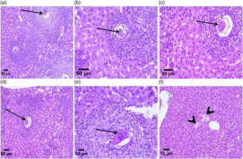
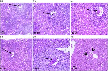
The examination of liver sections from infected untreated and DMSO-treated mice showed multiple large granulomas surrounding viable ova, with marked inflammation of the parenchyma (fig. 3a, b). As a consequence of both PPQ-8 treatment doses administration, smaller and well-circumscribed granulomas with degenerated or even absent ova, and improved liver inflammatory condition were observed (fig. 3c, d). Also, both treatment regimens resulted in a statistically significant reduction in both granuloma counts (75.4% and 89.1%, respectively) and diameter (26.2% and 47.4%, respectively), compared to the untreated group, as shown in table 4.
Histopathological study of liver sections of mice infected with Schistosoma mansoni euthanized ten weeks post infection. (a) Infected non-treated mice showing multiple irregularly outlined granulomas [Haematoxylin and Eosin (H&E) ×100]. (b) With higher magnification, there is mixed inflammatory cellular infiltrate (H&E ×200) surrounding the ova. (c) DMSO-treated mice showing irregularly outlined granuloma with mixed inflammatory cellular infiltrate, mainly eosinophils, surrounding the ova (H&E ×200). (d) Infected mice treated with 20 mg/kg PPQ-8 showing well-circumscribed granuloma (H&E ×100). (e) Higher magnification revealed granulomatous inflammatory reaction surrounding ova with eosinophils at the area of portal tract (H&E ×400). (f) Infected mice treated with 40 mg/kg PPQ-8 showing liver tissue with no detectable major pathological abnormalities (no detected bilharzial ova, granuloma or inflammatory cells) apart from the swelling of some hepatocytes (arrowheads). Arrows point to ova.
Effect of compound PPQ-8 on granuloma count and diameter in Schistosoma mansoni-infected mice.

N, number of mice dead; DMSO, dimethyl sulfoxide.
Values are expressed as means ± standard deviation. Values between parentheses refer to the percentage of reduction compared with infected non-treated group.
a Analysis of variance test.
* Significant difference from infected non-treated and DMSO-treated groups at P < 0.001.
** Significant difference from PPQ-8 (20 mg/kg)-treated group at P < 0.05.
*** Significant difference from PPQ-8 (20 mg/kg)-treated group at P < 0.001.
Liver and spleen indices
The treatment of S. mansoni-infected mice with 20 or 40 mg/kg PPQ-8 induced a significant reduction in liver index by 23.3% and 32.8%, respectively, compared to infected untreated mice (table 5). Also, the same drug regimens resulted in a significant reduction in spleen index by 37.5% and 45.3%, respectively, compared to non-treated mice (table 5).
Effect of compound PPQ-8 on liver index, spleen index and nitric oxide content in liver homogenates in Schistosoma mansoni-infected mice.

N, number of mice dead; DMSO, dimethyl sulfoxide.
Values are expressed as means ± standard deviation. Values between parentheses refer to the percentage of reduction compared with infected non-treated group.
a Analysis of variance test.
* Significant difference from infected non-treated and DMSO-treated groups at P < 0.001.
** Significant difference from PPQ-8 (20 mg/kg)-treated group at P < 0.001.
NO level
Interestingly, PPQ-8 given as 20 mg/kg or the double dose induced a significant decrease in NO levels by 22.5% and 47.2%, respectively, compared to the untreated mice group (table 5).
Discussion
Praziquantel is the drug used for controlling all forms of human schistosomiasis; however, the potential development of resistance is a major threat and the development of alternatives against Schistosoma species is mandatory (Cioli et al., Reference Cioli, Pica-Mattoccia and Basso2014). It is worth mentioning that both quinoline and quinoline derivatives exemplify a broad class of compounds, which are of great interest as they possess considerable biological and pharmacological potency (Rajesh, Reference Rajesh2018).
Recently, PPQ-8, a quinoline derivative, has been reported as a promising anti-Toxoplasma agent that probably acts by interfering with the apicoplast (Abd Elgawad et al., Reference Abd Elgawad, Alhusseiny and Taman2019). This result encouraged us to test its efficacy in mice infected with S. mansoni.
The present study showed that the oral administration of PPQ-8 as a single dose (20 or 40 mg/kg) resulted in significant reductions in total worm burden, male and female counts, with a more lethal effect on female worms. The reduction obtained in our study is less than that reported with mefloquine ‘a quinoline methanol’, but it is better than with other structurally related quinolines such as quinine and halofantrine (Keiser et al., Reference Keiser, Chollet and Xiao2009; Fahmy et al., Reference Fahmy, Rabia and Mansour2014); however, using different strains of S. mansoni should be considered.
It is interesting to note that adult female S. mansoni were more affected than males. Female worms’ specific drug susceptibility has been previously reported with artemisinins (Utzinger et al., Reference Utzinger, Chollet, Tu, Xiao and Tanner2002; Seif el-Din et al., Reference Seif el-Din, Al-Hroob and Ebeid2011; Li et al., Reference Li, Wang and Qu2012), compound naphthoquine phosphate and PTB-Iso (El-Beshbishi et al., Reference El-Beshbishi, Taman and El-Malky2013a; El Bialy et al., Reference El Bialy, Taman and El-Beshbishi2013), which may indicate sex-specific interference of the drug with the target or different drug target.
Schistosomes are blood-dwelling parasites, which feed on haemoglobin and release toxic heme. The aggregation of heme into insoluble HZ crystals is the first line of defence against the toxicity of the released heme (Oliveira et al., Reference Oliveira, Timm and Machado2002). The therapeutic effect of quinolines in schistosomes has been linked to interference with heme crystallization ( Oliveira et al., Reference Oliveira, d'Avila and Tempone2004; Corrêa Soares et al., Reference Corrêa Soares, Menezes and Vannier-Santos2009; Keiser et al., Reference Keiser, Chollet and Xiao2009). Although the detailed mechanism of action of PPQ-8 remains to be investigated, PPQ-8 has a quinoline ring, which raises the possibility that its schistosomicidal effect may be due to a reduction of HZ content in the gut, as detected by bright field microscopy. Subsequently, oxidative stress due to HZ reduction and increased concentration of free heme will be a leading cause of worm death. The prominent lethal effect on females is referred to their capability to ingest more red blood cells than male worms, and at a faster rate (Lawrence, Reference Lawrence1973).
Our results also showed that PPQ-8 treatment significantly reduced hepatic and intestinal egg load, which are a dose-dependent effect due to female death. Also, changing the oogram pattern in the form of decreasing immature eggs, especially with the higher dose, was reported, which could be due to the drug's lethal effect, mainly on female worms. The effect on mature eggs could be explained by the immunomodulatory effect of the drug on the developing eggs; however, the increased levels of dead eggs may be caused by the drug oxidant effect (El-Ahl et al., Reference El-Ahl, Hegazi and Mahmoud2013).
Since egg-induced immunopathology is the main cause of morbidity in schistosomes (Hoffmann et al., Reference Hoffmann, Wynn and Dunne2002), so, agents that significantly decrease ova production or increase their destruction would simultaneously reduce disease pathology and transmission. The current study reported that PPQ-8 significantly reduced the hepatic granuloma count and size with the presence of degenerated eggs, as a consequence of worm death and reduction in egg production.
In chronic S. mansoni infection, granuloma formation, portal hypertension, tissue reaction to worm-released metabolites and vascular changes lead to hepatomegaly and splenomegaly (Freitas et al., Reference Freitas, Barbosa, Fernandes and Andrade1999). Here, we reported reduction in liver and spleen indices with PPQ-8 treatment. This might be attributed to a reduction of egg deposition and an increase in the number of dead eggs, with a consequent reduction in granuloma count and the amelioration of hepatic pathology (Berhe et al., Reference Berhe, Myrvang and Gundersen2008), which lead to decreased portal hypertension and improvement of splenomegaly (Wang et al., Reference Wang, Zhou and Zheng2006). The ameliorating effect could also be due to the expected anti-inflammatory effect of PPQ-8 due to the quinoline ring (Upadhyay et al., Reference Upadhyay, Dodia, Khunt, Chaniara and Shah2018).
Schistosomiasis is associated with the liberation of free radicals and disturbance in the cellular anti-oxidant system. As the infection is settled, the host is subjected to oxidative stress or increased free radical formation (Tielens, Reference Tielens1994). The parasite is exposed to reactive oxygen species (ROS) produced by the immune system of the host. ROS leads to the release of toxic oxygen radicals, mainly hydrogen peroxide and superoxide radical anion during the respiratory burst (Aragon et al., Reference Aragon, Imani and Blackburn2008). The generation of oxygen-derived free radicals may be an initial non-specific defence reaction of the host toward parasitic infection, which is antagonized by the parasite anti-oxidant defence mechanism (Aragon et al., Reference Aragon, Imani and Blackburn2008).
In the present study, elevation in the level of NO in the control group was observed in schistosome-infected mice due to increased oxidative stress with excess tissue damage, and failure of the parasite anti-oxidant defence mechanisms to prevent the formation of excessive free radicals. With PPQ-8 treatment, reduction in NO level was detected, mostly due to a reduction in egg production, the main stimulus for NO formation (Seif el-Din et al., Reference Seif el-Din, Al-Hroob and Ebeid2011), with improvement of the stress condition.
The exact mechanism of anti-oxidant activity of PPQ-8 is not clear. PPQ-8 is structurally related to quinoline; hence, its anti-oxidant effect could be explained by stimulation of the anti-oxidant enzymes or the inhibition of lipid peroxidation. Early research reported that the injection of 20 mg/kg of chloroquine to rats is followed by an increase in the activity of super oxide dismutase (SOD) and catalase (CAT) enzymes (Magwere et al., Reference Magwere, Naik and Hasler1997). Moreover, Fahmy et al. (Reference Fahmy, Rabia and Mansour2014) recorded increased levels of CAT and amelioration of the oxidative status of S. mansoni-infected mice treated with mefloquine.
Schistosomes are covered with the tegument, representing a protective layer against host defence mechanisms. Although mefloquine has been reported to cause extensive damage of the adult worm tegument (Manneck et al., Reference Manneck, Haggenmuller and Keiser2010), PPQ-8 treatment didn't cause any observed tegumental alternation in worms recovered from mice, probably due to different drug targets.
The schistosome tegument is a lipid-rich membrane that serves as an effective barrier to most hydrophilic molecules (Skelly & Alan Wilson, Reference Skelly and Alan Wilson2006), which can be suitable anti-schistosomal agents. According to the calculated lipophilicity (clogP) of PPQ-8, which equals 3.25, we suggested that PPQ-8 is relatively lipophilic and more likely to permeate the cell membranes and, hence, the tegument (Banks, Reference Banks2009; Yang & Hinner, Reference Yang and Hinner2015) to interact with its specific target.
In conclusion, this was a preliminary study conducted to test if PPQ-8 has an effect on S. mansoni or not; therefore, we used a small number of animals and, consequently, only two controls were employed – infected untreated and infected DMSO-treated mice – so that the antischistosomal activity recorded in this study was due to the drug not something else. Schistosoma mansoni-infected mice treated with PZQ has been done before in our laboratory (El-Beshbishi et al., Reference El-Beshbishi, Taman and El-Malky2013a) and we found that PZQ had more of a reduction in total and female worm reductions when compared to PPQ-8. Yet, the results presented here showed that PPQ-8 has schistosomicidal, ovicidal and anti-oxidant effects, besides amelioration of hepatic pathology, giving support to its potential as a promising anti-schistosomal agent. Further in vitro and in vivo studies are needed to understand its mechanism/s of action, and record its activity on other human schistosomes, as well as its efficacy in combination with PZQ. In respect to the need to develop a cost-effective and cost-benefit drug therapy, it will be interesting to test the anti-malarial activity of PPQ-8, since in most parts of Africa malaria is co-endemic with schistosomes.
Acknowledgements
We are grateful to Prof Dr Mohamed Sobh, Former Director of the Medical Experimental Research Center (MERC), Faculty of Medicine, Mansoura University, and to Dr Aya Abd El-Hamid, MERC, for their help with the animal experiments.
Conflicts of interest
None.
Ethical standards
All experimental procedures were performed following the institutional guidelines of the Ethical Committee of Medical Research, Faculty of Medicine, Mansoura University, in accordance with the National Institute of Health guide for the care and use of laboratory animals.