





Introduction
The Diplostomoidea Poirier, 1886, comprises a diverse assemblage of trematode taxa, and, following recent taxonomic revision, several formally recognized families have been synonymized with the Diplostomidae Poirier, 1886, retaining it as one of the most speciose families within the superfamily (Achatz and Tkach, Reference Achatz and Tkach2025). Members of the Diplostomidae are characterized by complex heteroxenous life cycles involving molluscan first intermediate hosts and a broad range of vertebrate second intermediate and definitive hosts (Locke et al., Reference Locke, McLaughlin, Dayanandan and Marcogliese2010a; Blasco-Costa and Locke, Reference Blasco-Costa and Locke2017; Achatz et al., Reference Achatz, Dmytrieva, Kuzmin and Tkach2019a).
Similar to many other digenean lineages, diplostomid research is hampered by persistent gaps in life cycle resolution, whereby known adult stages often cannot be linked to their intermediate host stages, and conversely, many well-characterized larval stages from gastropod, fish and amphibian hosts remain unassociated with adult forms (Blasco-Costa and Locke, Reference Blasco-Costa and Locke2017; Achatz and Tkach, Reference Achatz and Tkach2025). The southern African region is no exception, however, targeted investigations and collaborative research efforts over the past decade have increasingly facilitated the elucidation and linkage of previously unknown or cryptic diplostomid life cycle stages within aquatic ecosystems (see Chibwana et al., Reference Chibwana, Nkwengulila, Locke, McLaughlin and Marcogliese2015; Achatz et al., Reference Achatz, Martens, Kudlai, Junker, Boe and Tkach2022c; Hüsken et al., Reference Hüsken, Truter, Schwelm, Sures, Smit and Malherbe2026).
Integrated studies combining morphological and molecular data have substantially increased knowledge of diplostomid metacercariae infecting fish hosts in southern Africa (Moema et al., Reference Moema, King, Rakgole and Baker2013, Reference Moema, King and Rakgole2019; Hoogendoorn et al., Reference Hoogendoorn, Smit and Kudlai2019, Reference Hoogendoorn, Smit and Kudlai2020; Achatz et al., Reference Achatz, Martens, Kudlai, Junker, Boe and Tkach2022c). In contrast, records of adults from definitive vertebrate hosts remain comparatively sparse. Within the region, sexual adult Diplostomidae are represented by a species of Tylodelphys Diesing, 1850 infecting darters, and species of Afroproterodiplostomum Achatz, Chermak, Junker et Tkach, 2022, Neodiplostomum Railliet, 1919, Neofibricola Achatz, Martens, Kudlai, Junker, Boe et Tkach, 2022, and Pseudoneodiplostomum Dubois, 1936, infecting the Nile crocodile Crocodylus niloticus Laurenti, 1768 (see King and van As, Reference King and van As1997; Tkach et al., Reference Tkach, Achatz, Pulis, Junker, Snyder, Bell, Halajian and de Vasconcelos Melo2020; Achatz et al., Reference Achatz, Chermak, Junker and Tkach2022a, Reference Achatz, Martens, Kudlai, Junker, Boe and Tkach2022c, Reference Achatz, Pulis, Woodyard, Rosser, Martens, Weinstein, Fecchio, McAllister, Carrión bonilla and Tkach2022d).
While diplostomids infecting crocodilians have been reported predominantly from adult specimens recovered from definitive hosts, knowledge of intermediate host stages remains particularly scarce. To date, life cycle information is only known for Neofibricola smiti Achatz, Martens, Kudlai, Junker, Boe et Tkach, 2022, with adults infecting the small intestine of C. niloticus and metacercariae known from the lungs of Müller’s clawed frog Xenopus muelleri (Peters, 1844) (Achatz et al., Reference Achatz, Martens, Kudlai, Junker, Boe and Tkach2022c). However, the first intermediate host and cercarial stages of N. smiti have remained unknown.
To address the absence of complete life cycle data for N. smiti and improve understanding of transmission pathways in crocodilian-infecting diplostomids, the present study aimed to identify and characterize the previously unknown intermediate life cycle stages of this species. Specifically, morphological and molecular data were used to link sporocysts and cercariae from a planorbid snail host and a metacercaria from a clariid fish host to adults previously described from C. niloticus and metacercariae recovered from X. muelleri. By elucidating the complete life cycle of N. smiti, this study provides the first molecularly characterized life cycle for a crocodilian-infecting diplostomid, establishes the first confirmed first intermediate host association for such a species, and expands current knowledge of host use and transmission dynamics within the Diplostomidae.
Materials and methods
Collection of hosts and parasites
Snails
A total of 88 snails of the genus Bulinus Müller, 1781, were collected in October 2024 from Magic Dam, Limpopo, South Africa (23°05’22.1” S, 30°23’01.4” E). In the laboratory, snails were maintained individually in beakers containing water from the collection site and exposed to artificial light for 3 consecutive days to induce cercarial shedding. Visual inspection for trematode cercariae in the water column was conducted daily using a Zeiss Stemi 305 stereomicroscope (Zeiss, Oberkochen, Germany). Following the 3-day shedding period, all snails were crushed between glass dishes and dissected to examine for prepatent (i.e. non-shedding) trematode infections. Live cercariae and sporocysts were investigated immediately using a Nikon i80 light microscope (Nikon, Tokyo, Japan) and subsequently preserved in 4% formaldehyde solution or 96% molecular grade ethanol for subsequent morphological and molecular analyses.
Fish
Fifteen individuals of Clarias gariepinus (Burchell, 1822) [mean total length 404 mm (SD 97.7), mean total mass 638 g (SD 401.3)] were collected in November 2018 from KuShokwe Pan in the Phongolo River and floodplain, Ndumo Game Reserve, KwaZulu-Natal, South Africa (26°52’12” S, 32°12’58” E), using rod and reel. Following collection, fish were maintained in aerated water from the collection site until humanely killed by percussive stunning followed by cervical transection. Each individual was subsequently subjected to a comprehensive parasitological screening for metazoan ecto- and endoparasites following the protocols described in Scholz et al. (Reference Scholz, Kuchta, Mašová, Kostadinova, Scholz, Vanhove, Smit, Jayasundera and Gelnar2018a). In addition to parasitological screening, biological material and associated data were collected for complementary ecological and ecotoxicological investigations, thereby maximizing the scientific value derived from each specimen and minimizing the need for additional host sampling, consistent with principles later formalized by Smit et al. (Reference Smit, Selbach, Wepener, Hilken and Sures2025). Results from these complementary investigations have been reported elsewhere (e.g. Svitin et al., Reference Svitin, Truter, Kudlai, Smit and du Preez2019; Acosta et al., Reference Acosta, Netherlands, Retief, De Necker, Du Preez, Truter, Alberts, Gerber, Wepener, Malherbe, Smit and Kideghesho2021; Truter et al., Reference Truter, Acosta, Weyl and Smit2021, Reference Truter, Hadfield and Smit2023; Fikiye et al., Reference Fikiye, Smit, Van As, Truter and Hadfield2023). A single metacercaria of N. smiti was recovered during the screening, relaxed in hot saline and subsequently stored in 96% molecular grade ethanol for morphological and molecular analyses (see Scholz et al., Reference Scholz, Řehulková, Kuchta, Scholz, Vanhove, Smit, Jayasundera and Gelnar2018b).
Morphological analyses
Sporocysts, cercariae and the metacercaria were examined using a Nikon Ni Eclipse compound microscope (Nikon, Tokyo, Japan) equipped with differential interference contrast optics and associated NIS-Elements BR 4.60 imaging software. Whole mounts of cercariae were prepared by staining with Mayer’s haematoxylin, dehydrating through a graded ethanol series, clearing in methyl salicylate and mounting in Canada balsam (see Yong et al., Reference Yong, Cutmore, Miller, Wee and Cribb2016). Specimens selected for scanning electron microscopy (SEM) were rinsed in tap water for 1 h to remove residual formaldehyde and dehydrated in a graded ethanol series followed by a graded hexamethyldisilazane series. Individual cercariae were mounted on aluminium stubs, sputter-coated with gold-palladium using a SPI MODULE™ Sputter Coater (SPI-Supplies, West Chester, United States of America) and photographed using a Phenom Pro Desktop Scanning Electron Microscope (ThermoFisher Scientific, South Africa).
The individual metacercaria stored in 96% molecular grade ethanol was rehydrated in water prior to examination. Subsequently, a temporary wet mount was prepared by placing the specimen in a drop of water on a microscope slide and flattened by applying light coverslip pressure.
Measurements of developmental stages were obtained using ImageJ image analysis software (Schneider et al., Reference Schneider, Rasband and Eliceiri2012) and are presented in micrometres (μm) as length × width unless otherwise indicated. Photomicrographs of sequenced specimens were retained as photohologenophores following Pleijel et al. (Reference Pleijel, Jondelius, Norlinder, Nygren, Oxelman, Schander, Sundberg and Thollesson2008). Terminology used in the description of the metacercaria follows Achatz et al. (Reference Achatz, Martens, Kudlai, Junker, Boe and Tkach2022c).
Molecular analyses
DNA extraction and PCR amplification
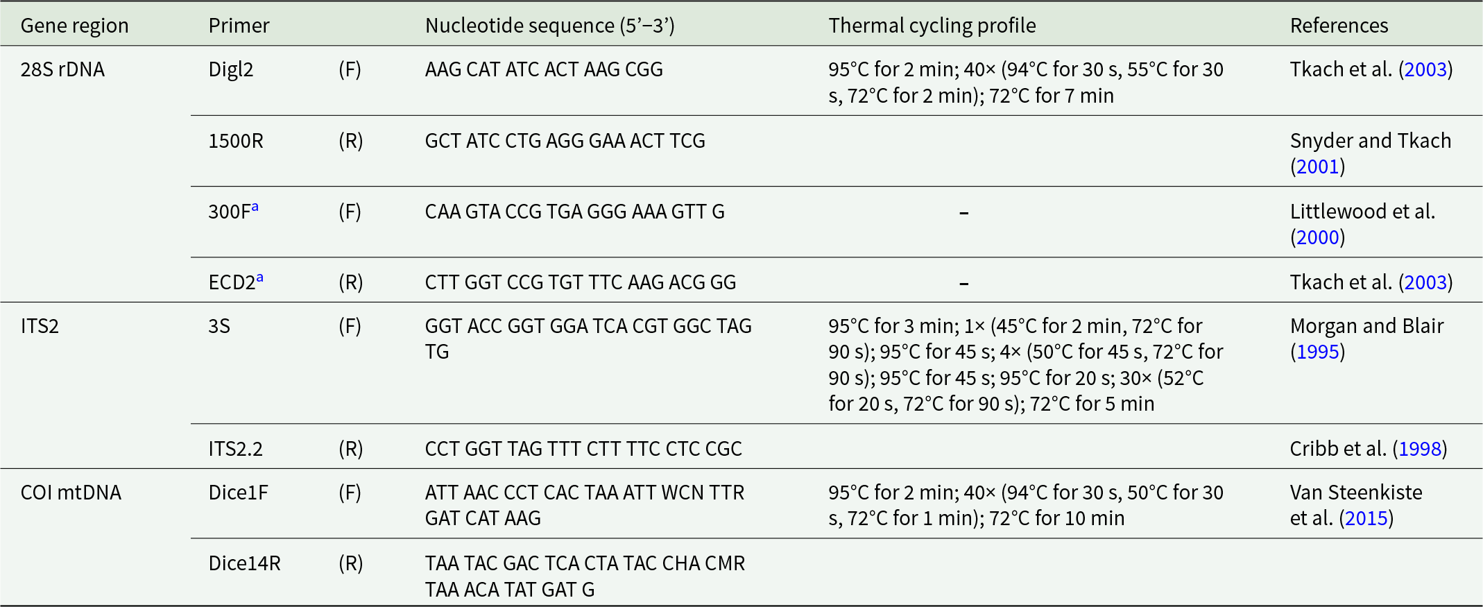
Deoxyribonucleic acid (DNA) was extracted using the PCRBIO Rapid PCR Kit (PCRBiosystems, Analytical Solution, Randburg, South Africa). For DNA extraction from the sporocysts and cercariae volume adjustments to 10 μL of 5 × PCRBIO Rapid Extract Buffer A and 5 μL of 10 × PCRBIO Rapid Extract Buffer B were made, respectively, and a final reaction dilution with 200 μL instead of 900 μL molecular grade water to obtain DNA at a higher concentration. DNA extraction from the metacercaria followed the manufacturer’s protocol, with a final reaction dilution of 450 μL instead of 900 μL molecular grade water. Primers used and thermal cycling conditions for the polymerase chain reaction (PCR) amplifications of the respective gene regions are provided in Table 1.
List of primers and thermal cycling conditions used for polymerase chain reaction amplification of the respective nuclear and mitochondrial markers for all ontogenetic stages presented in the present study

Table 1 Long description
List of primers and thermal cycling conditions used for polymerase chain reaction amplification of the respective nuclear and mitochondrial markers for all ontogenetic stages presented in the present study.
a Internal primers used only for sequencing.
Phylogenetic analyses
Newly generated sequences were visually inspected, and consensus sequence assembly was done using Geneious Prime® 2026.0.1 (Kearse et al., Reference Kearse, Moir, Wilson, Stones-Havas, Cheung, Sturrock, Buxton, Cooper, Markowitz, Duran, Thierer, Ashton, Meintjes and Drummond2012). All sequences were subjected to a BLAST search (https://blast.ncbi.nlm.nih.gov/Blast.cgi) to confirm identity and identify congeners for inclusion in the respective phylogenetic inferences of the 28S rDNA, ITS2 rDNA and COI mtDNA regions. The final list of species and details on each are presented in Supplementary Table S1. The alignment of each gene region was performed under the default parameters of MAFFT v.7.490 (Katoh et al., Reference Katoh, Misawa, Kuma and Miyata2002; Katoh and Standley, Reference Katoh and Standley2013) and final alignments were trimmed using GBlocks v.0.91b under the least stringent criteria (Castresana, Reference Castresana2000; Dereeper et al., Reference Dereeper, Guignon, Blanc, Audic, Buffet, Chevenet, Dufayard, Guindon, Lefort, Lescot, Claverie and Gascuel2008). Optimal phylogenetic model selection under the Akaike Information Criterion was performed in jModeltest v2.1.10 (Guindon and Gasceul, Reference Guindon and Gascuel2003; Darriba et al., Reference Darriba, Taboada, Doallo and Posada2012) and ModelFinder (Kalyaanamoorthy et al., Reference Kalyaanamoorthy, Minh, Wong, von Haeseler and Jermiin2017). Software packages available in the online computational CIPRES portal (Miller et al., Reference Miller, Pfeiffer and Schwartz2010) were used. The Bayesian Inference (BI) analyses of the 28S rDNA and COI mtDNA gene regions implementing the GTR + I + G model were performed in MrBayes v3.2.7a (Ronquist et al., Reference Ronquist, Teslenko, van der Mark, Ayres, Darling, Höhna, Larget, Liu, Suchard and Huelsenbeck2012). Each BI analysis was run for 10 million generations (ngen = 10 000 000), with 2 chains with 4 MCMC chains and a sample frequency of 1000 and discarding 25% of all sampled trees for each dataset. The ML analysis of the 28S rDNA region was inferred with 1000 bootstrap pseudoreplicates implemented in RAxML-HPC v.8 (Stamatakis, Reference Stamatakis2014). ML analysis of the COI mtDNA region was performed using IQ-TREE v.3.1.2 (Minh et al., Reference Minh, Schmidt, Chernomor, Schrempf, Woodhams, von Haeseler and Lanfear2020), implementing model GTR + F + I + R2 with 1000 bootstrap pseudoreplicates. Final trees were visualized in FigTree v.1.4.4 (http://tree.bio.ed.ac.uk/software/figtree/) and edited using Adobe Illustrator. The uncorrected p-distances for each alignment were generated in MEGA X (Kumara et al., Reference Kumar, Stecher, Li, Knyaz and Tamura2018) and number of base pair differences were determined in Geneious Prime® 2026.0.1.
Results
Taxonomic summary
Family Diplostomidae Poirier, 1886
Genus Neofibricola Achatz, Martens, Kudlai, Junker, Boe et Tkach, 2022
Neofibricola smiti Achatz, Martens, Kudlai, Junker, Boe et Tkach, 2022
Type host: Crocodylus niloticus Laurenti, 1768 (Reptilia: Crocodylidae).
First intermediate host: Bulinus sp. (Gastropoda: Planorbidae) (present study).
Second intermediate host: Xenopus muelleri (Peters, 1844) (Amphibia: Pipidae) (Achatz et al., Reference Achatz, Martens, Kudlai, Junker, Boe and Tkach2022c), Clarias gariepinus (Burchell, 1822) (Siluriformes: Clariidae) (present study).
Site of infection (definitive host): Small intestine.
Site of infection (first intermediate host): Not identified.
Site of infection (second intermediate host): Encysted in lungs (X. muelleri); free in eye viscera (C. gariepinus, present study).
Type locality: Crocodile River, Mpumalanga, South Africa (25°27’ S, 31°58’ E).
Other localities: Inflow of Lake Nyamithi (Phongolo River), KwaZulu-Natal, South Africa (26°53’59” S, 32°15’46” E) (Achatz et al., Reference Achatz, Martens, Kudlai, Junker, Boe and Tkach2022c). Present study – Magic Dam, Limpopo, South Africa (23°05’22.1” S, 30°23’01.4” E); KuShokwe Pan (Phongolo River), KwaZulu-Natal, South Africa (26°52’12” S, 32°12’58” E).
Sporocysts and cercariae (Figures 1A–C, 2A–F, and 4)
Infection rate: Prevalence (P) = 1.2% (1/88 snails)
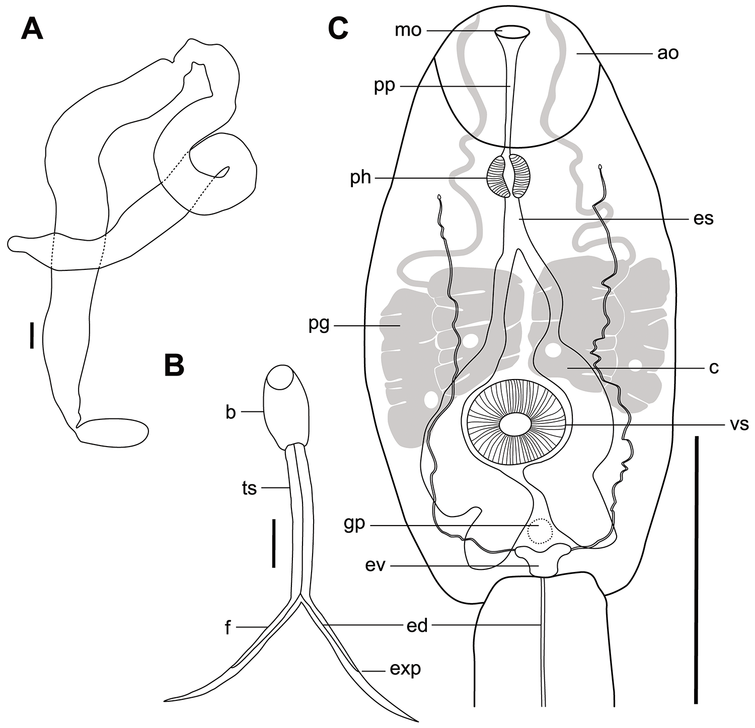
Schematic drawings of Neofibricola smiti Achatz, Martens, Kudlai, Junker, Boe et Tkach, 2022, from Bulinus sp. A sporocyst; B whole cercaria; C cercarial body. ao – anterior organ; b – body; c – caeca; ed – excretory duct; es – oesophagus; ev – excretory vesicle; exp – excretory pore; f – furca; gp – genital primordium; mo – mouth opening; pg – penetration gland cell; ph – pharynx; pp – prepharynx; ts – tail stem; vs – ventral sucker. Scale bars: 100 µm.

Figure 1 Long description
Schematic drawings of Neofibricola smiti Achatz, Martens, Kudlai, Junker, Boe et Tkach, 2022 from Bulinus sp. (A) sporocyst; (B) whole cercaria; (C) cercarial body. ao – anterior organ; b – body; c – caeca; ed – excretory duct; es – oesophagus; ev – excretory vesicle; exp – excretory pore; f – furca; gp – genital primordium; mo – mouth opening; pg – penetration gland cell; ph – pharynx; pp – prepharynx; ts – tail stem; vs – ventral sucker. Scale bars: 100 µm.
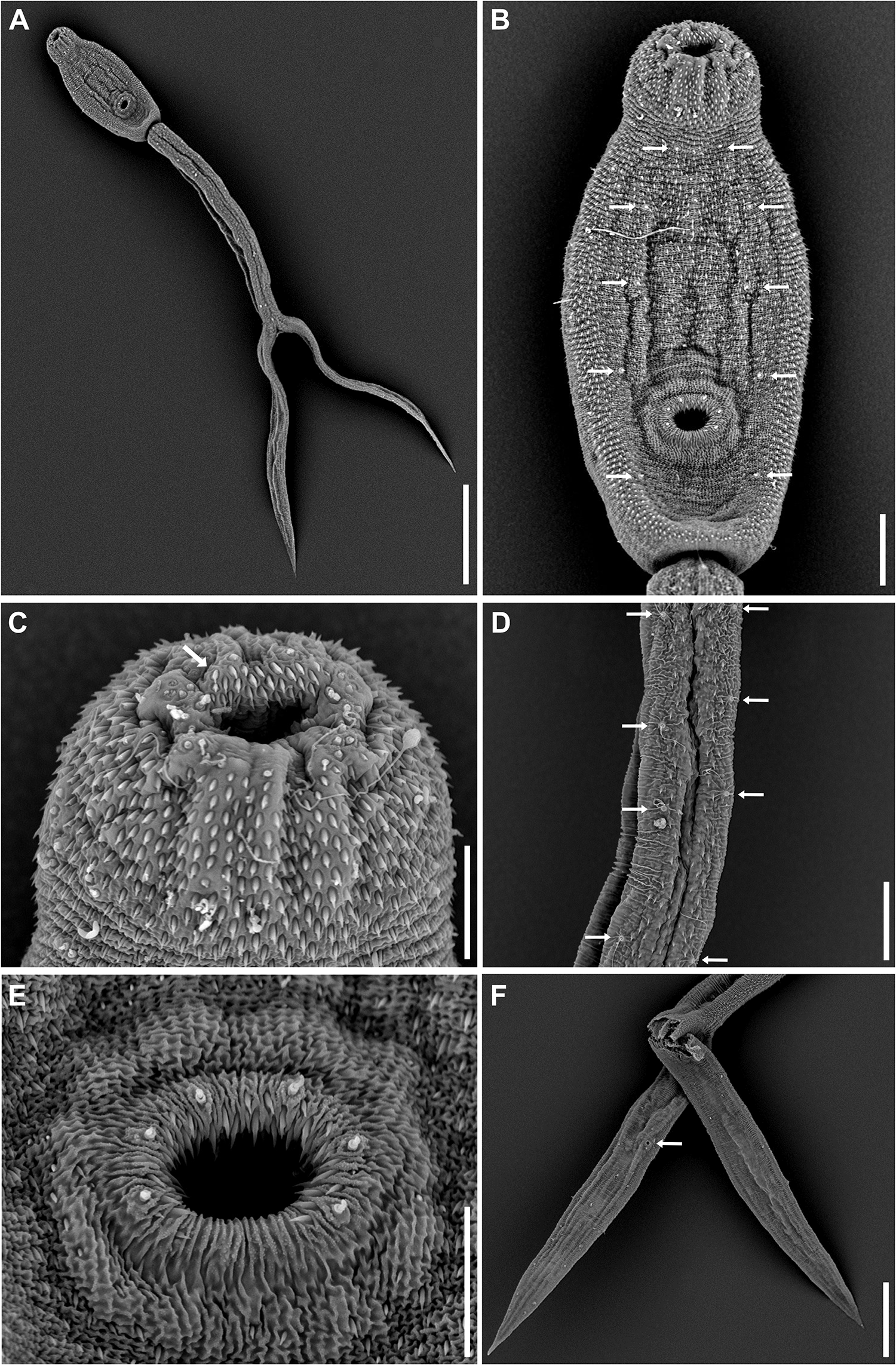
Scanning electron micrographs of Neofibricola smiti Achatz, Martens, Kudlai, Junker, Boe et Tkach, 2022 cercariae from Bulinus sp. (A) Whole cercaria, ventral view; (B) cercarial body, ventral view, papillae with sensilla arranged in pairs (white arrows); (C) anterior organ and mouth opening, with multiple rows of pre-oral spines (white arrow); (D) tail stem, ventral view, papillae with sensilla arranged in pairs (white arrows); (E) ventral sucker; (F) furcae, dorsolateral view, with excretory pore (white arrow); foreground furca broken during specimen preparation. Scale bars: 100 µm (A); 20 µm (B, D, and F); 10 µm (C and E).

Figure 2 Long description
Scanning electron micrographs of Neofibricola smiti Achatz, Martens, Kudlai, Junker, Boe et Tkach, 2022 cercariae from Bulinus sp. (A) Whole cercaria, ventral view; (B) cercarial body, ventral view, papillae with sensilla arranged in pairs (white arrows); (C) anterior organ and mouth opening, with multiple rows of pre-oral spines (white arrow); (D) tail stem, ventral view, papillae with sensilla arranged in pairs (white arrows); (E) ventral sucker; (F) furcae, dorsolateral view, with excretory pore (white arrow); foreground furca broken during specimen preparation. Scale bars: 100 µm (A); 20 µm (B, D, and F); 10 µm (C and E).
Representative DNA sequences: 28S rDNA – one sequence 1149 bp (cercariae) (GenBank PZ724619), one sequence 1141 bp long (sporocyst) (GenBank PZ724620); ITS2 – one sequence 490 bp (cercariae) (GenBank PZ724624); COI mtDNA – one sequence 833 bp (cercariae) (GenBank PZ724627), one sequence 832 bp (sporocyst) (GenBank PZ724628)
Voucher material: Supplementary Figure S1.
Description
Morphometrics and morphology based on a single live sporocyst, 3 live cercariae. Morphological features of cercariae also studied and described based on 6 haematoxylin-stained permanent mounts and 7 specimens used in SEM (see Supplementary Table S2). Sporocysts long, thread-like, 1905 × 199, measured at widest part (Figure 1A). Cercariae of longifurcate-pharyngeate (strigea) type sensu Schell (Reference Schell and Schell1985). Body elongate-oval, 220–230 × 92–107 (226 × 101), shorter than tail stem (tail stem length/body length = 1.42–1.49) (Figure 1B). Anterior organ subspherical, 42–55 × 44–52 (49 × 49), mouth opening ventro-subterminal. Pre-pharynx long, distinct, narrow; pharynx well-developed, round to oval, 17–18 × 16–19 (18 × 18); oesophagus short, bifurcating in first third of the body. Intestinal caeca well-developed, wide, with lobate walls, terminating near posterior end of the body. Ventral sucker post-equatorial, round, 36–39 × 36–39 (37 × 37), smaller than anterior organ (anterior organ width/ventral sucker width = 1.24–1.42). Penetration gland cells 2 pairs (4), large, similar in size, with large nucleus and fine granular content, anterior to ventral sucker, overlap caeca. Genital primordium a compact mass of cells, anterior to excretory vesicle, indistinct. Excretory vesicle small (Figure 1C). Tail stem 321–344 (329) long, 44–51 (48) wide at base; furcae 352–354 (353) long, 24–33 (27) wide at base (tail stem length/furcae length 0.91–0.97), without fin-folds. Caudal excretory duct passes through tail stem, excretory pores at mid-length of furcae (Figures 1B and 2F). Anterior organ covered with posteriorly directed spines; pre-oral spines arranged in multiple rows (Figure 2C). Body covered with spines of dissimilar size, bigger spines on ventral and ventro-lateral body, 2 long, smaller spines on dorsal body (<1). Dorsal body with papillae with long sensilla arranged in pairs (Figure 2B). Ventral sucker with c. 38 spines, with 6 papillae with sensilla on the outer margin (Figure 2E). Tail stem and furcae with minute spines, with size and density of spines decreasing towards terminal extremity of furcae (Figure 2D and F).
Metacercariae (Figures 3 and 4)
Synonym: Neodiplostomum Type I (sensu Truter et al., Reference Truter, Hadfield and Smit2023).
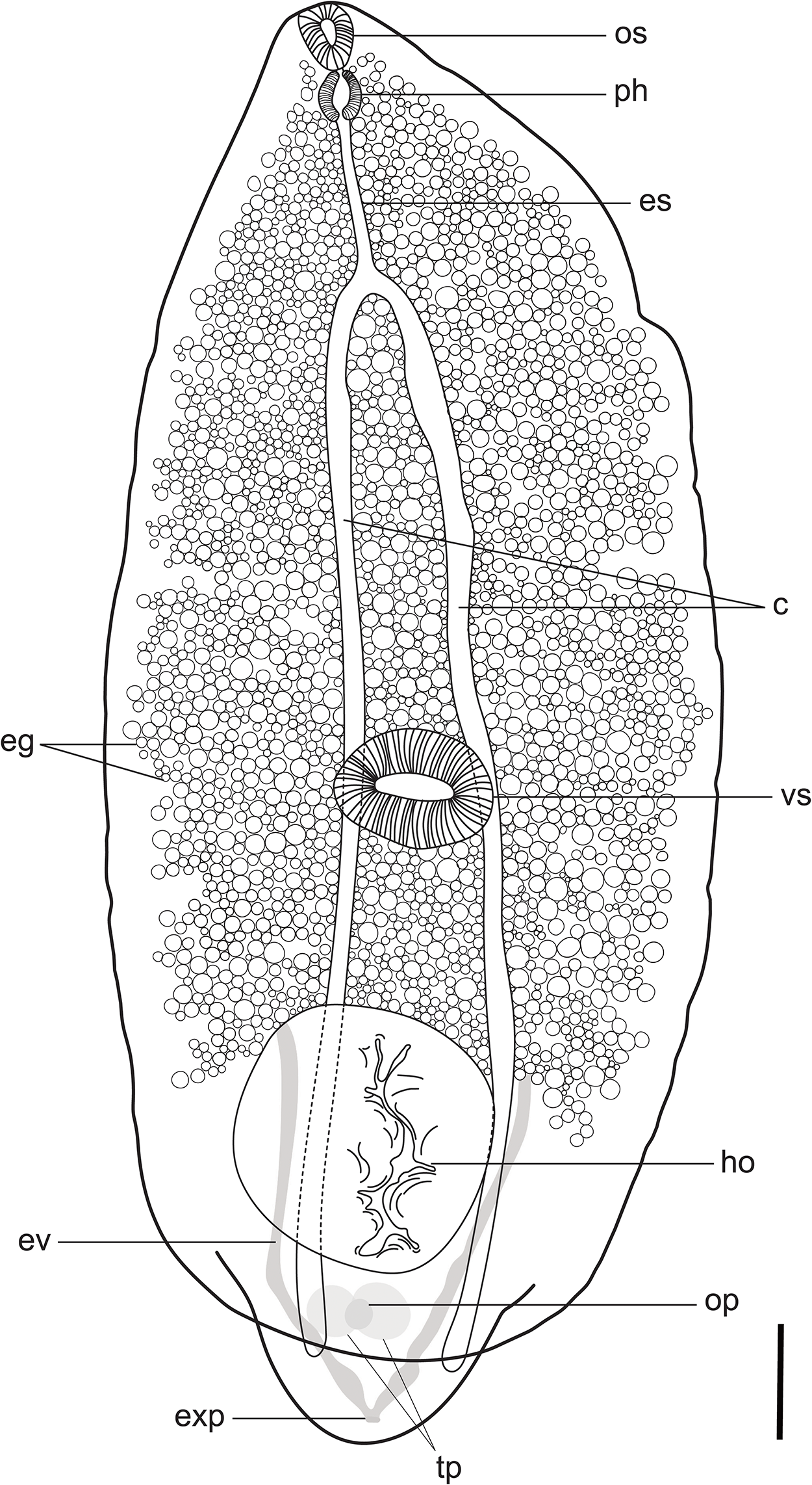
Schematic drawing of Neofibricola smiti Achatz, Martens, Kudlai, Junker, Boe et Tkach, 2022, metacercaria from the eye viscera of Clarias gariepinus (Burchell, 1822). c – caeca; eg – excretory granules; ep – oesophagus; ev – excretory vesicle; exp – excretory pore; ho – holdfast organ; op – ovary primordium; os – oral sucker; ph – pharynx; tp – testes primordium; vs – ventral sucker. Scale bar: 100 µm.

Figure 3 Long description
Schematic drawing of Neofibricola smiti Achatz, Martens, Kudlai, Junker, Boe et Tkach, 2022 metacercaria from the eye viscera of Clarias gariepinus (Burchell, 1822). c – caeca; eg – excretory granules; ep – oesophagus; ev – excretory vesicle; exp – excretory pore; ho – holdfast organ; op – ovary primordium; os – oral sucker; ph – pharynx; tp – testes primordium; vs – ventral sucker. Scale bar: 100 µm.
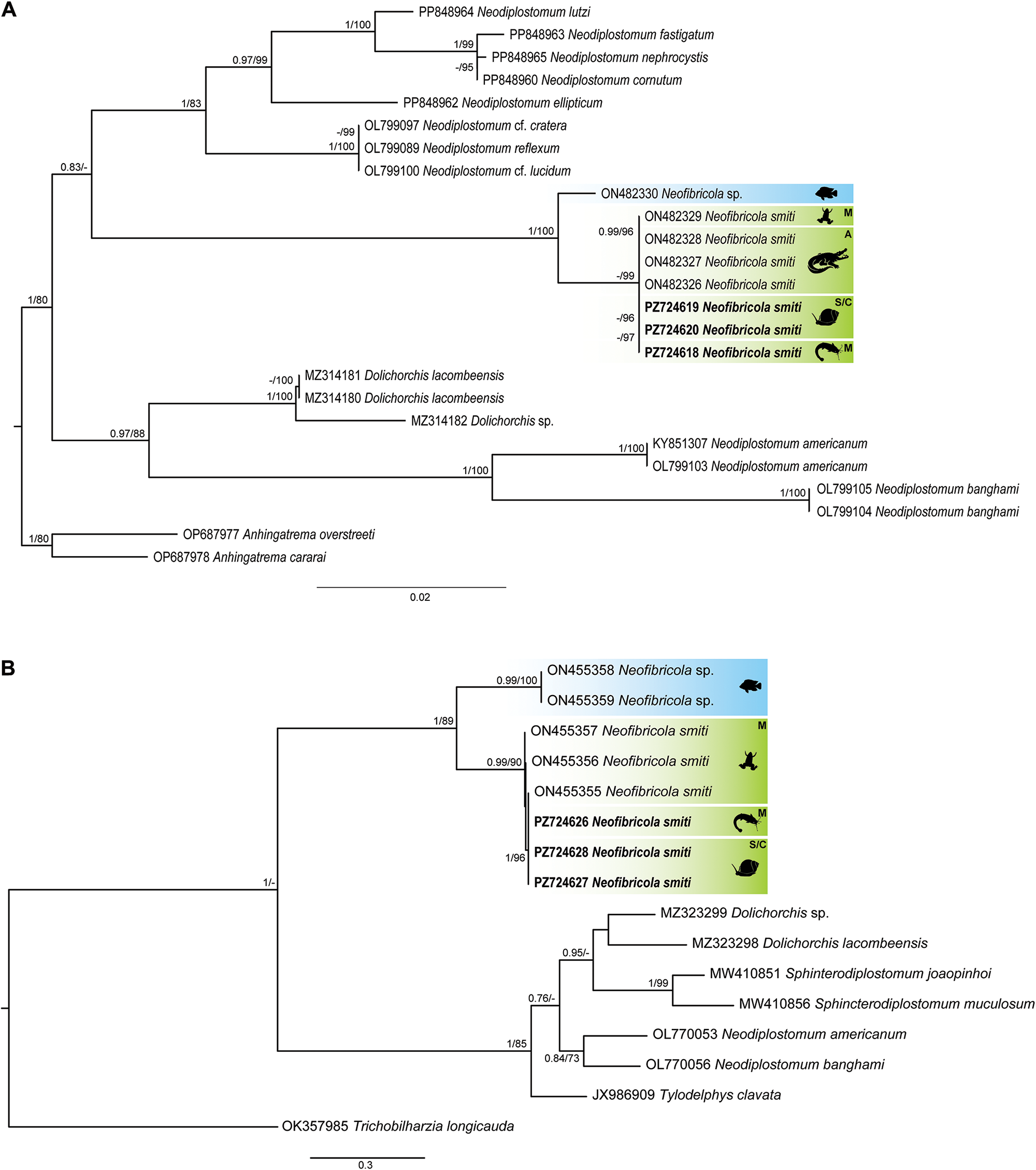
Phylogenetic tree based on the 1208 bp and 488 bp alignments of the 28S rDNA (A) and COI mtDNA (B) regions. Tree topology presented based on maximum likelihood analyses. Nodal support values indicate posterior probabilities from the BI (>70) and bootstrap values from ML (>70) analyses. A – adults; S/C – sporocysts/cercariae and M – metacercariae.

Figure 4 Long description
Phylogenetic tree based on the 1208 bp and 488 bp alignments of the 28S rDNA (A) and COI mtDNA (B) regions. Tree topology presented based on maximum likelihood analyses. Nodal support values indicate posterior probabilities from the BI (>70) and bootstrap values from ML (>70) analyses. A, adults; S/C, sporocysts/cercariae and M, metacercariae.
Infection rate: P = 5% (1/15); Intensity of infection (IF) = 1.
Representative DNA sequences: 28S rDNA – one sequence 1182 bp (GenBank PZ724618); COI mtDNA – one sequence 670 bp (GenBank PZ724626).
Description
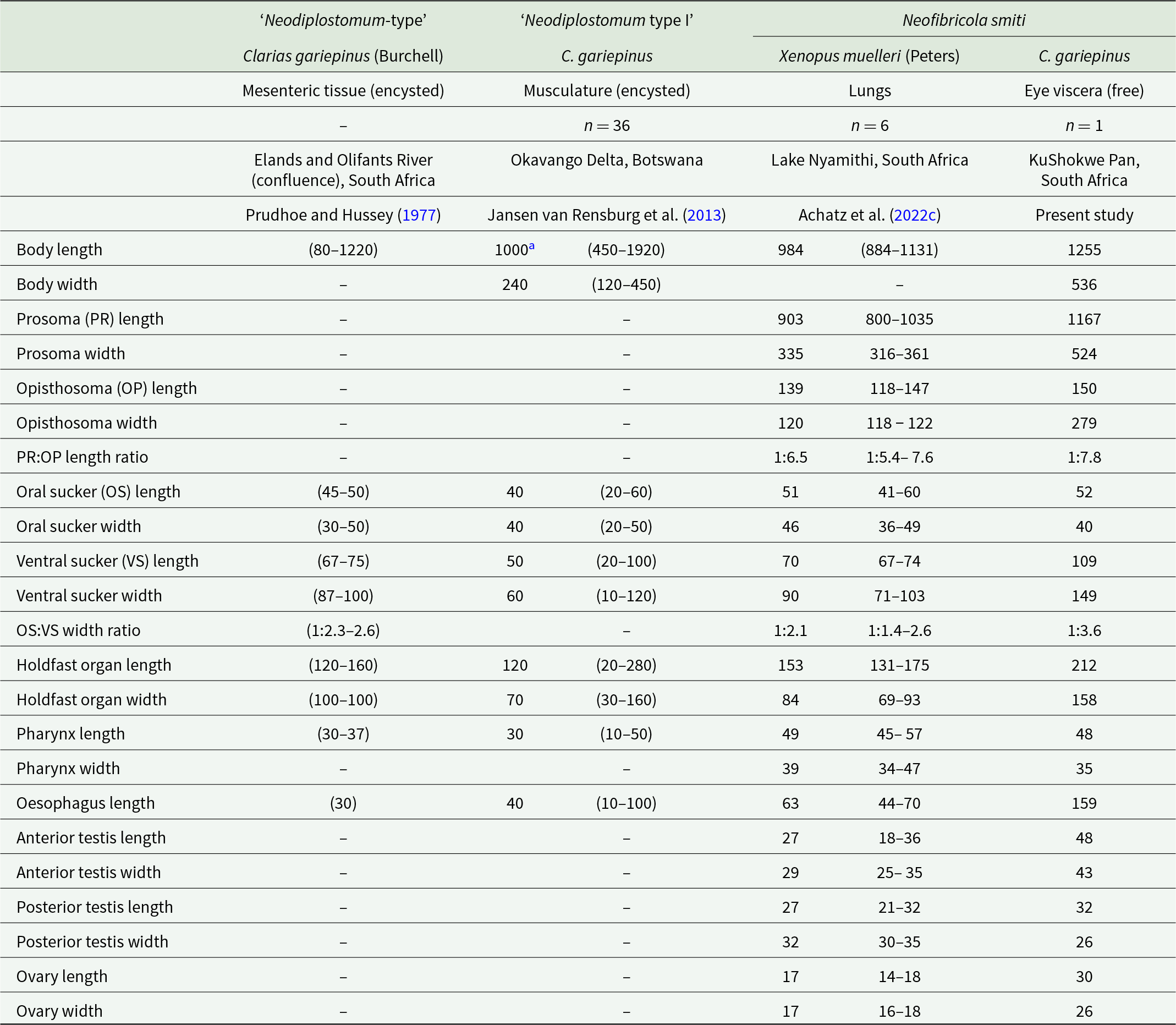
Measurements and description based on a single individual studied in a drop of water under light coverslip pressure. Body 1255 × 536, consist of distinct prosoma and opisthosoma (Figure 3). Prosoma elongate-oval, dorsoventrally flattened, 1167 × 524, maximum width anterior to ventral sucker. Opisthosoma elongate, conical, 150 × 279. Prosoma:opisthosoma length ratio 1:78. Pseudosuckers absent. Oral sucker subterminal, elongate-oval 52 × 40 (Figure 3). Ventral sucker transversely oval, 109 × 149, larger than oral sucker. Oral sucker:ventral sucker width ratio 1:3.6. Distance from ventral sucker to anterior end of prosoma 687. Distance from posterior end of prosoma 445. Prepharynx absent or short. Pharynx muscular, well developed elongate oval 48 × 35. Pharynx:oral sucker length ratio 1:1.1. Oesophagus medium to long, narrow 159. Caeca long and slender, extend to the posterior end of the opisthosoma. Genital primordia located in anterior half of opisthosoma. Holdfast organ large, in posterior part of prosoma, elongate-oval, 212 × 158. Holdfast organ length equal to ∼18% of prosoma length. Distance from holdfast organ to ventral sucker 169 (Figure 3). Testes primordia side-by-side, contiguous testis subspherical 48 × 43, posterior testis transversely oval, 32 × 26. Ovary primordium subspherical, entire, smaller than testes, 30 × 26, between testes or overlaps anterior testis ventrally. Excretory vesicle not observed. Excretory granules dense (numerous) throughout prosoma, present in approximately two-thirds of prosoma anterior to holdfast organ (Figure 3). Excretory pore terminal.
Remarks
The single metacercaria obtained from C. gariepinus has the general body shape of neodiplostomulum-type and conforms to metacercariae reported as ‘Neodiplostomum-type’ and ‘Neodiplostomum type I’ from the mesenteric tissue and musculature of C. gariepinus at the confluence of the Elands and Olifants Rivers, South Africa (Prudhoe and Hussey, Reference Prudhoe and Hussey1977) and Okavango Delta, Botswana (Jansen van Rensburg et al., Reference Jansen van Rensburg, Van As and King2013), and to N. smiti reported from the lungs of X. muelleri in South Africa (Achatz et al., Reference Achatz, Martens, Kudlai, Junker, Boe and Tkach2022c). Morphometrics are presented in Table 2. The metacercaria from C. gariepinus in the present study falls within the upper range of the total body length of metacercariae reported from C. gariepinus at the confluence of the Elands and Olifants Rivers, South Africa and Okavango Delta, Botswana and are larger than the specimens reported from X. muelleri in Lake Nyamithi, South Africa. The prosoma and opisthosoma length and width of specimens from C. gariepinus in the present study is longer and wider that those from X. muelleri and has a similar prosoma:opisthosoma length ratio. The specimen from the present study has a similar oral sucker size (length × width) to those reported from the other 3 localities, and a similar ventral sucker size (length) to the larger specimens of ‘Neodiplostomum type I’ from Botswana but a larger ventral sucker width and oral sucker:ventral sucker width ratio than any of the specimens reported from other localities and hosts (see Table 2). Holdfast organ size (length × width) overlaps between all specimens reported for this morphotype. Pharynx size (length) reported for ‘Neodiplostomum-type’ and ‘Neodiplostomum type I’ is smaller than reported for N. smiti from X. muelleri and for C. gariepinus in the present study, while there is overlap between the specimen from the present study and those reported from X. muelleri. Testes and ovary primordia in the specimen from the present study are similar in shape, but larger in size than reported for specimens from X. muelleri. The ovary is smaller than the testes, while the arrangement of the testes and ovary differ in being side-by-side and the ovary overlaps testes at a lateral position in C. gariepinus, opposed to the tandem testes with ovary positioned between testes or ventrally to the anterior testes in specimens from X. muelleri.
Morphometrics of the metacercariae of Neodiplostomum-type and Neofibricola smiti Achatz, Martens, Kudlai, Junker, Boe et Tkach, 2022 from respective intermediate hosts reported from Southern Africa

Table 2 Long description
Morphometrics of the metacercariae of Neodiplostomum-type and Neofibricola smiti Achatz, Martens, Kudlai, Junker, Boe et Tkach, 2022 from respective intermediate hosts reported from southern Africa.
a Value erroneously reported as 100 µm in Jansen van Rensburg et al. (Reference Jansen van Rensburg, Van As and King2013).
Molecular identification
A total of 3 sequences (one each from the metacercaria from C. gariepinus, and the sporocysts and cercariae from Bulinus sp.) were generated for the 28S rDNA gene region. One sequence was generated for the ITS2 region from cercariae in Bulinus sp., and 3 sequences for the COI mtDNA gene region of the sporocysts and cercariae from Bulinus sp. and the metacercaria from C. gariepinus. In all resulting phylogenetic analyses of the 28S rDNA and COI mtDNA gene regions, sequences of the metacercaria, cercariae and sporocysts generated in the present study cluster with N. smiti sister to Neofibricola sp. Tree topologies for the BI and ML from the phylogenetic inferences of both 28S rDNA and COI mtDNA were identical.
The final 28S rDNA alignment (Figure 4A) comprised 1208 bp, with sequences obtained from C. gariepinus (metacercaria) and Bulinus sp. (sporocysts and cercariae) identical to those of N. smiti from X. muelleri (metacercaria) and C. niloticus (adults). No intraspecific divergence was observed between ontogenetic stages of N. smiti from X. muelleri or C. niloticus, however, there is interspecific divergence of 1.1% and 12 bp between the newly obtained sequences and Neofibricola sp. from O. mossambicus (see Supplementary Table S3). The final ITS2 rDNA alignment was 490 bp long, with the newly obtained sequence from cercariae (ex. Bulinus sp.) having an intraspecific divergence of 0.7% (3–4 bp difference) to N. smiti (ex. C. niloticus and ex. X. muelleri) and interspecific divergence of 1.6–2.3% and 7–10 bp difference to Neofibricola sp. from O. mossambicus.
The final COI mtDNA alignment constituted 488 bp (Figure 4B), sequences obtained for the sporocysts and cercariae from Bulinus sp. and the metacercaria from C. gariepinus were identical but had 1–5 bp differences to the isolates from X. muelleri, while there is 14.9% divergence and 72–76 bp difference to Neofibricola sp. (ex. O. mossambicus) (see Supplementary Table S4).
Discussion
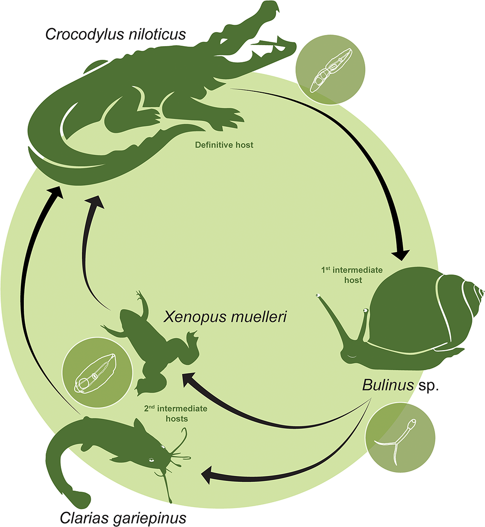
The present study identifies Bulinus sp. as the first intermediate host of N. smiti and expands the known range of second intermediate hosts through the discovery of a metacercaria in C. gariepinus, thereby providing the first fully elucidated and molecularly verified life cycle for a crocodilian-infecting diplostomid (Figure 5). These findings contribute to the growing body of research resolving and characterizing diplostomoid life cycles through the integration of morphological and molecular data (e.g. Locke et al., Reference Locke, McLaughlin, Lapierre, Johnson and Marcogliese2011; Ritossa et al., Reference Ritossa, Flores and Viozzi2013; Chibwana et al., Reference Chibwana, Nkwengulila, Locke, McLaughlin and Marcogliese2015; Blasco-Costa et al., Reference Blasco-Costa, Poulin and Presswell2016; Rosser et al., Reference Rosser, Alberson, Khoo, Woodyard, Pote and Griffin2016; López-Hernández et al., Reference López-Hernández, Locke, Melo, ÉM and Pinto2018; Achatz et al., Reference Achatz, Bell, Melo, Fecchio and Tkach2021; Schwelm et al., Reference Schwelm, Georgieva, Grabner, Kostadinova and Sures2021; Pyrka et al., Reference Pyrka, Kanarek, Gabrysiak, Jeżewski, Cichy, Stanicka, Żbikowska, Zaleśny and Hildebrand2022; Izrailskaia et al., Reference Izrailskaia, Besprozvannykh and Shchelkanov2024; Sokolov et al., Reference Sokolov, Vlasenkov, Bugmyrin, Kalmykov and Lebedeva2024; Hüsken et al., Reference Hüsken, Truter, Schwelm, Sures, Smit and Malherbe2026). Such integrative approaches have substantially advanced the understanding of diplostomoid diversity while highlighting the complexity of host associations, transmission pathways and life cycles strategies (Blasco-Costa and Locke, Reference Blasco-Costa and Locke2017; Achatz and Tkach, Reference Achatz and Tkach2025). Nevertheless, fully completed and verified life cycle reconstructions remain unavailable for the majority of known taxa.
Life cycle of Neofibricola smiti Achatz, Martens, Kudlai, Junker, Boe et Tkach, 2022 in South Africa, revealing transmission from a gastropod first intermediate host through fish and amphibian second intermediate hosts to the crocodilian definitive host.

Figure 5 Long description
Life cycle of Neofibricola smiti Achatz, Martens, Kudlai, Junker, Boe et Tkach, 2022 in South Africa, revealing transmission from a gastropod first intermediate host through fish and amphibian second intermediate hosts to the crocodilian definitive host.
This lack of life cycle resolution is particularly pronounced among trematodes infecting crocodilians, for which most species remain known exclusively from adult specimens recovered from definitive hosts (Moravec, Reference Moravec2001; Tellez, Reference Tellez2013; Achatz et al., Reference Achatz, Pulis, Junker, Binh, Snyder and Tkach2019b; Tkach et al., Reference Tkach, Achatz, Pulis, Junker, Snyder, Bell, Halajian and de Vasconcelos Melo2020, Reference Achatz, Chermak, Junker and Tkach2022a). However, existing linkages of developmental stages of crocodilian-infecting diplostomids have already revealed considerable variation in transmission strategies, including progenesis in metacercariae in Crocodilicola pseudostoma (Willemoes-Suhm, 1870) and the incorporation of paratenic hosts in Archaeodiplostomum overstreeti Achatz, Pulis, Junker, Snyder, Bell, Halajian et de Vasconcelos Melo, 2020 (formerly assigned to the Proterodiplostomidae) parasitizing Alligator mississippiensis (Daudin, 1802) (Hernández-Mena et al., Reference Hernández-Mena, García-Varela and Pérez-Ponce de León2017; Negrelli et al., Reference Negrelli, Vieira, Abdallah and Azevedo2020; Tkach et al., Reference Tkach, Achatz, Pulis, Junker, Snyder, Bell, Halajian and de Vasconcelos Melo2020). In the latter species, free immature metacercariae have been reported from the stomach of snake species Nerodia fasciata (Linnaeus, 1766) and encysted metacercariae in the mesenteries of Nerodia cyclopion (Duméril, Bibron et Duméril, 1854), with both species occurring sympatrically with the definitive host (Tkach et al., Reference Tkach, Achatz, Pulis, Junker, Snyder, Bell, Halajian and de Vasconcelos Melo2020). Paratenic host associations have also been documented in several other diplostomid genera (e.g. Seo, Reference Seo1990; Chai and Shin, Reference Chai and Shin2002; Shin et al., Reference Shin, Kim, Kim, Han, Park, Nawa, Kook, Lee and Chai2006; Achatz et al., Reference Achatz, Dmytrieva, Kuzmin and Tkach2019a; Bełcik et al., Reference Bełcik, Cencek, Korpysa-Dzirba, Samorek-Pieróg, Karamon, Sroka, Zdybel, Skubida and Bilska-Zając2025), suggesting that paratenic host use may be more widespread among diplostomids than currently recognized.
The present findings expand the known host associations of N. smiti, with metacercariae now documented from both amphibian and fish hosts. Furthermore, metacercariae of the presumed congeneric Neofibricola sp. were previously reported from the body cavity of O. mossambicus (Achatz et al., Reference Achatz, Martens, Kudlai, Junker, Boe and Tkach2022c). Collectively, these records suggest that members of Neofibricola may utilize a broader range of second intermediate hosts than currently recognized, spanning both amphibians and fishes, a pattern also observed in other digenean taxa (Johnson et al., Reference Johnson, Sutherland, Kinsella and Lunde2004; Chibwana et al., Reference Chibwana, Nkwengulila, Locke, McLaughlin and Marcogliese2015; Calhoun et al., Reference Calhoun, Leslie, Riepe, Achatz, McDevitt-Galles, Tkach and Johnson2020). Alternatively, the occurrence of N. smiti in amphibian and fish hosts may reflect different ecological roles within the transmission cycle rather than a broad second intermediate host range. Diplostomid trematodes infecting the eyes of fish hosts are often characterized by a high degree of site specificity (Locke et al., Reference Locke, McLaughlin, Dayanandan and Marcogliese2010a, Reference Locke, McLaughlin and Marcogliese2010b; Blasco-Costa et al., Reference Blasco-Costa, Faltýnková, Georgieva, Skírnisson, Scholz and Kostadinova2014, Reference Locke, Al-Nasiri, Caffara, Drago, Kalbe, Lapierre, McLaughlin, Nie, Overstreet, Souza, Takemoto and Marcogliese2015; Padrós et al., Reference Padrós, Knudsen and Blasco-Costa2018), whereas N. smiti has thus far been reported as encysted metacercariae in the lungs of X. muelleri and as free metacercariae in the eye viscera of C. gariepinus. These marked differences in infection site and developmental presentation may indicate distinct ecological roles for the 2 host groups within the life cycle. One possibility is that either amphibians or fishes serve as principal second intermediate or paratenic host in the life cycle. Such a scenario would be consistent with trophic interactions within the systems, as crocodilians are known to consume catfish more frequently than amphibians (Bruton, Reference Bruton1979; Wallace and Leslie, Reference Wallace and Leslie2008). Noticeably, low prevalence and intensity of infection were observed among both snail and fish hosts, respectively. While this could be attributed to naturally low prevalences of individual trematode species, as commonly documented in first intermediate snail hosts (see Esch et al., Reference Esch, Curtis and Barger2001), the single metacercaria in the eye viscera of C. gariepinus could be indicative of the complex ecological factors and interactions resulting from spatial and temporal heterogeneity (i.e. varying transmission reflecting seasonality or host density), potentially low encounter rates between cercariae and catfish due to habitat preference, overdispersion or aggregation of parasite distributions (Thieltges et al., Reference Thieltges, Jensen and Poulin2008; Poulin, Reference Poulin2013).
Further life cycle resolution among crocodilian-associated diplostomids will be necessary to determine whether broad intermediate host ranges, paratenic host use and alternative transmission strategies are characteristic of Neofibricola and related taxa. Ultimately, the respective roles of X. muelleri and C. gariepinus in the life cycle of N. smiti can only be resolved through targeted experimental studies. In particular, experimental assessment of the infectivity and transmission potential of metacercariae from amphibian to fish hosts and subsequently to crocodilian definitive hosts would provide critical insight into the transmission dynamics of this species.
Supplementary material
The supplementary material for this article can be found at https://doi.org/10.1017/S0031182026102650.
Data availability statement
Genetic sequences generated during the present study deposited into the GenBank online repository: 28S rDNA (GenBank PZ724618–20); ITS2 (GenBank PZ724624) and COI mtDNA (GenBank PZ724626–28).
Acknowledgements
Members of the Water Research Group, Marcel Kruger and Chelsea Withfield, are thanked for their assistance in collection of hosts and parasitic material. Mr Willem Landman, NWU Advance Microscopy Laboratory, is thanked for assistance with scanning electron microscopy. This is publication number 1066 from the NWU Water Research Group.
Author contributions
M.T.: Writing – original draft, visualization, investigation, conceptualization and writing – review & editing. A.H.: Writing – original draft, visualization, investigation, formal analysis and writing – review & editing. J.S.: Writing – review & editing, investigation and conceptualization. B.S.: Writing – review & editing, and supervision. W.M.: Writing – review & editing, supervision and resources. N.J.S.: Conceptualization, writing – review & editing, supervision and resources. M.T. and A.H. contributed equally.
Financial support
This research is part of the larger REFRESH project funded by the Foundational Biodiversity Initiative Programme (FBIP) of the National Research Foundation of South Africa (REFRESH—FBIP-211006643719; Grant no. 138573). A.H. was supported by a research grant from the DAAD-Stiftung (Treuhänder DAAD e.V.). M.T. was funded by the North-West University Postdoctoral fellowship programme and an NRF-South African Research Chairs Initiative, Inland Fisheries and Freshwater Ecology grant (Grant no. 110507). We also acknowledge the use of infrastructure and equipment provided by the NWU-WRG and the NRF-SAIAB Research Platforms and the funding channelled through the NFR-SAIAB Institutional Support system. Opinions, findings, conclusions and recommendations expressed in this publication are that of the authors, and the NRF accepts no liability whatsoever in this regard.
Competing interests
The authors declare that they have no known competing financial interests or personal relationships that could have appeared to influence the work reported in this paper. N.J.S., as Guest Editor for this Special Issue, was excluded from all stages of the editorial and peer-review process for this manuscript.
Ethical standards
All procedures contributing to this work comply with the ethical standards of the relevant national and institutional guides on the care and use of animals. Ethical approval for the use of animals for research purposes was obtained from the North-West University AnimCare Research Ethics Committee (NWU-00159-18-A5). Temporary holding of fish and humane killing followed protocols stipulated in SOP NWU-00272-17-A5 and NWU-00267-17-A5. Permits for the collection of animals for the purpose of research were obtained from the Ezemvelo KZN Wildlife (KwaZulu-Natal, permit Nos. OP 1075/2017, OP 1582/2018).


 Open access

