One of the aphorisms of the late and great Alexander Nadas, acknowledged as the father of Paediatric Cardiology, was that, if the observer diagnosed common arterial trunk or tetralogy of Fallot every time he or she noted the presence of a right-sided aortic arch, the diagnosis would be correct in the overwhelming majority of cases. It is certainly the case that the right-sided aortic arch, defined as the ascending aorta crossing the right bronchus before descending to pass through the diaphragm, is found most frequently in the setting of congenital cardiac malformations. It is also an expected feature when there is overall mirror-imagery of the body organs, including the atrial appendages. When the aortic arch is right-sided in such situations, it is well recognised that the arterial duct, or its ligamentous remnant, is usually a left-sided structure, which arises from the base of the brachiocephalic artery. The brachiocephalic artery, of course, is also left-sided when there is mirror-imaged branching from the ascending aorta. The left-sided arterial duct in this setting, however, is anteriorly located relative to the left bronchus. More importantly, it does not pass behind the oesophagus. When there is mirror-imaged branching of the aorta, nonetheless, the arterial duct would intuitively be expected to be right-sided. If patent, it would then be expected to arise as the last branch of the right-sided aortic arch, extending anteriorly to join the right pulmonary artery. It is also well described, however, that a persistently patent arterial duct, or its ligamentous remnant, can take origin from the right-sided aortic arch, but then extend in a retro-oesophageal manner to terminate at the left pulmonary artery. As explained in the account provided by McElhinney et al,Reference McElhinney, Hoydu, Gaynor, Spray, Goldmuntz and Weinberg
1
this is one of the variants of the lesions that are grouped together as “vascular rings”. The retro-oesophageal location of the vascular structure, or its ligamentous remnant, then has the potential to obstruct passage of nutrients down the oesophagus, producing so-called “dysphagia lusoria”, which translates literally as problematic swallowing due to a “prank of nature”. Such vascular rings are often encountered when the heart itself is also congenitally malformed, but can also be present, again as emphasised by McElhinney et al,Reference McElhinney, Hoydu, Gaynor, Spray, Goldmuntz and Weinberg
1
when the intracardiac anatomy is normal. In these latter situations, the anomalous location of the arch can reasonably be considered to be isolated.
It is such an example of an “isolated” right aortic arch that Jin et al2 discuss in a recent issue of the journal. They have then interpreted their findings in this case on the basis of analysis of an important archive of serially sectioned human embryos held in Madrid, Spain. The availability of such material, obtained from a large series of human embryos, is of inestimable value as we continue to seek to understand the morphogenesis of congenital cardiac malformations. It is axiomatic that we will never understand the abnormal arrangements until we have a firm grasp of normal findings. It is also the case, nonetheless, that understanding the sequences of development by studying serially sectioned histological material is far from easy. Without the aid of three-dimensional reconstruction, it is difficult even for the investigators themselves to build a comprehensive picture of the overall arrangement. If multiple panels of two-dimensional images are then presented so as to convey this information to the reader, understanding becomes more complicated. This is now the situation regarding the information provided by Jin et al. Their account is not made any easier by their use of words such as “bulbus”. We have made remarkable strides over the past two decades in understanding the anatomical changes that take place during formation of the heart. We now know that the outlet component of the ventricular loop will become the morphologically right ventricle. It is better, therefore, to describe the developing right ventricle, rather than retaining the archaic “bulbus”. During development, the outflow tract then extends from the developing right ventricle to the margins of the pericardial cavity, where its cavity becomes confluent with the aortic sac. The arteries arising from the aortic sac, which initially extend in a symmetrical manner through the pharyngeal mesenchyme before uniting posteriorly to form the descending aorta, usually become re-modelled during normal development to form the unilateral aortic arch and the arterial duct. Excellent three-dimensional reconstructions of the process of re-modelling were provided long since by Congdon.Reference Congdon
3
More importantly, the accuracy of the study by Congdon was endorsed very recently by an investigation published by the group from Amsterdam. These investigators used Amira software to reconstruct the pharyngeal arch arteries at various stages of their development, again using sections of human embryos.Reference Rana, Sizarov, Christoffels and Moorman
4
It would have helped in understanding, therefore, had Jin et al made reference to these earlier studies, or even cited our own published descriptions of reconstructions,Reference Bamforth, Chaudhry and Bennett
5
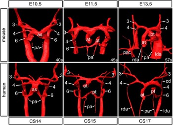
made from both human and mouse embryos (Fig 1).
Figure 1
The panels show reconstructions of the arteries coursing through the pharyngeal arches at various stages of development in the mouse (upper panels) and man (lower panels). The initially bilaterally symmetrical systems have become re-modelled to form the aortic arch and the arterial duct, derived from the left-sided fourth and sixth arch arteries, respectively, by the time the mouse embryo is at the end of the 13th day of development (E13.5) and the human embryo is at Carnegie stage 17. The numbers show the arch arteries related to the third and the sixth pharyngeal arches. as=aortic sac; at=aortic trunk; cd=carotid duct; lda=left descending aorta; pa=pulmonary arteries; pt=pulmonary trunk; rda=right descending aorta; psc=primitive subclavian artery.
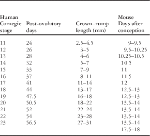
It would also have made the findings easier to assimilate had Jin et al described the temporal sequence of changes in terms of the so-called Carnegie staging for human embryos, rather than relying on the crown–rump length of the embryos themselves. Tables do exist to permit correlations to be made between crown–rump length and also the timing of the changes noted in the developing mouse heart; therefore, inclusion of this material would also have been of great help (Table 1). The greatest drawback of the study of Jin et al, however, is that it makes no mention of the concept of the so-called “hypothetical double aortic arch”. Initially postulated by the doyen of cardiac pathologists, Jesse Edwards,Reference Edwards
6
it is widely acknowledged that this concept provides an explanation for all vascular rings. McElhinney et alReference McElhinney, Hoydu, Gaynor, Spray, Goldmuntz and Weinberg
1
based their interpretations on a concept for development that is comparable with Edwards’ hypothetical double arch, although they did not credit Edwards for introducing this important concept.
Table 1
The Carnegie stages for human embryos correlated with post-ovulatory age, crown–rump length, and the age of comparable mouse embryos.
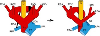
In the hypothetical double arch, there is persistence of arches crossing both bronchuses. The two arches join together posteriorly to form a neutrally positioned descending aorta. A common carotid artery and a subclavian artery arise in turn from the cranial surface of each arch, whereas a persistently patent arterial duct arises on each side from the caudal surfaces. The double arch can then be interrupted at any point between the origins of the vessels, providing a rational explanation for all the known variants of vascular rings, including the case as described by Jin et al (Fig 2). Analyses of this type show that, when the left-sided arterial duct arises from the right-sided aortic arch, it must incorporate the posterior part of the left-sided branch of the double arch as it extends in a retro-oesophageal manner (Fig 3 – left-hand panel). The double arch itself is then interrupted between the anterior and posterior components of the left-sided branch, but anterior to the origin of the left-sided duct from its inferior surface. It follows that the interruption itself must take place subsequent to the cranial migration of the left seventh intersegmental artery, which becomes the left subclavian artery (Fig 3 – right-hand panel). Such interpretation, as readily understood on the basis of Edwards’ stellar concept,Reference Edwards
6
provides a simple and rational explanation for the existence of an isolated right aortic arch with retro-oesophageal left-sided arterial duct. The explanation as illustrated in Figures 2 and 3 is the same as offered by McElhinney et al.Reference McElhinney, Hoydu, Gaynor, Spray, Goldmuntz and Weinberg
1
It is a mystery, therefore, why Jin et al should suggest that McElhinney et al described the arterial duct in this setting as being “right-sided”. This adds nothing but more confusion to an account that is already overly confusing. We find no evidence for the claim made by Jin et al that McElhinney et alReference McElhinney, Hoydu, Gaynor, Spray, Goldmuntz and Weinberg
1
introduced this inappropriate terminology.
Figure 2
The cartoons show Edwards’ concept that the hypothetical double aortic archReference Edwards
6
provides a simple and rational explanation to account for the retro-oesophageal origin of the left-sided arterial duct in the setting of a right aortic arch. The left-hand panel shows the hypothetical arch system with division of the segment between the origin of the left-sided arterial duct (LDA) from the underside of the left-sided arch and the origin more anterior to the left common carotid (LCC) and the left subclavian arteries (LSA) from the cranial aspect of the left arch. The right arch also gives rise to an arterial duct caudally (RDA) and right common carotid and subclavian arteries (RCC; RSA) cranially. The right-hand panel shows how the situation becomes re-modelled to produce the arrangement described by Jin and associates.Reference Jin, Yamada, Kim, Rodríguez-Vázquez, Murakami and Arakawa
2
AO=aorta; LPA=left pulmonary artery; O=oesophagus; PT=pulmonary trunk; RPA=right pulmonary artery.
Figure 3
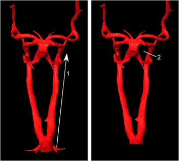
We have used the situation as demonstrated by reconstruction of the pharyngeal arch arteries from our human embryo at Carnegie stage 14 (bottom left-hand panel of Fig 1) to show how it is necessary for the seventh segmental artery, which will become the left subclavian artery, to migrate cranially (left-hand panel – 1), and for the left-sided dorsal aorta then to become interrupted proximal to the origin of the third and fourth arch arteries (right panel – 2). This then leaves a retro-oesophageal segment of the left arch comprised of the posterior component of the left-sided dorsal aorta and the left sixth arch, with the latter forming the left-sided arterial duct.






 You have
Access
One of the aphorisms of the late and great Alexander Nadas, acknowledged as the father of Paediatric Cardiology, was that, if the observer diagnosed common arterial trunk or tetralogy of Fallot every time he or she noted the presence of a right-sided aortic arch, the diagnosis would be correct in the overwhelming majority of cases. It is certainly the case that the right-sided aortic arch, defined as the ascending aorta crossing the right bronchus before descending to pass through the diaphragm, is found most frequently in the setting of congenital cardiac malformations. It is also an expected feature when there is overall mirror-imagery of the body organs, including the atrial appendages. When the aortic arch is right-sided in such situations, it is well recognised that the arterial duct, or its ligamentous remnant, is usually a left-sided structure, which arises from the base of the brachiocephalic artery. The brachiocephalic artery, of course, is also left-sided when there is mirror-imaged branching from the ascending aorta. The left-sided arterial duct in this setting, however, is anteriorly located relative to the left bronchus. More importantly, it does not pass behind the oesophagus. When there is mirror-imaged branching of the aorta, nonetheless, the arterial duct would intuitively be expected to be right-sided. If patent, it would then be expected to arise as the last branch of the right-sided aortic arch, extending anteriorly to join the right pulmonary artery. It is also well described, however, that a persistently patent arterial duct, or its ligamentous remnant, can take origin from the right-sided aortic arch, but then extend in a retro-oesophageal manner to terminate at the left pulmonary artery. As explained in the account provided by McElhinney et al,Reference McElhinney, Hoydu, Gaynor, Spray, Goldmuntz and Weinberg 1 this is one of the variants of the lesions that are grouped together as “vascular rings”. The retro-oesophageal location of the vascular structure, or its ligamentous remnant, then has the potential to obstruct passage of nutrients down the oesophagus, producing so-called “dysphagia lusoria”, which translates literally as problematic swallowing due to a “prank of nature”. Such vascular rings are often encountered when the heart itself is also congenitally malformed, but can also be present, again as emphasised by McElhinney et al,Reference McElhinney, Hoydu, Gaynor, Spray, Goldmuntz and Weinberg 1 when the intracardiac anatomy is normal. In these latter situations, the anomalous location of the arch can reasonably be considered to be isolated.
It is such an example of an “isolated” right aortic arch that Jin et al2 discuss in a recent issue of the journal. They have then interpreted their findings in this case on the basis of analysis of an important archive of serially sectioned human embryos held in Madrid, Spain. The availability of such material, obtained from a large series of human embryos, is of inestimable value as we continue to seek to understand the morphogenesis of congenital cardiac malformations. It is axiomatic that we will never understand the abnormal arrangements until we have a firm grasp of normal findings. It is also the case, nonetheless, that understanding the sequences of development by studying serially sectioned histological material is far from easy. Without the aid of three-dimensional reconstruction, it is difficult even for the investigators themselves to build a comprehensive picture of the overall arrangement. If multiple panels of two-dimensional images are then presented so as to convey this information to the reader, understanding becomes more complicated. This is now the situation regarding the information provided by Jin et al. Their account is not made any easier by their use of words such as “bulbus”. We have made remarkable strides over the past two decades in understanding the anatomical changes that take place during formation of the heart. We now know that the outlet component of the ventricular loop will become the morphologically right ventricle. It is better, therefore, to describe the developing right ventricle, rather than retaining the archaic “bulbus”. During development, the outflow tract then extends from the developing right ventricle to the margins of the pericardial cavity, where its cavity becomes confluent with the aortic sac. The arteries arising from the aortic sac, which initially extend in a symmetrical manner through the pharyngeal mesenchyme before uniting posteriorly to form the descending aorta, usually become re-modelled during normal development to form the unilateral aortic arch and the arterial duct. Excellent three-dimensional reconstructions of the process of re-modelling were provided long since by Congdon.Reference Congdon 3 More importantly, the accuracy of the study by Congdon was endorsed very recently by an investigation published by the group from Amsterdam. These investigators used Amira software to reconstruct the pharyngeal arch arteries at various stages of their development, again using sections of human embryos.Reference Rana, Sizarov, Christoffels and Moorman 4 It would have helped in understanding, therefore, had Jin et al made reference to these earlier studies, or even cited our own published descriptions of reconstructions,Reference Bamforth, Chaudhry and Bennett 5 made from both human and mouse embryos (Fig 1).
The panels show reconstructions of the arteries coursing through the pharyngeal arches at various stages of development in the mouse (upper panels) and man (lower panels). The initially bilaterally symmetrical systems have become re-modelled to form the aortic arch and the arterial duct, derived from the left-sided fourth and sixth arch arteries, respectively, by the time the mouse embryo is at the end of the 13th day of development (E13.5) and the human embryo is at Carnegie stage 17. The numbers show the arch arteries related to the third and the sixth pharyngeal arches. as=aortic sac; at=aortic trunk; cd=carotid duct; lda=left descending aorta; pa=pulmonary arteries; pt=pulmonary trunk; rda=right descending aorta; psc=primitive subclavian artery.
It would also have made the findings easier to assimilate had Jin et al described the temporal sequence of changes in terms of the so-called Carnegie staging for human embryos, rather than relying on the crown–rump length of the embryos themselves. Tables do exist to permit correlations to be made between crown–rump length and also the timing of the changes noted in the developing mouse heart; therefore, inclusion of this material would also have been of great help (Table 1). The greatest drawback of the study of Jin et al, however, is that it makes no mention of the concept of the so-called “hypothetical double aortic arch”. Initially postulated by the doyen of cardiac pathologists, Jesse Edwards,Reference Edwards 6 it is widely acknowledged that this concept provides an explanation for all vascular rings. McElhinney et alReference McElhinney, Hoydu, Gaynor, Spray, Goldmuntz and Weinberg 1 based their interpretations on a concept for development that is comparable with Edwards’ hypothetical double arch, although they did not credit Edwards for introducing this important concept.
The Carnegie stages for human embryos correlated with post-ovulatory age, crown–rump length, and the age of comparable mouse embryos.
In the hypothetical double arch, there is persistence of arches crossing both bronchuses. The two arches join together posteriorly to form a neutrally positioned descending aorta. A common carotid artery and a subclavian artery arise in turn from the cranial surface of each arch, whereas a persistently patent arterial duct arises on each side from the caudal surfaces. The double arch can then be interrupted at any point between the origins of the vessels, providing a rational explanation for all the known variants of vascular rings, including the case as described by Jin et al (Fig 2). Analyses of this type show that, when the left-sided arterial duct arises from the right-sided aortic arch, it must incorporate the posterior part of the left-sided branch of the double arch as it extends in a retro-oesophageal manner (Fig 3 – left-hand panel). The double arch itself is then interrupted between the anterior and posterior components of the left-sided branch, but anterior to the origin of the left-sided duct from its inferior surface. It follows that the interruption itself must take place subsequent to the cranial migration of the left seventh intersegmental artery, which becomes the left subclavian artery (Fig 3 – right-hand panel). Such interpretation, as readily understood on the basis of Edwards’ stellar concept,Reference Edwards 6 provides a simple and rational explanation for the existence of an isolated right aortic arch with retro-oesophageal left-sided arterial duct. The explanation as illustrated in Figures 2 and 3 is the same as offered by McElhinney et al.Reference McElhinney, Hoydu, Gaynor, Spray, Goldmuntz and Weinberg 1 It is a mystery, therefore, why Jin et al should suggest that McElhinney et al described the arterial duct in this setting as being “right-sided”. This adds nothing but more confusion to an account that is already overly confusing. We find no evidence for the claim made by Jin et al that McElhinney et alReference McElhinney, Hoydu, Gaynor, Spray, Goldmuntz and Weinberg 1 introduced this inappropriate terminology.
The cartoons show Edwards’ concept that the hypothetical double aortic archReference Edwards 6 provides a simple and rational explanation to account for the retro-oesophageal origin of the left-sided arterial duct in the setting of a right aortic arch. The left-hand panel shows the hypothetical arch system with division of the segment between the origin of the left-sided arterial duct (LDA) from the underside of the left-sided arch and the origin more anterior to the left common carotid (LCC) and the left subclavian arteries (LSA) from the cranial aspect of the left arch. The right arch also gives rise to an arterial duct caudally (RDA) and right common carotid and subclavian arteries (RCC; RSA) cranially. The right-hand panel shows how the situation becomes re-modelled to produce the arrangement described by Jin and associates.Reference Jin, Yamada, Kim, Rodríguez-Vázquez, Murakami and Arakawa 2 AO=aorta; LPA=left pulmonary artery; O=oesophagus; PT=pulmonary trunk; RPA=right pulmonary artery.
We have used the situation as demonstrated by reconstruction of the pharyngeal arch arteries from our human embryo at Carnegie stage 14 (bottom left-hand panel of Fig 1) to show how it is necessary for the seventh segmental artery, which will become the left subclavian artery, to migrate cranially (left-hand panel – 1), and for the left-sided dorsal aorta then to become interrupted proximal to the origin of the third and fourth arch arteries (right panel – 2). This then leaves a retro-oesophageal segment of the left arch comprised of the posterior component of the left-sided dorsal aorta and the left sixth arch, with the latter forming the left-sided arterial duct.