Introduction
Since the first report of live birth resulting from in vitro fertilization (IVF) (Steptoe and Edwards, Reference Steptoe and Edwards1978), assisted reproductive technologies (ART) have evolved rapidly. It is estimated that 1 to 3% of children born in developed countries are conceived through ART. In the most recent worldwide report by the International Committee for Monitoring ART, more than 237,000 babies were born through ART within a single year (Sullivan et al., Reference Sullivan, Zegers-Hochschild, Mansour, Ishihara, de Mouzon, Nygren and Adamson2013). However, efficiency remains relatively low: in a report comprising 584,835 IVF cycles, the total birth rate was 22.5% and an estimated 4.45 cycles were required to obtain a live birth (Sunkara et al., Reference Sunkara, LaMarca, Polyzos, Seed and Khalaf2016).
In addition to low efficiency, the incidence of multiple pregnancies historically represented a major concern, given their maternal, neonatal, social and financial implications (Strauss et al., Reference Strauss, Winkler, Middendorf, Kumper, Herber-Jonat and Schulze2008). Earlier in the evolution of ART, multiple pregnancy rates reached approximately 20% due to the transfer of multiple embryos (Kupka et al., Reference Kupka, Ferraretti, de Mouzon, Erb, D’Hooghe, Castilla, Calhaz-Jorge, De Geyter and Goossens2014). With the adoption of vitrification and elective single embryo transfer, however, these rates have dropped substantially and now average below 5% in many programmes (Smeenk et al., Reference Smeenk, Wyns, De Geyter, Kupka, Bergh, Cuevas Saiz, De Neubourg, Rezabek, Tandler-Schneider, Rugescu and Goossens2023).
Improvements in embryo selection may facilitate the identification of embryos with higher implantation potential and shorten the time to pregnancy; however, current evidence does not support a significant impact on cumulative live birth rates (Ahlström et al., Reference Ahlström, Lundin, Lind, Gunnarsson, Westlander, Park, Thurin-Kjellberg, Thorsteinsdottir, Einarsson, Åström, Löfdahl, Menezes, Callender, Nyberg, Winerdal, Stenfelt, Jonassen, Oldereid, Nolte, Sundler and Hardarson2022; Bhide et al., Reference Bhide, Chan, Lanz, Alqawasmeh, Barry, Baxter, Gonzalez Carreras, Choudhury, Cheong, Chung, Collins, Cong, Doidge, Heighway, Patel, Pardo, Rattos, Wright, Dodds, Perez, Khan and Thangaratinam2024). Among emerging technologies, time-lapse incubation systems (TLS) have been introduced as an additional approach to embryo assessment, enabling continuous developmental monitoring while maintaining stable culture conditions (Aparicio-Ruiz et al., Reference Aparicio-Ruiz, Romany and Meseguer2018). TLS captures sequential images across multiple focal planes (Lemmen et al., Reference Lemmen, Agerholm and Ziebe2008, Wong et al., Reference Wong, Loewke, Bossert, Behr, De Jonge, Baer and Reijo Pera2010), offering detailed insights into fertilization and early embryonic behaviour (Meseguer et al., Reference Meseguer, Herrero, Tejera, Hilligsoe, Ramsing and Remohi2011). Although morphokinetic annotation retains some subjectivity, continuous monitoring reduces inter-observer variability and minimizes the likelihood of missing key developmental events compared to conventional static observation.
Despite the availability of TLS, routine embryo selection in many ART laboratories still relies on traditional light-microscopy assessments performed at specific time points, typically on days 2, 3 and 5 of development (Alpha Scientists in Reproductive and Embryology, 2011). This intermittent approach remains inherently limited and may fail to capture dynamic events relevant to developmental competence.
Other technological strategies, such as preimplantation genetic testing (PGT), have also been proposed to refine embryo selection. PGT allows assessment of chromosomal constitution prior to transfer and may complement morphokinetic information, although its widespread application is influenced by cost, invasiveness and laboratory variability.
Many studies have evaluated factors that influence embryonic morphokinetics, and several have examined the effect of controlled ovarian stimulation (COS). Although COS increases the number of oocytes retrieved, high gonadotropin doses may negatively affect oocyte quality (Rubio et al., Reference Rubio, Mercader, Alamá, Lizán, Rodrigo, Labarta, Melo, Pellicer and Remohí2010). Mild stimulation has been associated with a higher proportion of morphologically good-quality embryos (Baart et al., Reference Baart, Martini, Eijkemans, Van Opstal, Beckers, Verhoeff, Macklon and Fauser2007; Heijnen et al., Reference Heijnen, Eijkemans, De Klerk, Polinder, Beckers, Klinkert, Broekmans, Passchier, Te Velde, Macklon and Fauser2007), and most studies investigating gonadotropin effects have focused on dose variation rather than hormonal composition. The quality of the oocyte is strongly linked to embryo developmental potential (Baart et al., Reference Baart, Martini, Eijkemans, Van Opstal, Beckers, Verhoeff, Macklon and Fauser2007), and the follicular endocrine environment can influence these outcomes.
Studies have also shown that the pace of early embryonic divisions may reflect underlying developmental competence. Moderate acceleration in cleavage has been associated with improved developmental potential, whereas excessively rapid or markedly delayed mitotic cycles may indicate instability. Shebl et al. (Reference Shebl, Haslinger, Kresic, Enengl, Reiter, Oppelt and Ebner2021) demonstrated that embryos exhibiting either very fast or very slow cell cycles have reduced live birth potential, suggesting that optimal timing of early divisions may reflect chromosomal and metabolic robustness.
The recombinant follicle-stimulating hormone (r-hFSH) and luteinizing hormone (r-hLH), individually or combined, are among the most commonly used gonadotropins for COS. Despite their distinct roles, they act synergistically during folliculogenesis, from recruitment to ovulation (Raju et al., Reference Raju, Chavan, Deenadayal, Gunasheela, Gutgutia, Haripriya, Govindarajan, Patel and Patki2013). A particular challenge in ART stimulation protocols is the suppression of endogenous gonadotropin release by GnRH antagonists (Mochtar et al., Reference Mochtar, Danhof, Ayeleke, Van der Veen and van Wely2017). Although exogenous FSH generally supports follicular development, some patients do not respond adequately to FSH alone, likely due to insufficient LH activity (Hill et al., Reference Hill, Levens, Levy, Ryan, Csokmay, DeCherney and Whitcomb2012). Evidence from randomized trials and meta-analyses shows that supplementation with r-hLH may benefit certain patient subgroups, particularly women of advanced maternal age (AMA) (reviewed in Alviggi et al., Reference Alviggi, Conforti, Esteves, Andersen, Bosch, Bühler, Ferraretti, De Placido, Mollo and Fischer2018). However, morphokinetic data from AMA patients undergoing COS with r-hFSH:r-hLH remain scarce.
The objective of this study was to describe embryonic morphokinetic patterns in AMA women stimulated with r-hFSH with or without r-hLH. Secondary outcomes included implantation, clinical and continued pregnancy rates, gonadotropin doses, stimulation duration, oocyte yield and maturation, fertilization rate and safety parameters such as ovarian hyperstimulation syndrome (OHSS) and miscarriage.
Materials and methods
Study design
This single-centre, non-interventional, retrospective descriptive research study was performed in a private university–affiliated IVF centre between March 2019 and May 2020. Kinetic data were analyzed in 1028 embryos, individually cultured in a TLS incubator (EmbryoScope +, Unisense Fertilitech, Aarhus, Denmark) until day five of development, derived from 136 patients undergoing their first or second intracytoplasmic sperm injection (ICSI) cycle. Timing of specific events from the point of insemination was determined using time-lapse imaging. Patients were split into two groups according to the protocol used for COS: the GONAL-f group, in which COS was performed using r-hFSH only, and the Pergoveris group, in which COS was performed using r-hFSH:r-hLH. The allocation to each stimulation protocol was not randomized; instead, it reflected routine clinical practice, where the choice between Pergoveris and GONAL-f was based on the treating physician’s discretion and individual patient characteristics. This approach aligns with the non-interventional, retrospective nature of the study, as treatment decisions were made independently of the research protocol. All patient consultation and treatment data were extracted from the clinic’s digital database. Morphokinetic events and ICSI outcomes were described for both groups.
Based on an initial feasibility test on the clinic’s database, 136 ICSI cycles of AMA patients (defined as women aged 35–40 years) in the database matched the inclusion criteria and were included in the retrospective descriptive analysis (n = 64 in the GONAL-f group and n = 72 in the Pergoveris group).
Inclusion criteria
To be eligible for inclusion in this study, each patient or cycle had to meet the following inclusion criteria: premenopausal women between 35 and 40 years of age, undergoing COS with either GONAL-f (300 IU) or Pergoveris 2:1 ratio (300 IU) for ICSI, which is a standard practice as per physician’s decision for these profiles of patients. Infertile women diagnosed with tubal infertility or unexplained infertility, or with partners diagnosed with male infertility factors, eligible for ICSI using fresh sperm from partner ejaculation; the study cycle was the first, second or third cycle of COS for ICSI, but in the presence of multiple cycles per patient, only the last one was included in the study; use of GnRH antagonist to prevent premature LH surge; use of recombinant human Chorionic Gonadotropin (r-hCG) to induce final oocyte maturation; culture of all embryos to D5; regular menstrual cycles every 25–35 days; body mass index between 18.5–24.9, calculated according to the following formula: body weight (kg)/(height × height (m2); presence of both ovaries; no clinically significant illness or pelvic and/or uterine abnormality; normal cervical cytology; serum FSH within normal limits (≤10 mU/ml).
Exclusion criteria
To be eligible for inclusion in this study, the patient may not present with any of the following exclusion criteria: known stage III–IV endometriosis; known history of recurrent pregnancy loss (defined as two consecutive losses after ultrasound confirmation of pregnancy, excluding ectopic pregnancy and before week 24 of pregnancy); known abnormal karyotype of the subject or his partner; any known clinically significant systemic disease (e.g., insulin-dependent diabetes); known inherited or acquired thrombophilia; known active arterial or venous thromboembolism, severe thrombophlebitis or a history of these events; known porphyria; known tumours in the ovaries, breasts, uterus, adrenal gland, pituitary or hypothalamus that would make the use of gonadotropins contraindicated; known moderate or severe impairment of kidney or liver function; undiagnosed vaginal bleeding; current known pelvic inflammatory diseases; hypersensitivity to any active ingredient or excipient in the medical products used for COS; or severe male factor infertility (including azoospermia or use of surgically retrieved sperm).
All patients signed a written informed consent form in which they agreed to share the outcomes of their cycles for research purposes, and the study was approved by the local Institutional Review Board.
Controlled ovarian stimulation and laboratory procedures
In the r-hFSH alfa group, the stimulus was initiated with the administration of 300 IU r-hFSH (GONAL-f ®, Merck KGaA, Darmstadt, Germany). In the Pergoveris group, the stimulus was started with the administration of 300 IU r-hFSH + 150 IU r-hLH (Pergoveris®, Merck Serono S.p.A, Bari, Italy). Ovarian stimulation was accompanied by serum oestradiol measurements (E2, pg/mL) and serial transvaginal ultrasounds.
For both groups, pituitary block was performed by administering the Gonadotropin Releasing Hormone antagonist analogue (GnRH, Cetrotide ©, Merck KGaA, Darmstadt, Germany), when at least two follicles with a diameter ≥14 mm were visualized. The administration of recombinant human Chorionic Gonadotropin (r-hCG, Ovidrel®, Merck KGaA, Darmstadt, Germany), used to induce final follicular maturation and luteinization, was done when at least three follicles reached ≥17mm in diameter. Oocyte recovery was performed 36 to 37 h after the administration of r-hCG through transvaginal follicular aspiration guided by ultrasound, the patient being subjected to sedation.
Oocyte handling
After recovery, oocytes were incubated in culture medium (Global® for Fertilization, Cooper Surgical, Trumbull, CT, USA) supplemented with 10% (Protein supplement, Cooper Surgical) and covered with paraffin oil (Paraffin Oil, Cooper Surgical) for 4 h in a controlled atmosphere at 37ºC and 7.5% CO2. The cells of the cumulus-corona complex were removed by exposure for 30s to the 4-(2-hydroxyethyl)-1-piperazineethanesulfonic acid (HEPES)-buffered medium containing 80 IU/mL of the hyaluronidase enzyme (Hyaluronidase, Fujifilm Irvine Scientific, Santa Ana, CA, USA). Subsequently, the remaining cells were carefully removed by mechanical denudation. The denuded oocytes were then evaluated in a stereomicroscope for their degree of nuclear maturation. Oocytes that had extruded the first polar body were classified as mature and were submitted to ICSI. The number and rates of immature (GV and MI) and mature (MII) oocytes were recorded.
Semen analysis and preparation
Semen samples were collected in the laboratory by masturbation. After liquefaction for 30 minutes, semen samples were evaluated for sperm count and motility. Sperm count and motility assessment were performed by following the instructions of the count chamber manufacturer (Leja® slide, Gynotec Malden, Nieuw-Vennep, the Netherlands).
Sperm samples were prepared using a two-layered density gradient centrifugation technique (50 and 90% Isolate, Fujifilm Irvine Scientific).
Intracytoplasmic sperm injection
Intracytoplasmic sperm injection was performed according to Palermo et al. (Reference Palermo, Joris, Devroey and Van Steirteghem1992). Sperm was selected at 400 x magnification using an inverted Nikon Eclipse TE 300 microscope and injected into the oocytes in a micro-injection dish prepared with buffered medium (Global w/HEPES, Cooper Surgical) covered with paraffin oil (Paraffin oil P.G., Cooper Surgical), on an inverted microscope heated stage (37.0°C ± 0.5°C).
Embryo culture
Injected oocytes were individually cultured in a 16 well culture dish (Embryoslide, Vitrolife, Gothenburg, Sweden), in 360 μl of continuous single-culture media (Global® total®, Cooper Surgical), overlaid with 1.8ml of mineral oil (Paraffin oil P.G., Cooper Surgical) in a TL-monitored incubator (EmbryoScope +, Vitrolife) set at 37◦C with an atmosphere of 6% O2 and 7.2% CO2 until day five of embryo development. The incubator high-definition camera was set up to record embryos’ images, in eleven focal planes, every 10 minutes. Recorded kinetic markers were timing to pronuclei appearance (tPNa) and fading (tPNf), timing to two (t2), three (t3), four (t4), five (t5), six (t6), seven (t7) and eight cells (t8) and timing to start blastulation (tSB) and to blastulation (tB). Data generated from EmbryoScope + was analyzed using the EmbryoViewer software (Vitrolife). The incidences of multinucleation at 2- and 4-cell stages and of abnormal cleavage patterns (direct or reverse cleavage) were recorded for each embryo. Morphokinetic timings were manually annotated by trained embryologists using the EmbryoViewer™ software (Vitrolife), following the Istanbul consensus (Alpha Scientists in Reproductive Medicine and ESHRE Special Interest Group of Embryology, 2011). The EmbryoScope™ system provides continuous automated image acquisition, while event identification is performed manually within the software. Quality assurance was ensured by independent verification of all annotations by at least two embryologists.
Fertilization rates, defined as the number of normally fertilized oocytes (zygotes presenting two pronuclei and two polar bodies) divided by the number of injected oocytes × 100, were recorded for every patient. Blastocyst formation was defined according to the Gardner and Schoolcraft classification (Gardner and Schoolcraft, Reference Gardner and Schoolcraft1999). Embryos that had reached at least grade 3 (expanded blastocoel cavity), regardless of inner cell mass or trophectoderm quality, were considered blastocysts. Blastocyst development rates, defined as the number of full blastocysts onwards divided by the number of obtained zygotes × 100, were recorded for every patient.
Clinical follow-up
Embryo selection for transfer was performed based on KIDScore rank. The KIDScore algorithm integrates morphokinetic parameters (e.g., timing of t2, t3, t5, start of blastulation and blastulation), as well as morphological indicators, to provide a ranking of embryos based on implantation potential (Kirkegaard et al., Reference Kirkegaard, Agerholm and Ingerslev2012). Embryo transfer was performed on day 5 of embryo development and one or two embryos were transferred per patient depending on maternal age and embryo quality. Embryos that were not transferred were vitrified and kept in the embryo bank.
Women with a positive pregnancy test, performed 10 days after the embryo transfer and repeated two days later, had a transvaginal ultrasound scan 4 weeks later. Clinical pregnancy was diagnosed after fetal heartbeat was detected. The clinical pregnancy rate was calculated per transfer. Spontaneous abortion was defined as clinical pregnancy loss before 20 weeks. Continued pregnancy was defined when the pregnancy completed ≥20 weeks of gestation. Spontaneous abortion rate was calculated per clinical pregnancy.
OHSS is an adverse event of special interest during controlled ovarian stimulation. We record symptoms of OHSS and use the guideline from the Practice Committee of the American Society for Reproductive Medicine (Practice Committee of the American Society for Reproductive Medicine, 2016), to assess the severity (mild, moderate or severe) and timing of onset (early or late) of each OHSS case. OHSS rate was calculated per the number of patients undergoing COS.
Early OHSS is defined as OHSS starting ≤9 days after induction of final follicular maturation. Preventive interventions against early OHSS include cycle cancellation due to excessive ovarian response, induction of final follicular maturation with GnRH agonist, and dopamine agonist administration (the latter being considered preventive intervention in subjects with ≥20 follicles ≥12mm). Late OHSS is defined as OHSS starting >9 days after induction of final follicular maturation.
As the study is non-interventional based on secondary use of data reporting of suspected adverse reactions in the form of Individual Case Safety Reports is not required (Rede Pan-Americana de Harmonização da Regulamentação Farmacêutica, 2011).
Data analysis and statistics
Because this was a retrospective, descriptive study, the sample size reflected all consecutive cycles meeting inclusion criteria during the study period, and no formal sample size or power calculation was performed. Missing data were mostly due to characteristics or endpoints not being recorded and were assumed to be missing completely at random; therefore, a complete case analysis was conducted for each endpoint.
Given the exploratory nature of the study and the expected low incidence of outcomes such as OHSS and miscarriage, the analysis was not powered to detect between-group differences for these variables. Accordingly, these endpoints are presented descriptively, without inferential interpretation.
No statistical hypotheses were pre-specified, and analyses focused on describing morphokinetic, laboratory and clinical parameters within each stimulation protocol.
General principles
No statistical hypotheses were pre-specified since it is a descriptive study. The rhFSH + r-hLH and r-hFSH patients were analysed separately and no formal testing between the two COS protocols was performed.
Patient characteristics
Potential confounders such as female age, BMI, anti-müllerian hormone level, E2, P4, antral follicle count (AFC) and gonadotropin doses were described per COS protocol and overall using descriptive statistics (means, standard deviations [SD], median and quartiles (Q1 and Q3) or frequencies and percentages, whichever were applicable).
Because multiple embryos could originate from the same woman, we used generalized estimating equations (GEE) with patient ID as the cluster variable to account for intra-patient correlation when modeling embryo outcomes.
Analysis of the primary endpoint
Conditionally on the fact that the blastulation stage is reached, tB expressed in hours was estimated per COS protocol on patient level using a generalized estimated equation normal regression model. Only the COS protocol was in the model and an exchangeable working correlation matrix was used. tB for each COS protocol was reported with a 95% confidence interval (CI). If necessary, a transformation (e.g., logarithmic) was applied and the median time and 95% CI (back transformed) was reported for each COS protocol. In addition, the proportion of embryo’s that reached the blastulation stage was estimated using a generalized estimated equation logistic regression model with only the COS protocol in the model and using an exchangeable working correlation matrix.
Analysis of secondary endpoints
The other time to stages endpoints were analyzed similarly as the primary endpoint. The rate of fertilization, the marginal incidences of multinucleation on 2- and 4-cell stages and the incidences of abnormal cleavage patterns (direct or reverse cleavage), the mean blastocyst development rate per patient and, and mean implantation rate per embryo transfer were estimated per COS protocol using a generalized estimated equation logistic regression model with only the COS protocol in the model and using an exchangeable working correlation matrix. The number of gestational sacs was described per COS protocol by means of frequencies and percentages. The clinical pregnancy rate per ET, continued pregnancy rate per ET, OHSS rate and the miscarriage rate were estimated per COS protocol. The rates with 95% confidence intervals were reported. The start dose of gonadotropin dose was summarized per COS protocol by means of frequencies and percentages. The duration of stimulation and the daily and total doses of gonadotropin were summarized per COS protocol by means of mean and standard deviation. The number of mature and immature oocytes retrieved were described per COS protocol by means of median, quartiles and range. Data analysis was conducted using the Statistical Analysis System (SAS®) version 9.4 (SAS Institute Inc., Cary, NC, USA).
Ad-hoc analysis
Given the descriptive nature of the study, an ad-hoc analysis was conducted to provide a p-value only for those variables that showed significant differences between the groups, which will be mentioned exclusively in the discussion section of the study.
Results
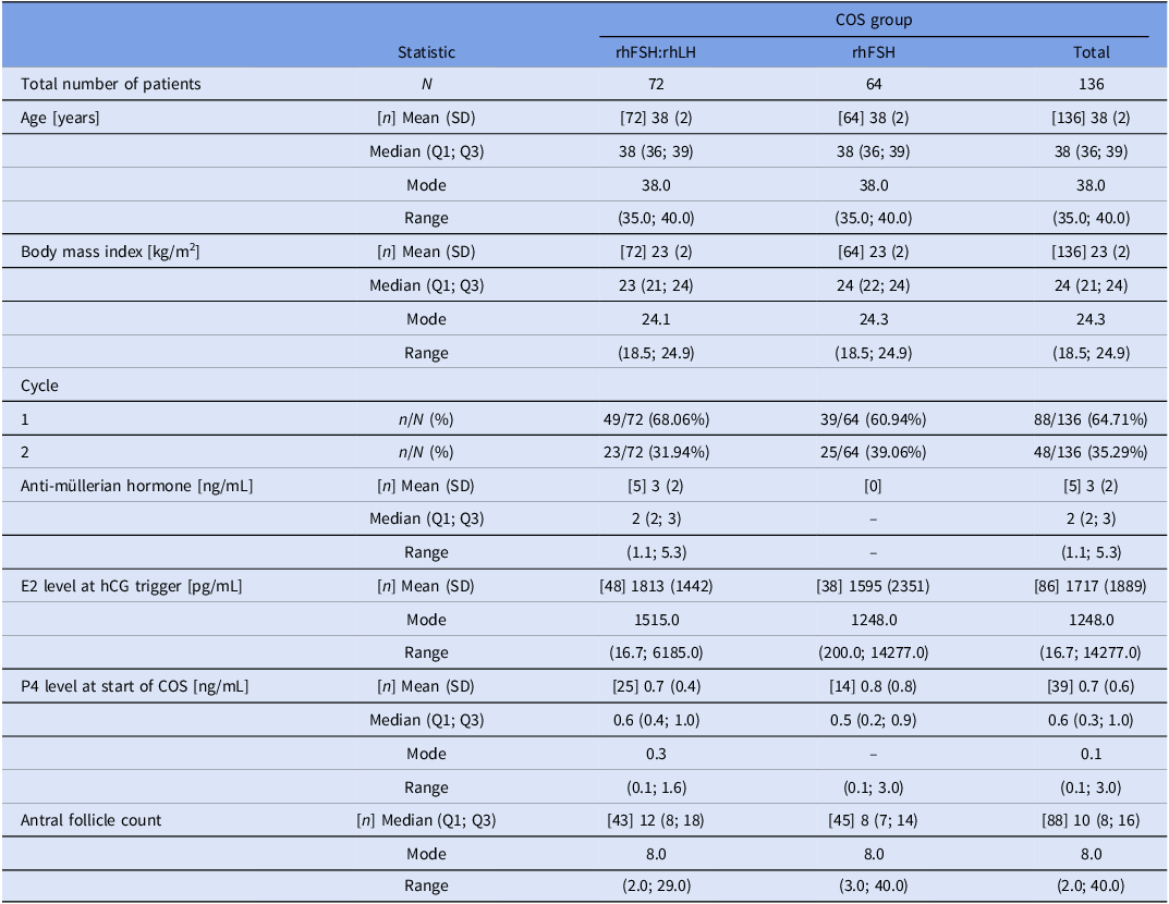
A total of 136 ICSI patients/cycles stimulated with either r-hFSH:r-hLH or r-hFSH alone for COS were included in this study. Baseline characteristics are shown in Table 1.
Baseline characteristics in study groups

Note: COS, controlled ovarian stimulation; rFSH, recombinant follicle-stimulating hormone; rLH, luteinizing hormone; E2, oestradiol; hCG, human chorionic gonadotropin; P4, progesterone; SD, standard deviation; Q1, first quartile; Q3, third quartile.
Semen quality parameters were comparable between groups. The mean sperm concentration was 45.2 ± 18.7 × 10⁶/mL in the r-hFSH:r-hLH group and 47.9 ± 20.3 × 10⁶/mL in the r-hFSH-only group (p = 0.422); progressive motility was 54.6 ± 9.8% vs. 55.3 ± 10.2% (p = 0.615); and normal morphology (strict Kruger criteria) was 4.5 ± 1.7% vs. 4.7 ± 1.8% (p = 0.581), respectively.
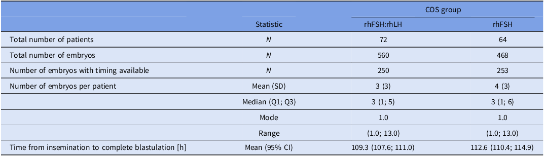
Results from the statistical analysis regarding the primary outcome are presented in Table 2. Embryos derived from patients stimulated with r-hFSH:r-hLH reached the blastocyst stage (tB) faster than those from the r-hFSH-only group (109.3 h vs. 112.6 h).
Primary endpoint analysis (tB) in study groups
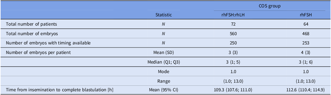
Note: Time from insemination to complete blastulation is estimated on embryo level. COS, controlled ovarian stimulation; rFSH, recombinant follicle-stimulating hormone; rLH, luteinizing hormone; E2, oestradiol; hCG, human chorionic gonadotropin; P4, progesterone; SD, standard deviation; Q1, first quartile; Q3, third quartile; CI, confidence interval.
Secondary outcomes are summarized in Tables 3.1A–6. All mean timings to reach specific morphokinetic milestones – tPNa, tPNf, t2 to t8, tM and tSB – were shorter in embryos derived from the r-hFSH: r-hLH group compared with those from the r-hFSH-only group (Table 3.1A, B and C).
Secondary endpoints analyses in study groups

Note: Time from insemination to a particular stage is estimated on embryo level. COS, controlled ovarian stimulation; rFSH, recombinant follicle-stimulating hormone; rLH, luteinizing hormone; SD, standard deviation; Q1, first quartile; Q3, third quartile; CI, confidence interval.
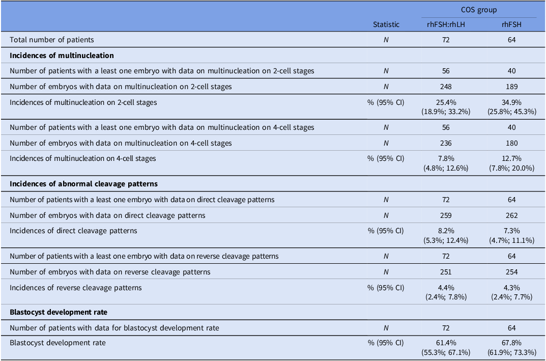
The incidences of multinucleation at the 2-cell and 4-cell stages were lower in embryos from the r-hFSH:r-hLH group, whereas direct and reverse cleavage patterns were more frequent in this group. The rate of blastocyst development was higher in women from the r-hFSH-only group (Table 4).
Incidences and rates

Note: COS, controlled ovarian stimulation; rFSH, recombinant follicle-stimulating hormone; rLH, luteinizing hormone; CI, confidence interval. Total embryos generated per group: 560 (r-hFSH:r-hLH) and 468 (r-hFSH). ‘N’ in this table refers to embryos with available data for each parameter.
The distribution of KIDScore categories did not differ significantly between stimulation protocols (χ2 test, p = 0.46). The proportions of embryos classified as <5, 5–6.9 and ≥7 were similar between groups (r-hFSH:r-hLH = 61.6, 18.0 and 20.4%; r-hFSH = 58.7, 19.3 and 22.0%, respectively), indicating that the morphokinetic differences observed were not reflected in the algorithm-based embryo classification.
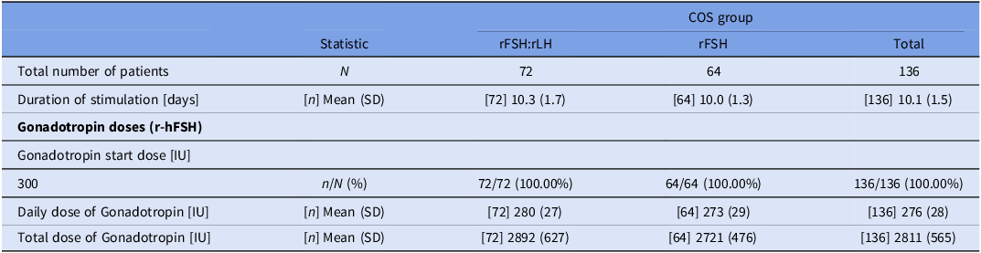
Stimulation characteristics are shown in Table 5. The mean duration of stimulation and both the daily and total gonadotropin doses were higher in the r-hFSH:r-hLH group.
Stimulation

Note: COS, controlled ovarian stimulation; rFSH, recombinant follicle-stimulating hormone; rLH, luteinizing hormone; SD, standard deviation; CI, confidence interval.
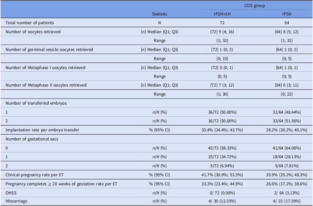
Other secondary endpoints are presented in Table 6. The r-hFSH:r-hLH group showed a higher number of retrieved and mature oocytes. Implantation, clinical and ongoing pregnancy rates were higher, whereas miscarriage and OHSS rates were lower compared with the r-hFSH-only group.
Other secondary endpoints

Note: COS, controlled ovarian stimulation; rFSH, recombinant follicle-stimulating hormone; rLH, luteinizing hormone; Q1, first quartile; Q3, third quartile; CI, confidence interval.
Discussion
Both the in vivo and in vitro reproductive potential are inversely correlated with maternal age (Klein and Sauer, Reference Klein and Sauer2001). Pregnancy chance progressively declines at 35 years, approaching zero after 45 (Cetinkaya et al., Reference Cetinkaya, Siano, Benadiva, Sakkas and Patrizio2013; Klipstein et al., Reference Klipstein, Regan, Ryley, Goldman, Alper and Reindollar2005). Maternal ageing is associated with a reduced number and quality of retrieved oocytes (Fitzgerald et al., Reference Fitzgerald, Zimon and Jones1998) and higher aneuploidy rates (Fitzgerald et al., Reference Fitzgerald, Zimon and Jones1998; Klein and Sauer, Reference Klein and Sauer2001), which negatively affect fertilization, embryo development, implantation and pregnancy outcomes (Munné et al., Reference Munné, Chen, Colls, Garrisi, Zheng, Cekleniak, Lenzi, Hughes, Fischer, Garrisi, Tomkin and Cohen2007).
Various gonadotropin combinations can be used to stimulate follicle growth. FSH and LH act synergistically in folliculogenesis during stimulated cycles. Improved outcomes have been reported with FSH and LH-like-action gonadotropins, particularly in AMA patients undergoing ICSI (Alviggi et al., Reference Alviggi, Conforti, Esteves, Andersen, Bosch, Bühler, Ferraretti, De Placido, Mollo and Fischer2018; Mochtar et al., Reference Mochtar, Danhof, Ayeleke, Van der Veen and van Wely2017). In a recent study from our group, r-hLH supplementation improved oocyte retrieval and ICSI outcomes (Setti et al., Reference Setti, Braga, Iaconelli and Borges2021).
The present study described embryonic morphokinetic data and ICSI outcomes in AMA patients stimulated with r-hFSH with or without r-hLH. We observed that the number of oocytes retrieved (p = 0.013), embryonic morphokinetics in terms of tPNa (p = 0.036), tM (p = 0.010), tSB (p = 0.006) and tB (0.022), as well as implantation rate (p < 0.001), were significantly higher with r-hLH supplementation.
r-hLH may favor ICSI outcomes by enhancing oocyte competence and endometrial receptivity. LH binds to receptors on ovarian target cells, initiating a cascade of molecular events (Aflatoonian et al., Reference Aflatoonian, Yousefnejad, Eftekhar and Mohammadian2012). It increases follicular insulin sensitivity and converts progesterone into androgens, which can be aromatized to estrogens, creating a hormonal environment that enhances follicular maturation and fertilization potential (Gutman et al., Reference Gutman, Barak, Maslovitz, Amit, Lessing and Geva2009). Cumulus cell apoptosis is lower in oocytes from r-hLH-supplemented cycles than from r-hFSH-only cycles (Ruvolo et al., Reference Ruvolo, Bosco, Pane, Morici, Cittadini and Roccheri2007). LH also reduces premature progesterone rise, improving endometrial receptivity (Hill and Propst, Reference Hill, Propst and Darwish2012). Together, these mechanisms may promote more competent oocytes and embryos with improved morphokinetic behaviour.
The observed differences in morphokinetic behaviour may result from the distinct endocrine milieu created by r-hLH. By increasing intra-follicular androgen production and aromatization to oestradiol, r-hLH contributes to optimal oocyte maturation and synchronization of nuclear and cytoplasmic development. LH activity also prevents premature luteinization and excessive progesterone rise, preserving endometrial receptivity (Hill and Propst, Reference Hill, Propst and Darwish2012). Reduced cumulus apoptosis (Ruvolo et al., Reference Ruvolo, Bosco, Pane, Morici, Cittadini and Roccheri2007) further supports oocyte metabolism, potentially explaining the faster and more coordinated embryonic divisions observed in the r-hFSH:r-hLH group.
Although stimulation in the r-hFSH:r-hLH (Pergoveris) group required higher total gonadotropin doses and a longer duration than r-hFSH alone, these parameters did not compromise oocyte or embryo quality. On the contrary, oocyte yield, maturation and embryonic developmental competence appeared enhanced, as reflected by earlier cleavage timings and higher implantation rates observed in this study. This aligns with evidence that while supraphysiologic gonadotropin exposure can be detrimental, LH addition in AMA women may enhance follicular responsiveness and competence (Rubio et al., Reference Rubio, Mercader, Alamá, Lizán, Rodrigo, Labarta, Melo, Pellicer and Remohí2010; Alviggi et al., Reference Alviggi, Conforti, Esteves, Andersen, Bosch, Bühler, Ferraretti, De Placido, Mollo and Fischer2018), highlighting the balance between stimulation intensity and oocyte quality.
Embryo morphokinetics allows characterization of cell divisions for embryo selection (Barberet et al., Reference Barberet, Bruno, Valot, Antunes-Nunes, Jonval, Chammas, Choux, Ginod, Sagot, Soudry-Faure and Fauque2019). Despite potential drawbacks of TLI systems (light, heat, magnet, lubricant exposure) (Chen et al., Reference Chen, Wei, Hu, Yuan and Liu2017), meta-analyses show improved pregnancy outcomes (Pribenszky et al., Reference Pribenszky, Nilselid and Montag2017). In this study, all morphokinetic stages occurred faster in embryos from r-hFSH:r-hLH cycles. Although these differences appear small, the higher clinical outcomes in this group may indicate a biological effect. This interpretation is consistent with findings from Shebl et al. (Reference Shebl, Haslinger, Kresic, Enengl, Reiter, Oppelt and Ebner2021), who demonstrated that both excessively rapid and markedly delayed mitotic cycles are associated with lower live birth rates, suggesting that optimal cleavage pacing reflects underlying developmental competence. Prior studies report that embryos reaching the blastocyst stage and implanting tend to cleave earlier (Meseguer et al., Reference Meseguer, Herrero, Tejera, Hilligsoe, Ramsing and Remohi2011; Dal Canto et al., Reference Dal Canto, Coticchio, Mignini Renzini, De Ponti, Novara, Brambillasca, Comi and Fadini2012; Kirkegaard et al., Reference Kirkegaard, Kesmodel, Hindkjær and Ingerslev2013). Shorter t4 and tSB intervals have been linked to euploid blastocysts (Minasi et al., Reference Minasi, Colasante, Riccio, Ruberti, Casciani, Scarselli, Spinella, Fiorentino, Varricchio and Greco2016). Thus, earlier cleavage may reflect greater developmental competence, though excessively rapid division can be detrimental (Chamayou et al., Reference Chamayou, Patrizio, Storaci, Tomaselli, Alecci, Ragolia, Crescenzo and Guglielmino2013). Recent large-scale analyses (Campbell et al., Reference Campbell, Smith, Barrie and Patrick2024) emphasize that morphokinetic data must be interpreted in context; moderate acceleration suggests competence, while extreme deviation may indicate abnormal development. Therefore, faster cleavage may correlate with higher implantation rates but should be interpreted cautiously. The selection of embryos for transfer was based on the incubator’s algorithm (KIDScore), which also considers such patterns.
Consistent with these findings, the distribution of KIDScore categories (<5, 5–6.9, and ≥7) did not differ significantly between stimulation protocols, suggesting that the faster morphokinetic profile observed in the r-hFSH:r-hLH group did not alter the overall embryo ranking within the algorithm. This supports the notion that subtle variations in cleavage or blastulation timing may occur within the same KIDScore strata and are not necessarily predictive of clinical competence.
Although specific morphokinetic variables (t2, t3, tSB) have been associated with implantation and euploidy potential (Meseguer et al., Reference Meseguer, Herrero, Tejera, Hilligsoe, Ramsing and Remohi2011; Minasi et al., Reference Minasi, Colasante, Riccio, Ruberti, Casciani, Scarselli, Spinella, Fiorentino, Varricchio and Greco2016), their isolated predictive value is limited. Embryo development is multifactorial; thus, integrated models like KIDScore, which combine morphokinetic and morphological data, show superior predictive performance. Campbell et al. (Reference Campbell, Smith, Barrie and Patrick2024) further highlighted that such integrated approaches better represent developmental competence than single-event predictors.
While morphokinetic annotation can vary between observers, TLI provides continuous monitoring, reducing missed developmental events compared to static observation. Inter-observer agreement is generally high when standardized criteria are applied (Barberet et al., Reference Barberet, Bruno, Valot, Antunes-Nunes, Jonval, Chammas, Choux, Ginod, Sagot, Soudry-Faure and Fauque2019; Kirkegaard et al., Reference Kirkegaard, Kesmodel, Hindkjær and Ingerslev2013; Meseguer et al., Reference Meseguer, Herrero, Tejera, Hilligsoe, Ramsing and Remohi2011).
Although time-lapse technology and morphokinetic algorithms have improved standardization and may reduce time to pregnancy, their effect on overall success remains debated. Recent randomized clinical trials, including the TILT study (Bhide et al., Reference Bhide, Chan, Lanz, Alqawasmeh, Barry, Baxter, Gonzalez Carreras, Choudhury, Cheong, Chung, Collins, Cong, Doidge, Heighway, Patel, Pardo, Rattos, Wright, Dodds, Perez, Khan and Thangaratinam2024) and Ahlström et al. (Reference Ahlström, Lundin, Lind, Gunnarsson, Westlander, Park, Thurin-Kjellberg, Thorsteinsdottir, Einarsson, Åström, Löfdahl, Menezes, Callender, Nyberg, Winerdal, Stenfelt, Jonassen, Oldereid, Nolte, Sundler and Hardarson2022), found no significant improvement in cumulative live birth rates compared to conventional culture. Consistent with these findings, the latest ESHRE guideline reports insufficient evidence that time-lapse culture improves live birth or other clinical outcomes versus conventional incubation (Apter et al., Reference Apter, Ebner, Freour, Guns, Kovacic, Le Clef, Marques, Meseguer, Montjean, Sfontouris, Sturmey and Coticchio2020). Its benefits appear mainly related to workflow optimization and standardization rather than enhanced efficacy. Hence, while morphokinetic assessment offers valuable insights, its role in improving cumulative outcomes remains uncertain.
Several studies have assessed the applicability and effectiveness of supplementing different LH forms and doses during COS (Humaidan et al., Reference Humaidan, Bungum, Bungum and Yding Andersen2004; Griesinger et al., Reference Griesinger, Schultze-Mosgau, Dafopoulos, Schroeder, Schroer, von Otte, Hornung, Diedrich and Felberbaum2005; Orvieto, Reference Orvieto2015; Levi-Setti et al., Reference Levi-Setti, Zerbetto, Baggiani, Zannoni, Sacchi, Smeraldi, Morenghi, De Cesare, Drovanti and Santi2019). Initially, hCG was used to provide LH activity in ART cycles; however, r-hLH has since been approved for clinical use, offering a more physiological and controllable source of LH bioactivity (Casarini et al., Reference Casarini, Santi, Brigante and Simoni2018). Adequate LH levels are crucial for optimal steroidogenesis, promoting conversion of progesterone to androgens within theca cells and subsequent aromatization to estrogens in granulosa cells (Hillier, Reference Hillier2001; Balasch and Fábregues, Reference Balasch and Fábregues2002). This balanced milieu supports nuclear and cytoplasmic maturation, proper spindle formation and mitochondrial function (Ruvolo et al., Reference Ruvolo, Bosco, Pane, Morici, Cittadini and Roccheri2007; Alviggi et al., Reference Alviggi, Conforti, Esteves, Andersen, Bosch, Bühler, Ferraretti, De Placido, Mollo and Fischer2018). r-hLH also reduces premature luteinization by stabilizing progesterone levels during the late follicular phase, improving endometrial receptivity and synchronization (Hill and Propst, Reference Hill, Propst and Darwish2012). Conversely, excessive LH exposure can be detrimental, leading to premature luteinization, follicular atresia or impaired oocyte maturation (Chappel and Howles, Reference Chappel and Howles1991; Hillier, Reference Hillier2000; Huirne et al., Reference Huirne, van Loenen, Schats, McDonnell, Hompes, Schoemaker, Homburg and Lambalk2005). Thus, maintaining LH within a physiological window appears essential to achieve optimal responsiveness, particularly in AMA women with reduced LH receptor sensitivity.
A key limitation of this study is the relatively small number of patients, which restricts the ability to draw conclusions for low-incidence outcomes such as OHSS and miscarriage. Although these events were documented for completeness, the study was not powered to compare them between stimulation protocols. Therefore, any numerical differences observed here must be interpreted strictly as descriptive trends rather than clinically actionable findings. As a single-centre analysis, the findings may not be generalizable to all ART settings, particularly given potential differences in laboratory workflows, stimulation preferences and time-lapse annotation protocols.
This retrospective, descriptive design also introduces inherent limitations. Because patient allocation to stimulation protocols was not randomized but based on clinical judgment, selection bias cannot be excluded; however, this approach reflects real-world ART practice. Only the most recent COS cycle per patient was included to minimize bias from previous cycle adjustments, though this may still limit generalizability.
We also acknowledge that embryo transfer decisions relied on KIDScore rankings generated by the EmbryoScope™ system, which incorporates morphokinetic variables that were analyzed as outcomes in this study. This introduces potential circularity. Although subgroup analyses stratified by KIDScore categories were not performed, future prospective studies could determine whether the observed kinetic differences persist independently of algorithm-driven classification.
The present findings may assist clinicians in choosing COS protocols for AMA patients. While descriptive by nature, this study helps characterize morphokinetic behaviour across stimulation strategies and provides hypotheses for prospective validation.
Acknowledgements
We thank Dr Kris Bogaerts, PhD, for his expertise and assistance throughout the study and for his help in performing the statistical analysis of the study.
Funding
This study was partially funded by Merck KGaA, Darmstadt, Germany, which played a role in supporting the research efforts.
Competing interests
All authors declare that there is no conflict of interest that could be perceived as prejudicing the impartiality of the research reported.