I. INTRODUCTION
The development of the Rietveld method (Rietveld, Reference Rietveld1967, Reference Rietveld1969) for structural refinement from powder diffraction data has been one of the most important achievements for structural chemistry and physics in the last few decades. The popularization of structure determination from powder diffraction data has additionally boosted the use of the Rietveld method since solving an unknown structure and refining its structure from powder diffraction data is nowadays possible in a significant percentage of the cases (David and Shankland, Reference David and Shankland2008).
One of the difficulties of structural analysis from powder diffraction data is the assessment of the accuracy of the resulting structural model and therefore the extraction of structural conclusions from the results. The inherent drawback of the powder method and peak overlap, produces a reduction of the amount of independent data reducing the quality of the structural model. Bénard et al. (Reference Bénard, Louër and Louër1991), Gascoigne et al. (Reference Gascoigne, Tarling, Barnes, Pygall, Benard and Louër1994) and Louër et al. (Reference Louër, Louër, Bétourné and Touboul1996) have compared structural results from single crystals and powder X-ray diffraction looking for a measure of the mentioned loss of accuracy of the model. Their general conclusion is that structures refined using conventional X-ray powder diffraction data are, on average, less accurate than those refined using single-crystal X-ray diffraction data, but this loss of accuracy is more marked for the compounds containing heavy atoms such as Cd5(OH)8(NO3)2·2H2O (Bérnard et al., 1991) or Zr(OH)2SO4·3H2O (Gascoigne et al., Reference Gascoigne, Tarling, Barnes, Pygall, Benard and Louër1994) than in the compound where most atoms have similar contributions to the total scattered intensity such as Li4B2O5·H2O (Louër et al., Reference Louër, Louër, Bétourné and Touboul1996).
An extensive case by case study of how powder and single crystals derived structures match would be required to carry out a general evaluation of this problem which is well beyond our interest and capabilities. We have studied the case of a metal–organic framework (MOF) compound using conventional and synchrotron X-ray diffraction data compared with single-crystal X-ray diffraction data.
The design and synthesis of self-assembled open framework coordination polymers have been of great interest in view of their intriguing variety of architectures and topologies (Moulton and Zaworotko, Reference Moulton and Zaworotko2001; Sisson et al., Reference Sisson, Shah, Bhosale and Matile2006; Ariga et al., Reference Ariga, Vinu, Hill and Mori2007; Maspoch et al., Reference Maspoch, Ruiz-Molina and Veciana2007; Férey, Reference Férey2008; Robson, Reference Robson2008). In particular, the so-called MOFs have found applications in the field of gas storage (Murray et al., Reference Murray, Dinca and Long2009), gas separation (Czaja et al., Reference Czaja, Trukhan and Müller2009), and molecular containers for polymerization reactions (Uemura et al., Reference Uemura, Yanai and Kitagawa2009).
In this context, heterometallic coordination polymers containing lanthanide ions present additional and interesting features, owing to the magnetic and luminescent properties of the metallic centres (Bünzli and Piguet, Reference Bünzli and Piguet2005; Shen et al., Reference Shen, Sun and Yan2008).
Polycarboxylic acids are extensively used as linkers in the construction of these materials. They provide the dimensionality of the network and tune (together with the coordination number of the metal ion) the pore size and shape. The flexible ligand 2,2′-oxydiacetic acid (H2oda) has been successfully used to prepare heteropolymetallic coordination compounds with general formula [Ln2M 3(oda)6] (M = bivalent cation) (reviewed by Kremer et al., 2008). The first report on these compounds contained the crystal structure of [Gd2Cu3(oda)6]·3H2O (Mao et al., Reference Mao, Song, Huang and Huang1997). Later, the series was extended with analogous compounds with Y, Pr, Nd, Sm, Eu, Tb, Dy, Er, Yb and finally, the first non-containing copper(II) complex, [La2Co3(oda)6(H2O)6]·12H2O was prepared (Domínguez et al., Reference Domínguez, Torres, Peluffo, Mederos, González-Platas, Castiglioni and Kremer2007). The synthesis is straightforward by a direct combination of the reactants in an aqueous solution. The complexes are isostructural (hexagonal crystal system and P6/mcc space group) and contain nine-coordinated Ln ions surrounded by nine O atoms from three independent oda ligands. The building block [Ln(oda)3] still has uncoordinated O-carboxylate donor atoms that are able to link [M(H2O)2] (M = Cu, Co) units. The structure contains large hexagonal channels (average diameter: 10.7 Å) along the crystallographic c-axis that houses the crystallization water molecules. The magnetic properties of the complexes were investigated, showing weak anti-ferromagnetic interactions M(II)–M(II) in the low-temperature range (Torres et al., Reference Torres, Peluffo, Domínguez, Mederos, Arrieta, Castiglioni, Lloret and Kremer2006).
The complex [{Mn(H2O)6}{MnGd(oda)3}2]·6H2O was prepared and fully characterized by Prasad et al. (Reference Prasad, Rajasekharan and Costes2007). It is synthesized by a similar procedure and follows the general formulae of the previous polynuclear complexes. However, the crystal structure (cubic crystal system, Fd3c space group) is completely different. The [Gd(oda)3] building blocks are still present, but the Mn(II) ions are not equivalent. One of them links the building blocks and forms a 3D structure with two types of cubic cavities, one filled by water molecules and the other filled by [Mn(H2O)6]2+ cations. This spatial arrangement provokes a ferromagnetic interaction between the Gd(III) and intra-network Mn(II) ions, whereas the hexaaquo Mn(II) appears to remain magnetically disordered. The reason is not clear for this different behaviour. The relative size of the f and d metal ions could be the key point. The understanding of this can be the basis to design new materials built upon the Ln–M–oda complexes such as recently prepared [{Mg(H2O)6}{MgLn(oda)3}2]·6H2O (Ln = Nd, Gd, Tb, Eu) compounds with the same structure (Chang et al., Reference Chang, Zhao, Gu, Liu, Wei, Liu, Mo and Yu2012).
With this in mind, we have undertaken a detailed structural study of a series of particular MOF compounds [{Ca(H2O)6}{CaLn(oda)3}2]·xH2O (Ln = lanthanoid) isostructural with previously mentioned [{Mn(H2O)6}{MnGd(oda)3}2]·6H2O, using the powder method since single crystals of sufficient quality for diffraction were unavailable. In these particular compounds, both Ln and Ca cations occupy special positions with fixed coordinates and additional systematic absence conditions, contributing to a reduced number of intensities. The light elements C, O and H, however, are in lower symmetry positions and they contribute to more reflections than heavy elements but reflections have also lower intensities. Still, it is possible to accurately determine light atom structural parameters if accurate intensities are available.
During the course of our Rietveld analysis, a number of powder diffraction datasets were collected in in-house conventional and synchrotron-based powder diffractometers. Problems related to sample degradation because of absorbed doses were detected and treated to obtain the best possible structural model. The results of these treatments are compared with conventionally treated powder diffraction data from both types of sources. Eventually, along this work, a suitable single crystal for X-ray diffraction was obtained for the Ln = Gd compound and the structure was solved and refined. The complete powder diffraction and single-crystal refinement results are included in this work to provide a wider comparison between different data and the refinement method and to assess in a better way the accuracy of the structural model extracted from Rietveld refinement of different kinds of datasets for this particular compound.
II. EXPERIMENTAL
A. Synthesis of [{Ca(H2O)6}{CaGd(oda)3}2].nH2O
All common laboratory chemicals were of reagent grade, purchased from commercial sources and used without further purification. Gd(NO3)3·6H2O and CaCl2·2H2O were used as metal sources. 2,2′-oxydiacetic acid was purchased from Fluka.
[{Ca(H2O)6}{CaGd(oda)3}2]·nH2O was prepared by mixing 10 mL of an aqueous solution containing 1.5 mmol of Ca2+ ion and 1.0 mmol of Gd3+ ion and 10 ml of another aqueous solution of 2,2-oxydiacetic acid (0.40 g, 3.0 mmol) with pH value adjusted to 7.0 with ammonium hydroxide. The clear final solution was allowed to evaporate slowly for 1–2 weeks to obtain crystals. Yield 50–70%. Although transparent, regular and well-developed faceted crystals were obtained by this procedure they were multiple twins making them unsuitable for single-crystal structural analysis and were crushed into fine powders for chemical and structural characterization. Eventually, one single crystal suitable for structural characterization was obtained and used for the experiment described below.
B. Chemical characterization
The infrared spectra, as KBr pellets, were obtained from a Bomen MB 102 FT-IR spectrophotometer. IR peaks associated with the oda ligand appear centred at: 1605, 1439 and 1124 cm−1.
Elemental analysis (C, H) was performed on a Carlo Erba EA 1108 instrument. Confirmation of sample purity and crystalline quality were performed by X-ray powder diffraction on crushed crystals of the as-prepared sample on a Rigaku ULTIMA IV diffractometer equipped with CuKα sealed tube source operating at 40 kV/30 mA power and diffracted beam curved Ge monochromator providing the Kα 1−2 doublet with λave = 1.5418 Å. Experiments were performed at 20(1)°C in θ–θ Bragg–Brentano geometry with the sample mounted horizontally on a glass holder at the centre of the 285 mm radius goniometer.
Thermal analysis was performed on a Shimadzu TGA-50 instrument with a TA 50I interface, using a platinum cell and nitrogen atmosphere. Experimental conditions were: temperature ramp rate, 0.5°C min−1; nitrogen flow rate: 50 mL min−1.
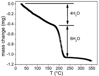
Figure 1 shows the thermogram of [{Ca(H2O)6}{CaGd(oda)3}2]·nH2O. In total 8.391 mg compound was balanced and temperature was increased up to 350 °C. Starting from room temperature, a slow but constant weight loss is observed, accounting for 0.419 mg or 4.99% of the initial mass from 25°C to 180 °C followed by a larger and faster weight loss of 0.638 mg or 7.60%, up to 210 °C, where the weight stabilizes. These two distinct weight loss processes correspond to the loosely bound crystallization and tightly bound structural water molecules corresponding to hexaaquocalcium(II) counter ion, respectively. Expected weight loss for [{Ca(H2O)6}{CaGd(oda)3}2]·4H2O in the first and second processes are 5.1% (4 water molecules) and 7.7% (6 water molecules), respectively. From this and C–H elemental analysis, the structural formula can be derived (experimental: C 21.5, H 3.5, H2O 12.6%; calculated: C 20.5, H 3.2, H2O 12.8%) for [{Ca(H2O)6}{CaGd(oda)3}2]·4H2O.
Thermogram of [{Ca(H2O)6}{CaGd(oda)3}2]·4H2O showing weight loss in two separate processes leading to the loss of four weakly bound crystallization and six tightly bound structural water molecules per formula unit.

C. X-ray diffraction data collection, processing and analysis
1. Conventional X-ray powder diffraction
Rietveld-quality conventional X-ray powder diffraction data collection was performed using the instrument described above in 0.02° steps from 8° to 149° in 2θ with a counting time of 30 s per step (total data collection time of ~60 h). This data was used for Rietveld analysis in the conventional way without further processing.
Rietveld structural refinement was performed using GSAS/EXPGUI suite of programs (Toby, Reference Toby2001; Larson and Von Dreele, Reference Larson and Von Dreele2004) starting with the final model obtained from single-crystal X-ray diffraction structural determination and refinement. A shifted Chebyschev polynomial background function and a modified TCHZ profile function (Thompson et al, Reference Thompson, Cox and Hastings1987) corrected for anisotropic broadening (Stephens, Reference Stephens1999) (type 4) were used for profile modelling. The March–Dollase preferential orientation correction (Dollase, Reference Dollase1986) was applied to account for a slight orientation of crystallites along 100 directions in the flat sample.
2. Synchrotron X-ray powder diffraction
Synchrotron X-ray powder diffraction data collection was performed at Argonne's Advanced Photon Source, 11BM-B beamline (Lee et al., Reference Lee, Shu, Ramanathan, Preissner, Wang, Beno, Von Dreele, Ribaud, Kurtz, Antao, Jiao and Toby2008; Wang et al., Reference Wang, Toby, Lee, Ribaud, Antao, Kurtz, Ramanathan, Von Dreele and Beno2008). The samples were loaded in ϕ = 0.8 mm polyimide capillaries and data were collected using ~30 keV energy radiation (λ = 0.410 07 Å). Twelve Si-111 crystal analyser-detector systems spaced nominally 2° apart are mounted on the diffractometer 2θ arm so that a 2° scan provides measurement of a 24° 2θ range. Data collection was performed covering the 0–44° 2θ range with a step of 0.001° and 0.1 s per step (total of ~66 min). Data processing was performed with the CMPR program (Toby, Reference Toby2005) as is described below. Each of the 12 detectors produced a partial pattern that is called a data bank in the subsequent discussion.
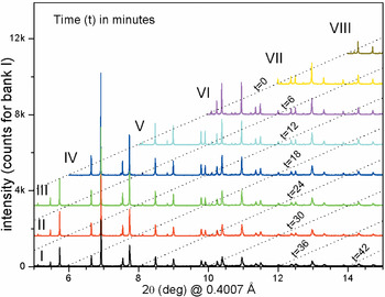
Figure 2 shows the resulting banks I–VIII (corresponding to detectors I–VIII) in the region 5–15° in 2θ of [{Ca(H2O)6}{CaGd(oda)3}2]·4H2O sample. Isochronous lines for the representation in Figure 2 can be built as diagonal dotted lines connecting the same time point in different banks. Each detector scans for 20° in 2θ, therefore, the beginning of e.g., bank VIII corresponds to 42 min of scan for detector I or 24 min for detector IV. It is evident from Figure 2 that the [{Ca(H2O)6}{CaGd(oda)3}2]·4H2O sample suffers from radiation damage since the intensity of each peak decreases with time (looking down at any fixed angle in Figure 2 or at selected peaks in Figure 3(a)) and additionally the positions of the peak shift towards higher 2θ with time as shown in Figure 3(a). This radiation damage goes, in general, undetected with single-detector instruments or is difficult to process in multiple detector datasets since each pattern represents the sample in different times and conditions and no two patterns have the same intensity for the same peak. The need for a special processing procedure for this sample is evident.
Consecutive scans of 11BM-B diffractometer for sample [{Ca(H2O)6}{CaGd(oda)3}2]·4H2O. Bank number is shown in roman figures. Dotted diagonal lines represent isochronous lines in the figure. Time increases rightwards for each pattern and downwards for each peak of the pattern.

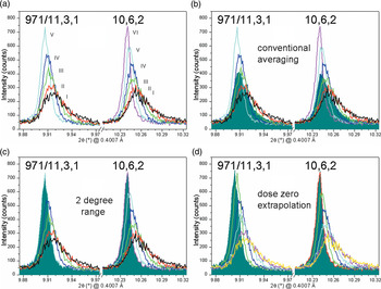
(a) Selected regions of the data banks I–VI showing evolution of peak intensity and position of 971/11, 3, 1 doublet and 10, 6, 2 peak with detector number. The 10, 6, 2 peak above 10° was scanned by six detectors (I–VI), one more than 971/11, 3, 1 peaks below 10°. (b) The result of averaging the intensities of the patterns for the same peaks shown in (a). (c) The result of selecting the first 2 degrees of data for each bank. (d) The result of extrapolation to doze zero of observed peaks. Note that small changes in relative intensities and peak positions are clearly visible among different processing methods.

a. Conventional data processing
Averaging of all banks was performed for each 2θ point to obtain one conventional dataset for this pattern. The peaks below 4° (including the most intense peak in the pattern) were scanned only twice and the average is performed within a sample irradiated for less than 12 min, whereas peaks in the region 18–20° were scanned 10 times by detectors 1–10 and the average corresponds to a sample irradiated for about 60 min. Most high-angle peaks correspond also to very long irradiation times therefore none of the banks contain significant diffraction information at the highest possible angle (44°) where intensity decay with 2θ adds to peak intensity decrease because of radiation damage. We may consider that this conventionally processed dataset is comparable in quality with the conventional X-ray diffraction dataset, collected with only one detector over a sample that decomposed under irradiation for ~60 h except for the fact that the peaks are narrower and have better counting statistics.
b. 2-degree range selection
To avoid all possible averaging of peaks with different intensities and positions after sample decay a different strategy was chosen to obtain the final dataset. Non-overlapping regions of each bank were selected in such a way that the whole pattern is covered and the timeframe of irradiation is reduced to its possible minimum. This corresponds to the superposition of the first 2° of data in each bank, or the fringe of data contained between the 0 and 6 min isochronous lines in Figure 2. In this way, a pattern is constructed with 12 2° segments covering from 0° to 24° in 2θ. In this new pattern, there is no data averaging and each peak has been included in its first scan. This dataset is not error free, since two peaks in the beginning and end of one 2° interval correspond to irradiation times that can be as long as 6 min away from each other.
c. Dose zero extrapolation
To obtain a dataset that would simulate the intensity of each peak at the starting point of data collection, where the sample's absorbed dose is zero, an extrapolation of the intensities and peak positions for each observed peak of the pattern in each bank was performed. The procedure that will be described in detail elsewhere (Jun Wang et al., unpublished results), consists in the assignment of a time flag and hkl (Miller) indices to each peak in each of the 12 banks obtained from each of the detectors. This is possible because the rate of peak shift and intensity reduction with time is small and no sign of a phase transition with absorbed dose is observed. Each peak is later integrated and its position determined. For the same group of hkl indices, a set of intensity vs. time and position vs. time tables can be constructed (containing between 2 and 10 entries each) and by a simple least-squares fit of the data an extrapolation of intensity and peak position to t = 0 is performed. A new pattern is constructed using the extrapolated intensity and position of each reflection. The peak shape of each extrapolated peak and the background around it is taken from the first entry of the table (the peak obtained at the lowest dose). This pattern contains ideally dose-corrected intensities but somehow time-dependent peak shapes that, as in the case of the 2° range pattern, degrade slowly but recover within 6 min timeframes (or 2° 2θ intervals).
Figure 3 shows the results of the three processing procedures for 9, 7, 1/11, 3, 1 doublet at 2θ ≈ 9.92° and 10, 6, 2 peak at 2θ ≈ 10.24° where the differences between peaks collected over the beginning of a 2° range (10, 6, 2) and at the end (9, 7, 1/11, 3, 1 doublet) are clearly visible and the differences in the result of the different processing methods are also clearly visible.
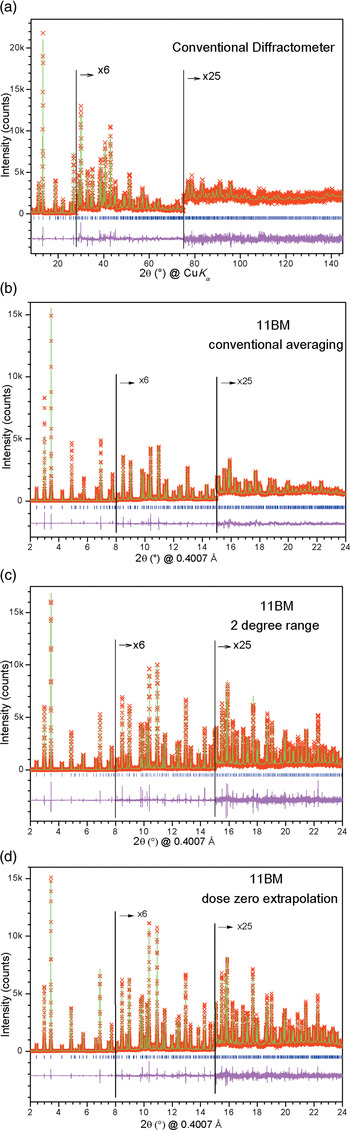
Rietveld structural refinements were performed using GSAS/EXPGUI suite of programs (Toby, Reference Toby2001; Larson and Von Dreele, Reference Larson and Von Dreele2004) on the three datasets as described for conventional powder diffraction data analysis. In these three cases, however, no preferential orientation correction was required. Initially, the structural model for [{Mn(H2O)6}{MnGd(oda)3}2]·6H2O compound (Prasad et al., Reference Prasad, Rajasekharan and Costes2007) was used changing Mn for Ca and allowing water content to refine obtained good fits of all the data confirming that [{Ca(H2O)6}{CaGd(oda)3}2]·4H2O is isostructural with the Mn-containing compound. Finally, the model obtained from single-crystal X-ray diffraction structural refinement was employed as a starting point to the refinement to allow direct comparison among coordinates and atom labels obtaining the same result. All the atoms were freely refined with isotropic atomic displacement parameters except for Gd and Ca in the dose zero extrapolated dataset where anisotropic atomic displacement parameters were allowed to refine. Hydrogen atoms bonded to C2 were not refined riding but at a fixed position with respect to C2 only. Figure 4 shows the Rietveld fits of the four powder patterns described above. High angle regions have been amplified to show the fit quality of the low intensity peaks in the four cases.
Rietveld fit of (a) conventional X-ray powder diffraction data (Rigaku ULTIMA IV diffractometer) and synchrotron X-ray powder diffraction data (11BM instrument at APS) after (b) conventional averaging, (c) 2-degree selection and (d) extrapolation to dose zero. Note that the expanded regions of the patterns show that at the highest diffraction angles (c) and (d) show the highest relative peaks and that fits of low angle data are the best for (d).

3. Single-crystal X-ray data collection and analysis
A small colourless block (0.13 × 0.14 × 0.20 mm) of the last batch of [{Ca(H2O)6}{CaGd(oda)3}2]·4H2O prepared was found suitable for single-crystal X-ray data collection and structural analysis. Data were collected at 20(1) °C on a Bruker APEX2 diffractometer, with graphite-monochromated MoKα radiation from a sealed tube source (λave = 0.710 73 Å) and CCD detector. In total, 84 917 intensities were integrated of which 824 were unique and 512 were observed (I > 2σI).
The structure was solved using the SHELXS program (Sheldrick, Reference Sheldrick2008) by the Patterson method and completed by ΔF recycling using SHELXL (Sheldrick, Reference Sheldrick2008). All non-H atoms were refined anisotropically in the last least-squares cycle. H atoms bonded to C2 carbon of oda were placed in geometrically suitable positions and refined riding on the parent C atom. H atoms from water molecules were not located on the final ΔF maps therefore were not included in the final model. The site occupation factor of the labile crystallization water molecule was allowed to refine in order to compare refinement results with thermogravimetric analysis.
CCDC 895623 contains the supplementary crystallographic data for this paper. These data can be obtained free of charge from the Cambridge Crystallographic Data Centre (http://www.ccdc.cam.ac.uk/data_request/cif).
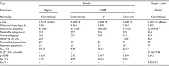
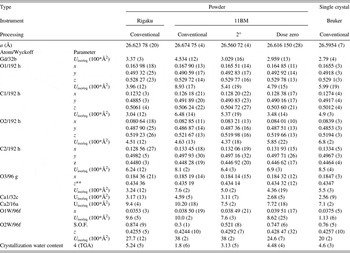
Table I contains final refinement details and residuals for the five different datasets used to analyse the structure of [{Ca(H2O)6}{CaGd(oda)3}2]·4H2O compound. Table II shows the atomic coordinates and isotropic or equivalent atomic displacement parameters for the five different datasets used to refine the structure.
Measurement conditions, data processing procedures and refinement fit indicators and residuals for different refinements of the structure of [{Ca(H2O)6}{CaGd(oda)3}2]·4H2O.

*Considering that the position of the maximum of two non-overlapping reflections are separated by more than 1 FWHM.
Final structural parameters of [{Ca(H2O)6}{CaGd(oda)3}2]·4H2O compound obtained from the different refinements performed (space group Fd3c #229).

*U iso/eq is the isotropic atomic displacement parameter or the equivalent isotropic atomic displacement parameter of an atom refined anisotropically.
**This coordinate is related to the x-coordinate by x = y + 1/4 and z = y + 1/2.
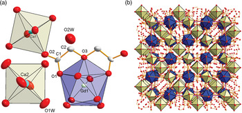
Figure 5 contains the final structural plot of one Gd(oda)3 unit and one of each type of Ca cations with their respective coordination spheres obtained from single-crystal X-ray diffraction data and the packing of the structure.
(a) The coordination polyhedra around Gd1, Ca1 and Ca2 in [{Ca(H2O)6}{CaGd(oda)3}2]·4H2O compound and one oxygen atom of crystallization water molecule. (b) Packing of [{Ca(H2O)6}{CaGd(oda)3}2]·4H2O showing alternation of Gd and Ca polyhedra in the cubic network with Ca(H2O)6 octahedra occupying half of the cubic holes and crystallization water molecule the other half but in an off-centre position.

III. RESULTS AND DISCUSSION
Looking at all the refinement results, the structure of [{Ca(H2O)6}{CaGd(oda)3}2]·4H2O can be described as formed by an anionic MOF of [Gd2Ca2(oda)6]2− units in an arrangement where Gd(III) and Ca(II) cations alternate in the corners of the cubes bridged by carboxylate units from the oda ligands. Gd1–Ca1 distance in the network is equal to one-quarter of the cell parameter or approximately 6.65 Å. Hexaaquocalcium(II) units occupy half of the centres of the cubes formed by the anionic network to neutralize the structure. Ca2 cations are equidistant from Gd1 and Ca1 centres at one-eighth of the body diagonal of the cubic cell (~5.76 Å). Six labile crystallization water molecules could occupy each of the empty cubes in the network, not in their centre but close to the hexaaquocalcium(II) cations probably to achieve stabilization by hydrogen bonding (that could not be confirmed because of the impossibility to determine the position of H atoms from our single-crystal data). Partial removal of these water molecules leads to a reduction in the expected stoichiometric amount of six crystallization water molecules per formula unit to the observed amount of four in thermogravimetric analysis or 1.8–5.2 molecules as obtained from the X-ray diffraction experiments (see bottom row of Table II). The Gd(III) cations show the expected 9-fold coordination geometry previously described for this kind of compound (Mao et al., Reference Mao, Song, Huang and Huang1997; Torres et al., Reference Torres, Peluffo, Domínguez, Mederos, Arrieta, Castiglioni, Lloret and Kremer2006; Domínguez et al., Reference Domínguez, Torres, Peluffo, Mederos, González-Platas, Castiglioni and Kremer2007; Prasad et al., Reference Prasad, Rajasekharan and Costes2007; Kremer et al., Reference Kremer, Torres and Domínguez2008) and the Ca(II) cations show two different coordination polyhedra, both regular octahedra, of carboxylate or water oxygen atoms. Carboxylate units of oda are linked to Gd1 and Ca2 atoms in an anti–anti configuration to maximize cation–cation distance.
The only significantly different result from the five refinements presented in this work and with the compound [{Mn(H2O)6}{MnGd(oda)3}2]·6H2O is precisely the crystallization water content. Several factors may play an important role in this difference: (1) different preparation conditions; (2) different behaviour of crystallization water molecules for different M(II) cations since crystallization water molecules are hydrogen-bonded to the hexaaquo M cation; (3) sample decay under irradiation or exposition to the atmosphere, related to the removal of this labile water molecule that varies its concentration with time; (4) different kinds of statistical errors associated with the different datasets allow for significant changes in the three parameters refined for this water molecule (one atomic position coordinate, one isotropic atomic displacement parameter and the site occupation factor). To assess these options, we would first like to discuss how the comparison between models will be performed.
The correct comparison of two structural models obtained from very different refinement procedures (as is the case of this study) may be a difficult problem, even considering that the estimated standard uncertainty (s.u.) of each of the refined variables can always be extracted from the least-squares procedures. Precision of the structural model (as measured by the s.u.) and accuracy of this model (understood as the deviation from the real structure) obtained from different determinations may be poorly described just by assessing s.u. values or R factors. This comparison is even harder when considering that the traditional single-crystal quality of fit indicators fail to give significant assessment of the result obtained from powder diffraction data. This occurs for a number of reasons mainly related by the differences in the datasets used for the determination and the procedure to obtain the observed diffraction intensities involved, leading to the use of different classes of quality indicators for both methods.
Only as an example, any parameter that includes the number of observed intensities is clearly defined for single-crystal data but is absolutely dependent on the way intensities are treated in powder diffraction. This comes from the presence of significant peak overlap in the one-dimensional projection of the three-dimensional diffraction space made in powder diffraction. Different ways of computing the number of observed reflections and the effective numbers for our refinements are included in Table I for our study. The number of observed intensities may be computed as the count of Bragg reflections present in the diffraction space scanned in the experiment. This count included under “Reflections unique/all” solely depends on the 2θ range of the data for powder diffraction and additionally the degree of redundancy (measure of equivalent reflections) of single-crystal data. No redundancy can be applied to powder data therefore the total number and the unique intensities roughly coincide with the number of independent intensities in the single-crystal data collection in the same 2θ range (provided 100% completeness is achieved in the latter). The number of metrically distinct reflections may be calculated for powder data, since many reflections not related to symmetry coincide exactly in any powder pattern because of lattice metrics (as is the case of peaks 971 and 11, 3, 1 with the same d-spacing = 2.32 Å in Figure 3, or any other group of reflections with identical h 2 + k 2 + l 2 sum in cubic structures as this one). This reduces the number of independent intensities by roughly 50% in the d > 1 Å range or less if a larger d-range is considered. This number is the same as the total number of independent reflections in single-crystal data since in that case metrically equivalent reflections never overlap. The number of non-overlapping reflections may be calculated after defining in some precise manner what are overlapping reflections, as done by Altomare et al. (Reference Altomare, Cascarano, Giacovazzo, Guagliardi, Moliterni, Burla and Polidori1995) or David (Reference David1999). We have not attempted such calculations but calculated the number of reflections that are away from each other by more than 1 full-width at half-maximum (FWHM) and used this number only to show the additional reduction of the number of independent observations in powder diffraction patterns. More precisely, we have determined the FWHM of reflections at d ≈ 1.5 Å (FWHM varies significantly with 2θ for conventional diffractometer but less for synchrotron data). For the Rigaku diffractometer used for conventional powder diffraction experiment and for 11BM synchrotron data processed for dose zero correction, the FWHM of the peaks was nearly 0.25° and 0.02°, respectively, leading to an additional ~5% reduction in the number of independent reflections for powder data. However, for conventional diffractometer data, the presence of the Kα 1–2 doublet reduces further the number of non-overlapping reflections since at high angles the splitting of both components of the Kα doublet produces a larger number of peaks. Once again there is no reduction of observed data points in single-crystal data since the data collection strategy (detector-sample distance) is adjusted to the cell dimensions to prevent peak overlap. Finally, the number of observed intensities using single-crystal criterion (I > 2σI) may be computed with an additional 1% or 2% reduction in the number of independent peaks as calculated for conventional and dose-corrected data. However, this observation criterion is not commonly used in powder diffraction, where the peak to background intensity ratio or the peak to background noise ratio may be more realistic approaches when intensity extraction is performed by automated procedures.
Calculation of the s.u. of coordinates may be regarded as equivalent in both procedures since atomic coordinates are variables in the least-squares refinement procedures used in both types of refinements. However, calculation of the s.u. of bond distances and angles is also affected by the different procedures applied in the refinement process. The cell parameters of the unit cell are not variables in a single-crystal structural refinement but they are separately calculated from a sub-set of all observed intensities selected to span a sufficient 2θ range with certain precision and included as constants with s.u. In a Rietveld refinement, the cell parameters must be adjusted together with the structural model using all the observed peaks in the pattern. This makes the s.u. of the lattice parameters in powder diffraction much lower than in single-crystal diffraction, owing to the larger number of data points used for the determination. Even so, selection of peak shape, zero point or other geometric parameters often applied in Rietveld refinements (such as anisotropic broadening, preferential orientation, etc.) may affect the values of the refined cell parameter well beyond the calculated s.u. Moreover, the variation of lattice parameters with temperature because of thermal expansion is, in general, significant if compared with the refined s.u. of lattice parameters. This leads to an artificially low s.u. in bond distances and angles extracted from powder diffraction Rietveld refinements.
In the absence of any thermal expansion data for our compound or similar ones, it is very difficult to estimate the magnitude of the last point discussed above. Even in ideal conditions, not considering that the sample subject of this study loses water at room temperature as shown in Figure 1, therefore may also change lattice parameters with time, we usually do not have data vs. temperature to estimate the thermal expansion coefficient (TEC). However, assuming that the sample shows a small TEC of the order of about 1 × 10⁻⁶ K−1 and considering that sample temperature control is reliable within 1 K, the standard uncertainty of the unit cell parameter (a) can be estimated to be approximately 2.6 × 10⁻⁴ Å. This is well in excess of the estimated s.u. of the cell parameters of all synchrotron X-ray powder diffraction data Rietveld refinements given in Table II and of the same order of magnitude of the s.u. of the lattice parameter in conventional X-ray powder diffraction data. Therefore, s.u. of bond distances and angles are somehow underestimated for powder diffraction results owing to the underestimation of the unit cell parameter s.u.
After this discussion, it seems that the comparison of the quality of the structural models obtained from different refinement procedures should not be based on the examination of s.u. provided by the refinement programs, as done by Louër et al. (Reference Louër, Louër, Bétourné and Touboul1996), but a different procedure should be used. In our case, there is an additional need to compare five such groups of coordinates which makes the task rather tedious unless global and systematic assessment of the structure quality is used.
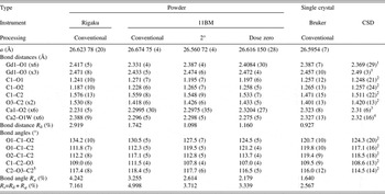
We have attempted to produce a global quality measure of the accuracy of the structural model by comparing individual bond lengths and angles observed in each model to average bond distances and angles of the same kind in similar compounds included in the Cambridge Structural Database (CSD) (Allen, Reference Allen2002). To avoid the comparison of individual parameters that may lead to different considerations by different observers, we have calculated a global R factor for each kind of parameter (bond length, bond angle and the combination of both). This R factor is defined in an identical manner of the conventional R factor in single-crystal X-ray diffraction where F obs and F calc are replaced by d obs and d CSD (for R d) or φ obs and φ CSD (for R φ being φ a bond angle) and is given in Table III as a percentage, with an additional R s parameter (the addition of R d and R φ) together with the observed bond distances and angles for the five different refinements and the average bond distances and angles extracted from CSD.
Cell parameters, bond distances and angles of [{Ca(H2O)6}{CaGd(oda)3}2]·4H2O compound obtained from the different refinements performed and average Gd–O, Ca–O, C–O and C–C bond distances and angles obtained for similar compounds in the CSD.

1Average from CSD entries: DOYZUN, FUHQUU, MAZJEC, NAOAGD, QUMYON, RONCIG, TUFLUB, VARREL, WEVQOD and YEZSEC.
2Average from CSD entries: BOHPUJ, DOTHEA, DOTWAL, ESUMOU, ESUMOU01, GERPUP, IYUYEG, MAZJAY, MAZJIG, NIJZIP, NIJZOV, NUVWOR, ODIMEU, QUMYON, QUMYUT, RONCIG, ULUPUM, ULUPUM01, URXDAC10, VARQUA, WANQEH, AVATAT, OTUFUF, FUFFIV, GISVIN, HAFRUB, KIBZUQ, QIZKOA, RIXBOQ, SUGJOT, TUHVUN, VEGWOT, XOXJEA, XOXJIE, FUHQUU, MAZJEC, NAOAGD, RONCIG, TUFLUB, WEVQOD, YEZSEC, DOYZUN, QUMYON and VARREL.
3Average from CSD entries: FUFFIV, GISVIN, HAFRUB, KIBZUQ, QIZKOA, RIXBOQ, SUGJOT, TUHVUN, VEGWOT, XOXJEA and XOXJIE.
4Average from CSD entries: DECDOE, EMETEW, GAYLOH, NOGLEA and RALHAO02.
5z − 1/4, 1 − y, x + 1/4.
The values of R d, R φ and R s parameters in Table III may be regarded as expected or obvious since they undoubtedly show that all powder diffraction structural models deviate more than the single-crystal model from the expected average bond distances and angles. They also show that conventional X-ray diffraction data performs in a poorer manner than synchrotron X-ray diffraction data in reproducing the expected structural model. However, they also indicate that, in this particular case, where evident sample deterioration was observed with time, the use of alternative strategies for processing the synchrotron X-ray diffraction data are worth the extra work since both 2 degrees and dose zero-corrected data show lower R factors than the conventionally processed data. Furthermore, the results may suggest that the dose zero correction gives an overall best model than simpler processing by taking the first 2 degrees of data of each detector, justifying its systematic implementation. Both 2 degrees and dose zero-corrected data provide structural models that may well compare with single-crystal data except for the crystallization water content of 2 degrees data that falls too low when compared with single crystal and TGA results.
This may be a key point in understanding the process through which this structure degrades with time and irradiation. The loss of loosely bound water molecules generates framework distortions that reduce the crystalline quality and its scattering power. This water loss is expected from the observation of significant water loss even at room temperature in the TGA and may probably affect different datasets in different ways since sample conditions and data collection times were different as described in Table II. It is an expected result that conventional X-ray diffraction data provide the lowest water content even in the case of significantly slower sample deterioration (probably because of the much lower dose received in the ~60 h period compared with the synchrotron radiation). The 2 degrees fit seems to be worse than the dose zero fit when visual inspection of the patterns is performed (see Figures 4(c) and (d)) with some of the high intensity peaks being poorly accounted for, which may be a consequence of the relative difference between peak intensities in this dataset because of slow sample degradation as explained above.
It is also interesting that in this particular compound the heavy atoms lie in high symmetry positions, leading to only two adjustable structural parameters for both Gd1 and Ca1 (anisotropic atomic displacement parameters) and only one for Ca2 (isotropic atomic displacement parameter), while all the light atoms (C, H and O) lie in general or less symmetrical positions with many adjustable parameters. In particular, in the Fd3c space group, positions 16a (Ca2), 32b (Gd1) and 32c (Ca2) have special additional systematic extinction conditions that make these three atoms only contribute to some of the diffraction peaks but not contribute at all to others. The extra conditions imposed by the high symmetry locations of these three atoms make only reflections with h, k and l even numbers and the three of them are either multiples of 4 or are not multiples of 4. This makes all reflections with h, k and l being odd numbers to receive no contribution from the heavy atoms of the structure. This allows for the refinement of light atoms in the structure with particularly high accuracy provided that the small intensities coming from these peaks are correctly determined. This is the case of the 2 degrees and dose-corrected datasets where all the small hkl peaks with odd indices are included in the refinement, but not in the case of the conventional datasets where either data averaging or high background hinder the small peaks, or the single-crystal dataset where the majority of low peaks are dubbed as unobserved for having I < 2σI and therefore low weighted in the final model (since the weighting scheme favours intense and well-fitted reflections over weaker or poorly fitted ones). Since water removal seems to be parallel with sample decay, it is expected that a decay correction, such as the one provided by dose zero data would account for this phenomena in a better way and achieve to represent the structure at the state before data collection, where water loss was not so significant. The correct determination of light atom coordinates and crystallization water content strongly justifies the application of this new data processing procedure that has been previously attempted to study macromolecular crystals and to account for radiation damage in electron crystallography but to the best of our knowledge not for powder diffraction structural studies of small molecules or metal–organic compounds.
IV. CONCLUSION
We have performed the structural refinement of the compound [Ca(H2O)6][{CaGd(oda)3}2]·4H2O with five different datasets: a conventional diffractometer with Bragg–Brentano geometry running on CuKα radiation, a synchrotron-based 12-detector high resolution diffractometer running at 30 keV radiation using three different data processing procedures to obtain the final datasets and a single-crystal X-ray diffraction data collection using MoKα radiation on a CCD-equipped diffractometer. The comparison of results shows that for this particular compound, where the coordinates of light atoms may be accurately determined even in the presence of heavy atoms, the two procedures implemented to process synchrotron data yield results that approach significantly to the degree of accuracy reached by single-crystal structural analysis. This strongly suggests that this kind of atypical data processing is worth being explored further looking for better accuracy in crystalline structures determined by powder diffraction using modern facilities.
ACKNOWLEDGEMENTS
The authors are indebted to Professor Jorge Castiglioni for performing the thermogravimetric experiment. L.S. is indebted to D. Louër for helpful discussion, with the Brazilian Synchrotron Light Source (LNLS) for providing beam time at D10B-XPD beam line to study these and related compounds whose results are not shown for the sake of brevity and with the National Synchrotron Light Source, Brookhaven National Laboratory for fully supporting a one-month visiting scientist position in 2009 to work with J.W. on the decay correction of the powder diffraction data. J.T. and C.K. are indebted to CSIC – Universidad de la República – Programa de Apoyo a Grupos de Investigación for financial support. L.S., R.F., J.T. and C.K. thank PEDECIBA and ANII (Uruguayan organizations) for financial support. Use of the Advanced Photon Source at Argonne National Laboratory was supported by the U.S. Department of Energy, Office of Science, Office of Basic Energy Sciences, under Contract No. DE-AC02-06CH11357. We acknowledge the Spanish Research Council (CSIC) for providing us with a free-of-charge license for the CSD (Allen, Reference Allen2002).