Introduction
In vitro embryo production (IVP) is an applied reproductive biotechnology which improves and accelerates the genetic selection. The success of IVP depends on the quality of oocytes (Swain and Pool, Reference Swain and Pool2008), the sperm used in fertilization (Hansen, Reference Hansen2006), and the in vitro culture conditions (pH, osmolarity, temperature and CO2 tension) (Lonergan and Fair, Reference Lonergan and Fair2008). These culture conditions can cause an increase in reactive oxygen species (ROS), which leads to oxidative stress and consequently cellular damage (Agarwal et al., Reference Agarwal, Maldonado Rosas, Anagnostopoulou, Cannarella, Boitrelle, Munoz, Finelli, Durairajanayagam, Henkel and Saleh2022). Therefore, to protect cells against the damage caused by excessive amount of ROS, the use of antioxidants in the culture medium is highly recommended.
Antioxidants are substances that can prevent, reduce, or repair the ROS-induced damage of a target biomolecule (Li et al., Reference Li, Jia and Trush2016). Among the antioxidants, turmeric has been studied in the assisted reproductive techniques for improving human granulosa cells (Moreira-Pinto et al., Reference Moreira-Pinto, Costa, Fonseca and Rebelo2020) or preventing ovarian failure (Melekoglu et al., Reference Melekoglu, Ciftci, Eraslan, Cetin and Basak2018). Turmeric (Curcuma longa Linn) is a member of the Zingiberaceae family and its powder is used extensively as a colouring and flavouring agent in curries and mustards. Moreover, it has traditionally been used for medical purposes, with a wide range of pharmacological activities such as antioxidant, anti-protozoal, anti-venom activities, anti-microbial, anti-malarial, anti-inflammatory, anti-proliferative, anti-angiogenic, anti-tumour and anti-ageing (for review see Amalraj et al., Reference Amalraj, Pius, Gopi and Gopi2016). The antioxidant activity of turmeric is exerted by the molecule curcumin (CUR), being studied in different type of tissue and animals (Amalraj et al., Reference Amalraj, Pius, Gopi and Gopi2016). In reproduction, its use effectively protected testicular cells in Wistar rats (Sahoo et al., Reference Sahoo, Roy and Chainy2008), cumulus cells in pigs (Qin et al., Reference Qin, Cao, Lai, Yang, Ge, Zhang, Cheng, Sun, Qin, Shen and Li2015), and buffalo embryos (Ritikita et al., Reference Ritika, Saini, Shavi, Ramesh, Selokar, Ludri and Singh2024) from oxidative damage caused by stress and free radicals, with curcumin modulating antioxidant defence and apoptotic pathways to enhance cellular viability. However, CUR intracellular activity is limited due to its low solubility in the culture medium, as it is a polyphenolic (Zhai et al., Reference Zhai, Brockmüller, Kubatka, Shakibaei and Büsselberg2020). Thus, to overcome the problems of poor solubility and low bioavailability, nanoparticle-based drug delivery approaches, in which curcumin is associated with metal nanoparticles such as zinc oxide, i.e. zinc oxide nanoparticle (ZnO(np)) have been proposed and reported in in vitro studies (Dhivya et al., Reference Dhivya, Ranjani, Rajendhran, Mayandi and Annaraj2018; Perera et al., Reference Perera, Dissanayake, Ranatunga, Hettiarachchi, Perera, Unagolla, De Silva and Pahalagedara2020). ZnO(np) is considered to be non-toxic and non-immunogenic as well and is a cost-effective starting material (Jiang et al., Reference Jiang, Pi and Cai2018). However, to our knowledge there are no studies evaluating the effect of zinc oxide nanoparticles functionalized with curcumin (ZnO(np)+CUR) on the IVP.
Thus, the aim of this study was to evaluate the effect of ZnO(np)+CUR addition during the in vitro maturation (IVM) of bovine oocytes on the in vitro embryo production and the cellular antioxidant response.
Materials and methods
Research ethics
This study adhered to ethical guidelines by evaluating the effects of ZnO(np)+CUR on in vitro embryo production (IVP) using bovine COCs from slaughterhouse ovaries, an ethically preferable by-product of the food industry. Additionally, the research was reviewed and approved by the Animal Ethics Committee, ensuring compliance with ethical standards.
Chemicals
Unless mentioned, all chemical reagents used are from Sigma Chemical Co. (St. Louis, MO, USA).
In vitro maturation
Embryo production was performed as previously described (Silva et al., Reference Silva, Lima, Morais, Lienou, Watanabe, Joaquim, Morais, Alves, Pereira, Santos, Alves, Padilha, Gastal and Figueiredo2022). Ovaries from post-mortem Nelore cows, typically slaughtered for beef production at ages ranging from 18 months to 3 years (healthy), were collected from a local slaughterhouse located 30 km from the laboratory. The ovaries were transferred in a 36ºC thermal bottle and transported to the laboratory within 2 to 3 h. Afterwards, the ovaries were kept in a water bath at 36ºC while the follicle aspiration was performed using a syringe and 18 g needle. The follicular fluid was analysed under a stereomicroscope to collect the cumulus-oocyte complexes (COCs) and the grade I and II COCs (classification of the international embryo technology society-IETS) were selected for in IVM for 22 h in microdroplets of 100 µL of IVM medium (TCM-199 - Ref.11150059; Gibco; Thermo Fisher Scientific, Inc., Waltham, MA, USA) supplemented with 10% fetal bovine serum (FBS), 25 mg/mL sodium bicarbonate, 22 mg/mL sodium pyruvate, 50 mg/mL amikacin, 5 mg/mL FSH, 50 mg/mL LH, and 0.1 mg/mL estradiol) covered by mineral oil. All structures were cultured with 5% CO2 and at 38.5ºC.
Preparation of zinc oxide nanoparticles functionalized with curcumin
The zinc oxide nanoparticles (Sigma 205532- ZnO(np)) were synthesized by the solvothermal method. In this method, the zinc acetate was dissolved in benzyl alcohol to obtain a 30 mM solution, which remained for 24 h at 150ºC in a suitable reactor for nanoparticles synthesis. The suspension was centrifuged, and separated solid was rinsed with absolute ethanol and deionized water. Subsequently, the solid was dried in an oven at 60°C for 24 h. The nanoparticles displayed a spherical shape with an average size of 5 nm, as characterized by transmission electron microscopy (TEM) (Figure 1).
(A) Transmission electron microscopy of ZnO nanoparticles (B) histogram of size distribution of nanoparticles.

The zinc oxide nanoparticles functionalized with curcumin (ZnO(np)+CUR), surface functionalization was synthesized by mixing the compounds in a common solvent. In this suspension, the CUR powder (Sigma - C1386) was dispersed in chloroform (0.0340 g/L). After the whole dispersion, ZnO(np) was added in CUR suspension with molar ratio of 1:1. The mixture then remained under magnetic stirring for a period of 4 h at room temperature. Then, the suspension was centrifuged, washed with deionized water and after with absolute ethanol until the supernatant became as clear as possible. The solid was dried in an oven at 60°C for 24 h. The surface modification of ZnO(np)+CUR was confirmed by means of absorption spectroscopy in the infrared region (Figure 2). In the spectrum, typical curcumin bands were observed in the 3300-3410 cm−1 region, which can be attributed to the vibrations of the free OH group (Del Socorro et al., Reference Del Socorro, Teves and Madamba2013). Moreover, the intense bands at 1640, 1645 and 1647 cm−1, related to the carbonyl group vibration, and the bands in the region 1340–1470 cm−1, attributed to the C = C vibrations of the aromatic ring, were also observed in the spectrum. The bands in the 1020–1155 cm−1 region refer to the stretching of the C − O group of ether, whereas the bands in about 710 and 960 cm−1 refer to the C − H of the alken groups. Finally, it was also observed a band in the region of 1400 cm−1 characteristic of the Zn–O vibration of the ZnO(np).
Infrared absorption spectrum of ZnO(np), CUR and ZnO(np)+CUR.

The ZnO(np)+CUR powdered was diluted in dimethyl sulfoxide (DMSO - Sigma D2650) (Huang et al., Reference Huang, Lan, Kang, Liu, Hsuuw, Chan and Huang2013), generating a stock solution at a concentration of 12.0x10⁻⁴ M and then stored at −8ºC. The stock solution was added to the IVM medium to obtain the final concentrations of 3 µM, 6 µM, 12 µM of ZnO(np)+CUR.
In vitro fertilization
After 22 h of maturation, the COCs were washed in in vitro fertilization (IVF) medium (TALP fertilization medium supplemented with 10 mg/mL heparin, 22 mg/ mL sodium pyruvate, 50 mg/mL amikacin, 6 mg/mL bovine serum albumin (BSA; fatty acid-free), 2 mM penicillamine, 1 mM hypotaurine, and 0.25 mM epinephrine) and transferred to a fertilization plate formed by 70 µL microdroplets covered by mineral oil. The drops were inseminated with Percoll-purified sperm (1 × 10⁶ sperm cells). In vitro fertilization was performed for 18 h using semen from a single Nelore bull, with proven fertility, obtained from a semen collection centre.
In vitro embryo culture
Eighteen hours after fertilization, the presumptive zygotes were removed from surrounding cells and sperm from successive pipetting. The presumptive zygotes were denuded, washed and then transferred to a culture plate composed of Charles Rosenkrans (CR2) modified medium supplemented with 50 mg/mL amikacin, 0.1 mM amino acids, 2.5% FBS, and 6mg/mL BSA, where they remained for 7 days. On the 2nd day of in vitro culture, the cleavage rates were evaluated and on the 7th day, blastocyst formation rates were recorded.
Evaluation of ROS production
Intracellular ROS levels were determined with DCFH-DA probe (10 µM) (Anjos et al., Reference Anjos, Aguiar, Sá, Souza, Cibin, Alves, Santos and Figueiredo2019). After the IVM, COCs were mechanically denuded at the stereo microscope and the oocytes were incubated with DCHF-DA 100 µL for 15 min, fixed in 4.0% paraformaldehyde and washed three times in PBS. Slides were assembled using glycerol and the fluorescent dye Hoechst 33342 (emission at 483 nm). DCF fluorescence intensity emission was recorded at 520 nm (with 480 nm excitation). The stained oocytes were immediately imaged using fluorescence microscopy on an EVOS system (ThermoFisher Scientific, Waltham, MA) with DAPI and GFP filters. Exposure time, gain, and area of exposure were adjusted based on the control treatment and remained consistent across all samples throughout the evaluation. Staining intensity was quantified using ImageJ analysis software. Fluorescence intensities were measured from RGB colour images, specifically quantifying the green channel corresponding to ROS levels (Silva et al., Reference Silva, Lima, Morais, Lienou, Watanabe, Joaquim, Morais, Alves, Pereira, Santos, Alves, Padilha, Gastal and Figueiredo2022). Additionally, we clarified that no image treatments or enhancements were applied to the captured images.
Biochemical assay
After IVM, groups of 45 COCs (3 replicates) were washed in PBS, frozen in liquid nitrogen and stored at −80ºC. On the day of analysis, the samples were kept on ice and vortexed for 5 min with 0.5% Triton X-100 for complete lysis. Total protein levels of the sample were confirmed by Bradford method. Also, levels of superoxide dismutase (SOD), catalase (CAT) and total antioxidant capacity were measured.
Superoxide dismutase activity
The SOD activity was measured according to Marklund and Marklund (Reference Marklund and Marklund1974), with modifications. The principle of this method is based on the inhibition of the reaction of the radical superoxide with pyrogallol. The COCs was mixed with 80 U/mL CAT and 24 mM pyrogallol, diluted in 50 mM Tris-HCl buffer with 1 mM EDTA, pH 8.2. The kinetic assay was monitored in 96-well microplates for 10 min at 420 nm (Molecular Devices). An analytical curve was constructed with SOD as standard to quantify the enzymatic activity of SOD.
Catalase activity
The CAT activity was measured according to Aebi (Reference Aebi1984), with modifications. The principle of this method is based on H2O2 decomposition. The COCs was incubated with 1% Triton X-100 for 15 min and then H2O2 solution (0.2%, in 10 mM phosphate buffer, pH 7.0) was added to the mixtures. The kinetic assay was monitored in 96-well microplates for 10 min at 240 nm (PerkinElmer LS 55).
Total antioxidant capacity
The Ferric Reducing Antioxidant Power (FRAP) assay was used to measure the total antioxidant capacity, to assess the total antioxidant capacity in COCs. The samples were incubated with 300 mM sodium acetate buffer (pH 3.6), 10 mM 2,4,6-tri(2-pyridyl)-s-triazine (TPTZ) and 20 mM ferric chloride (10:1:1) for 6 min at 37ºC (Franco et al., Reference Franco, Mota Alves, Ribeiro Zabisky, Justino, Martins, Saraiva, Goulart and Espindola2020) and the absorbance was measured at 593 nm (Molecular Devices, Menlo Park, CA, USA). An analytical curve was constructed with 6-hydroxy-2,5,7,8-tetramethylchroman-2-carboxylic acid) (trolox) as standard to quantify the total antioxidant capacity. The percentage of activity (PA) was obtained according to the following formula: PA(%) = [(A control–A sample) /(A control)] × 100. A control represents the absorbance value of the blank sample and A sample is the absorbance value of the samples/controls.
Experimental design
COCs (n = 1,625) were matured in vitro in the absence (0 µM) or presence of ZnO(np)+CUR (3 µM, 6 µM, 12 µM). COCs (n = 985) were destined for in vitro embryo production, totalling 201 in vitro produced embryos and 640 COCs were destined for oxidative stress assessments (ROS, FRAP, CAT, SOD).
Statistical analysis
Statistical data were analysed using Sigma Plot 11 software version 23.0 (Systat Software Inc., San Jose, CA, USA). The Shapiro–Wilk test was used to verify the normality of distribution, and the homoscedasticity of variance was verified by Levene’s test. The treatments were compared by PROC GLM, followed by Fisher’s Test (LSD), and the concentrations of ZnO(np)+CUR considered as fixed effects. Only the measurement of ROS showed non-parametric distribution, being analysed by the Kruskal–Wallis test. Data were presented as mean (± SEM), and statistical significance was established at p < 0.05.
Results
In vitro embryo production
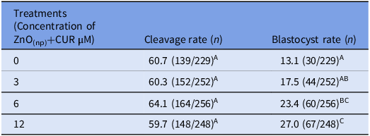
Cleavage and blastocyst rates are shown in Table 1. A total of 985 matured COCs were fertilized and 201 embryos were produced in vitro. IVM medium supplementation with ZnO(np)+CUR during the maturation of bovine oocytes had no effect on cleavage rates (p > 0.05). However, when compared to the control treatment, 6 µM and 12 µM ZnO(np)+CUR treatments showed higher rates of blastocyst production (p < 0.05). In addition, higher blastocyst production rates was observed for the treatment with 12 µM ZnO(np)+CUR compared to 3 µM ZnO(np)+CUR treatment (p < 0.05).
Influence of 0 µM, 3 µM, 6 µM or 12 µM ZnO(np)+CUR added to the bovine oocyte maturation medium on the cleavage and blastocyst rates

A, B, C Different letters in the same column denote significant differences (p < 0.05).
ROS production
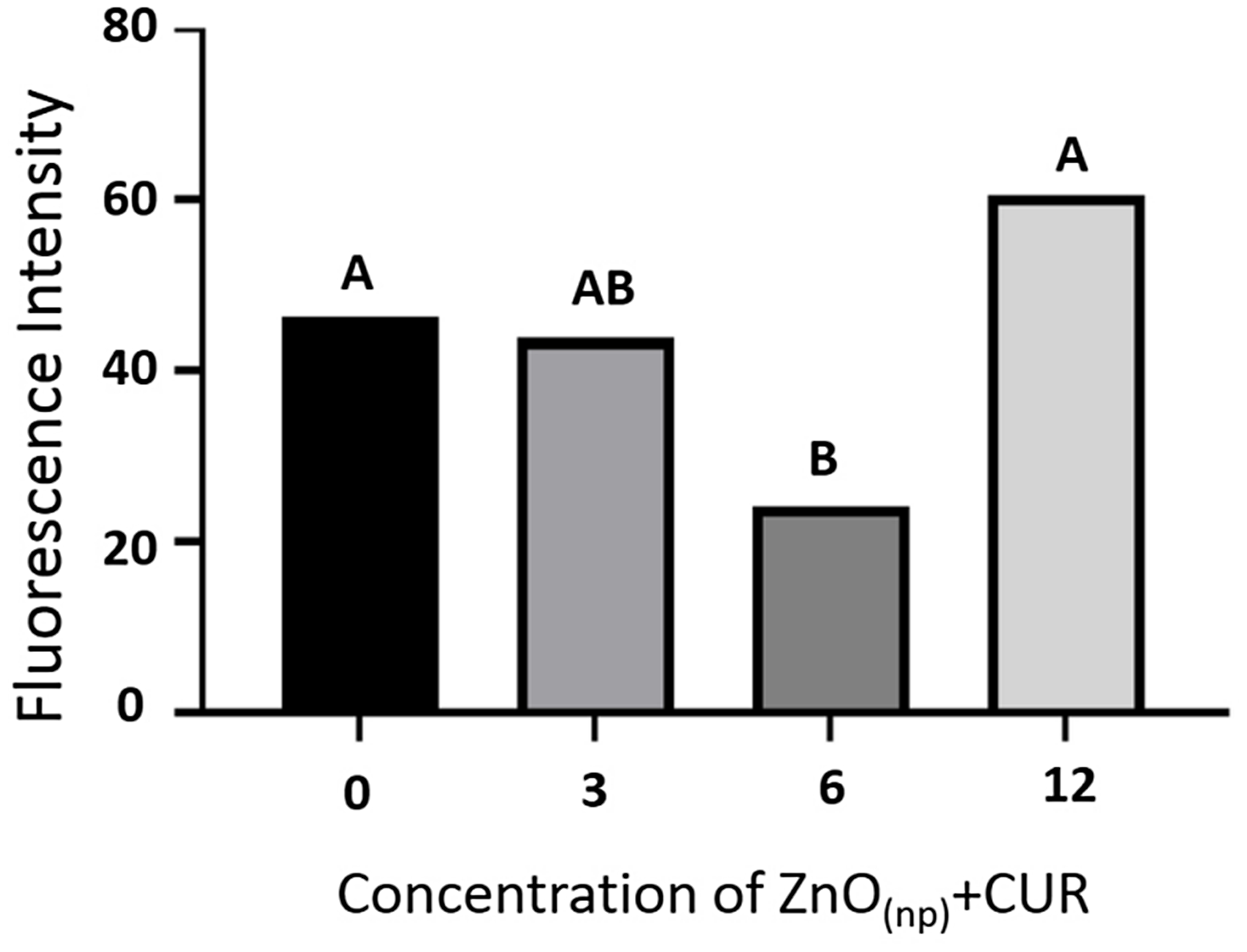
The levels of ROS are shown in Figure 3. A total of 100 oocytes were stained with DCFH and evaluated under fluorescence microscope. 6 µM ZnO(np)+CUR treatment presented lower ROS production (p < 0.05) than control and 12 µM ZnO(np)+CUR treatment.
Influence of 0 µM, 3 µM, 6 µM or 12 µM ZnO ZnO(np)+CUR added to the oocyte maturation medium on the ROS production. A,B Different letters denote significant differences (p < 0.05).

Intracellular antioxidant response
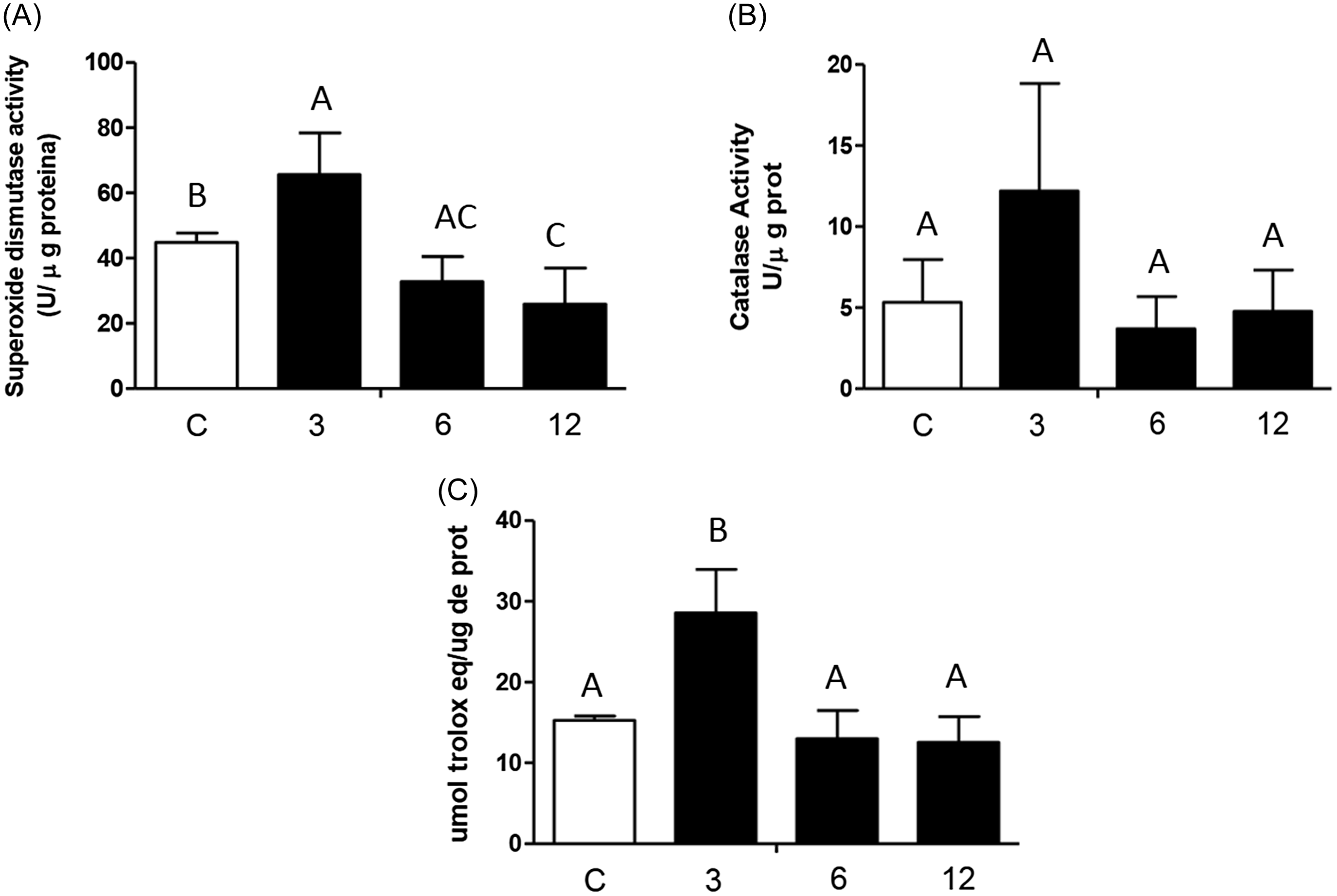
The SOD activity, CAT activity and FRAP levels are shown in Figures 4A, 4B and 4C, respectively. A total of 1,620 COCs were evaluated for this assay. The ZnO(np)+CUR 3 µM treatment presented the highest (P < 0.05) SOD activity (Figure 4A) and total antioxidant capacity (Figure 4C). SOD activity was significantly lower (p < 0.05) in the 12 µM ZnO(np)+CUR treatment when compared to the 3 µM ZnO(np)+CUR treatment and the control (Figure 4A). Finally, different from what was observed in FRAP and SOD, CAT activity was similar among the treatments (Figure 4B).
Influence of 0 µM, 3 µM, 6 µM or 12 µM ZnO(np)+CUR added to the oocyte maturation medium on SOD expression (A) and CAT expression (B) and total antioxidant capacity (C). A,B,C Different letters denote significant differences (p < 0.05).

Discussion
The present study investigated for the first time the effect of adding different concentrations (0 µM, 3 µM, 6 µM or 12 µM) of ZnO(np)+CUR during IVM of bovine oocytes and their impact on in vitro embryo production. The results showed that the evaluated parameters (embryo production, antioxidant capacity and SOD activity) were differentially affected by the concentration of ZnO(np)+CUR used.
The addition of ZnO(np)+CUR at the concentration of 3 µM in the IVM medium increased total oxidant capacity and SOD activity when compared to the control, showing that the CUR at this concentration was able to activate the antioxidant defence system. However, this treatment did not increase either the in vitro embryo production rate or reduce ROS levels. On the other hand, the addition of ZnO(np)+CUR at concentrations of 6 and 12 µM increased the blastocyst production rates when compared to the control. Ritika et al. (2024) reported that curcumin at 5 µM, when not associated with nanoparticles, was more effective than at 10 µM in mitigating heat stress and the effects of ROS in buffalo embryos cultured in vitro at 38.5°C. Additionally, curcumin has been shown to activate the PI3K/Akt signalling pathway both in vivo in mouse ovaries (Yan et al., Reference Yan, Dai, Fu, Zheng, Bao, Yin, Chen, Nie, Hao, Hou and Cui2018) and in vitro in cultured cells (Bai et al., Reference Bai, Zhao, Su, Chen, Cui, Sun and Zhang2022). The PI3K/Akt signalling pathway is a key mechanism to promote the oocyte nuclear maturation in bovine (El-Sheikh et al., Reference El-Sheikh, Mesalam, Khalil, Idrees, Ahn, Mesalam and Kong2023). Thus, we believe that the improved blastocyst rates observed at higher ZnO(np)+CUR concentrations may be linked to curcumin’s role in modulating oxidative stress and regulating the PI3K/Akt pathway. In porcine species, Namula et al. (Reference Namula, Sato, Wittayarat, Le, Nguyen, Lin, Hirata, Tanihara and Otoi2020) reported that curcumin at 0, 5, 10, 20, and 40 µM did not affect the cleavage rate but increased blastocyst production after the addition of 10 µM CUR. On the other hand, a study investigating the addition effect of CUR (5, 10 and 20 µM) during the IVM of mouse oocytes showed that CUR at concentrations of 5 and 10 µM did not affect any studied parameters (cleavage rate, blastocyst rate, number of cells/blastocyst). Nevertheless, CUR at a concentration of 20 µM increased apoptosis in embryonic cells (Chen and Chan, Reference Chen and Chan2012). Taking together, experimental results from literature suggested that the effect of the CUR is dose-dependent and may vary among species.
The results of the present study showed that the concentration of 6 µM ZnO(np)+CUR was able to decrease ROS production. The beneficial effect of CUR+NP on embryo production is probably due to its antioxidant action demonstrated in several studies using many cell types (for review see Amalraj et al., Reference Amalraj, Pius, Gopi and Gopi2016). CUR is known to reduce lipid peroxidation, eliminate superoxide and hydroxyl radicals (Reddy and Lokesh, Reference Reddy and Lokesh1994), as well as nitrogen dioxide (Unnikrishnan and Rao, Reference Unnikrishnan and Rao1995). Tvrdá et al. (Reference Tvrdá, Tušimová, Kováčik, Paál, Greifová, Abdramanov and Lukáč2016) tested the effect of CUR on bovine sperm subjected to an in vitro induced oxidative stress. These authors showed that CUR was able to stimulate the production of endogenous enzymes (SOD, CAT and glutathione peroxidase) and reduce lipid peroxidation. In vitro studies using 20 µM CUR showed a reduction in ROS production in fibroblasts after in vitro culture under zearalenone-induced stress (Perker et al., Reference Perker, Orta Yilmaz, Yildizbayrak, Aydin and Erkan2019). Thus, we suggested that the improvement in embryo production observed in the 6 µM treatment in the present study may be due to a reduction in ROS levels probably owing to an increase in glutathione system activity (not evaluated in the present study), since SOD and CAT activities were similar to control. Ritika et al. (2024) reported that curcumin at 5 µM was more effective than at 10 µM in mitigating heat stress and ROS effects in embryos cultured in vitro at 38.5°C, as evidenced by the significantly higher mRNA expression of heat shock proteins (HSP10, HSP60) and antioxidant-related genes (SOD2, SOD3, GPX1, GPX2) compared to controls. On the other hand, in the present study, the improvement in in vitro embryo production observed in the 12 µM CUR+NP treatment was not associated with a significant reduction in ROS levels, but rather with a significant reduction in SOD activity when compared to the control. SOD is the enzyme produced by cells which helps eliminating free radicals such as superoxide by catalysing the dismutation of superoxide into hydrogen peroxide and oxygen (Yasui and Baba, Reference Yasui and Baba2006). Some authors related high SOD activity as a response to cellular stress (Flores et al., Reference Flores, Souza, Betancourt, Teteltitla, González-Márquez, Casas, Bonilla, Ramírez-Noguera, Gutiérrez-Ruíz and Ducolomb2017). Thus, it is suggested that the 12 µM CUR treatment improved embryo production by maintaining the redox balance that is associated with the oocyte morphofunctional features (oocyte developmental competence).
In conclusion, the best outcome for in vitro embryo production was obtained when 6 and 12 µM ZnO(np)+CUR was added during IVM of bovine oocytes. However, dependent on ZnO(np)+CUR concentration, this improvement in in vitro embryo production was not associated with either the reduction of ROS production or SOD and CAT activities.
Acknowledgements
This work was supported by the Cearense Foundation of Scientific and Technological Support (FUNCAP - grant number: PR2-0101-00049.01.00/15) and the National Institute of Science and Technology in Theranostics and Nanobiotechnology-INCT-TeraNano (grant number: 465669/2014-0). FSE receive scholarship grants from FAPEMIG (PPM-00503-18) and CNPq (PQ–Research productivity, process no. 312812/2021-3) for research in nanostructured bioactive compounds.
Competing interests
The authors declare that there is no potential conflict of interest that can be perceived as prejudicing the impartiality of this study.