INTRODUCTION
Nephroselmis genus is one of the primitive green flagellates, which may shed light on the early evolution of green plants (Yamaguchi et al., Reference Yamaguchi, Suda, Nakayama, Pienaar, Chihara and Inouye2011). This genus was reported as the earliest-branching chlorophytic lineage based on a chloroplast multi-gene analysis (Turmel et al., Reference Turmel, Gagnon, O'Kelly, Otis and Lemieux2009; Leliaert et al., Reference Leliaert, Verbruggen and Zechman2011). Thus, this unicellular nanoflagellate, having several layers of organic body scales and flagellar scales (Melkonian, Reference Melkonian, Margulis, Corliss, Melkonian and Chapman1990; Nakayama et al., Reference Nakayama, Suda, Kawachi and Inouye2007; Bell, Reference Bell2008), might characterize what the ancient green flagellates might have resembled. The Nephroselmis genus was first identified by Stein (Reference Stein1878) and placed under the class Cryptophyceae. Later, it was noticed that pigments of this genus were more similar to species of chlorophytes (Chadefaud, Reference Chadefaud, Chadefaud and Emberger1960; Christensen, Reference Christensen, Böcher, Lange and Sørensen1962; Parke & Rayns, Reference Parke and Rayns1964). After the creation of the class Prasinophyceae (Chadefaud, Reference Chadefaud, Chadefaud and Emberger1960; Christensen, Reference Christensen, Böcher, Lange and Sørensen1962) under the phylum Chlorophyta, the Nephroselmis genus was included in this class (Moestrup & Ettl, Reference Moestrup and Ettl1979; Norris, Reference Norris and Cox1980; Moestrup & Throndsen, Reference Moestrup and Throndsen1988) and it is still placed within this clade by many investigators (Throndsen, Reference Throndsen and Tomas1996; Throndsen et al., Reference Throndsen, Hasle and Tangen2007; Turmel et al., Reference Turmel, Gagnon, O'Kelly, Otis and Lemieux2009; Leliaert et al., Reference Leliaert, Verbruggen and Zechman2011, Reference Leliaert, Smith, Moreau, Herron, Verbruggen, Delwiche and De Clerck2012). However, according to Nakayama et al. (Reference Nakayama, Suda, Kawachi and Inouye2007), the class Prasinophyceae is not appropriate for Nephroselmis since Prasinophyceae was typified by Prasinocladus (synonymous with Tetraselmis; Norris et al., Reference Norris, Hori and Chihara1980) and they have suggested that Nephroselmis be included in the class Nephroselmidophyceae (Nephrophyceae) Cavalier-Smith, Reference Cavalier-Smith and Lewin1993 (Guiry & Guiry, Reference Guiry and Guiry2015). Nevertheless, Bell (Reference Bell2008) reports that the name Prasinophyceae [originally termed ‘Prasinophycinées’ by Chadefaud (Reference Chadefaud, Chadefaud and Emberger1960)] is derived from ‘prasinos’, the Greek word for ‘green’, rather than from the genus Prasinocladus (Moestrup & Throndsen, Reference Moestrup and Throndsen1988), and the Nephroselmis genus has been placed within the class Nephrophyceae in recent systematics (Guiry & Guiry, Reference Guiry and Guiry2015). Nephrophyceae class involves a total of 11 genera and 28 species. There are 14 species accepted taxonomically belonging to the Nephroselmis genus (Guiry & Guiry, Reference Guiry and Guiry2015). Only one species out of 14, Nephroselmis marina, has been reported in the Adriatic Sea region of the Mediterranean Sea (Bell, Reference Bell2008; Guiry & Guiry, Reference Guiry and Guiry2015) so far. In the present study, Nephroselmis pyriformis (N. Carter) Ettl is identified for the first time from the Mediterranean Sea.
MATERIAL AND METHODS
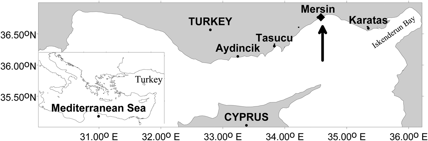
Samples were taken with a hand bucket from a pier in the north-eastern Mediterranean Sea coast (36°46′N 34°35′E, Figure 1) on 21 September 2013. Nephroselmis pyriformis was isolated from the surface waters and maintained in F/2 medium (circa 38% salinity) at 20°C with a 12 h:12 h (light:dark) cycle. After collection, water samples were put directly into two 100 ml Erlen-Meyer flasks containing F/2 medium in the laboratory. A few weeks later it was observed that other diatom, dinoflagellate and ciliate species had died in the flask and N. pyriformis dominated. The inoculation of this flask to another flask containing F/2 medium resulted in the dominance of a pennate diatom (naviculoid) and other types of unidentified flagellates. Micropipetting efforts carried out for N. pyriformis, in order to have a single-cell culture, were unsuccessful. Using a F/2-Si medium did not help isolate it to a single-cell culture. A Nikon TS-100-F inverted microscope and a Canon IXUS 115 HS Full HD camera, together with a Kameram 318CU model camera, attached to a Nikon Eclipse E100 model microscope were used to take photographic and video images.
Location map of the sampling region.

RESULTS AND DISCUSSION
The distribution area of Nephroselmis pyriformis is defined by Throndsen (Reference Throndsen and Tomas1996) as the coastal waters of the Atlantic, Arctic and Pacific. This species is reported in Britain, Denmark, Finland, Norway, West Greenland, Namibia, North and South America, Japan, Australia, New Zealand and Thailand (Bell, Reference Bell2008; Guiry & Guiry, Reference Guiry and Guiry2015 and references therein). There is no previous record of this species in the Mediterranean Sea, most likely due to difficulties in the identification of cells under a light microscope in preserved materials. It is reported that, whether fixed with iodine or formaldehyde, the identification of prasinophytes in preserved samples is most often inadequate and the usage of an electron microscope for samples fixed with osmic acid is suggested (Throndsen, Reference Throndsen and Tomas1996). However, identification to genus level in live samples would be fairly effective. Moreover, the different swimming patterns of cells observed under a light microscope allow many cells to be identified to the species level (Throndsen, Reference Throndsen and Tomas1996).
Characteristic features
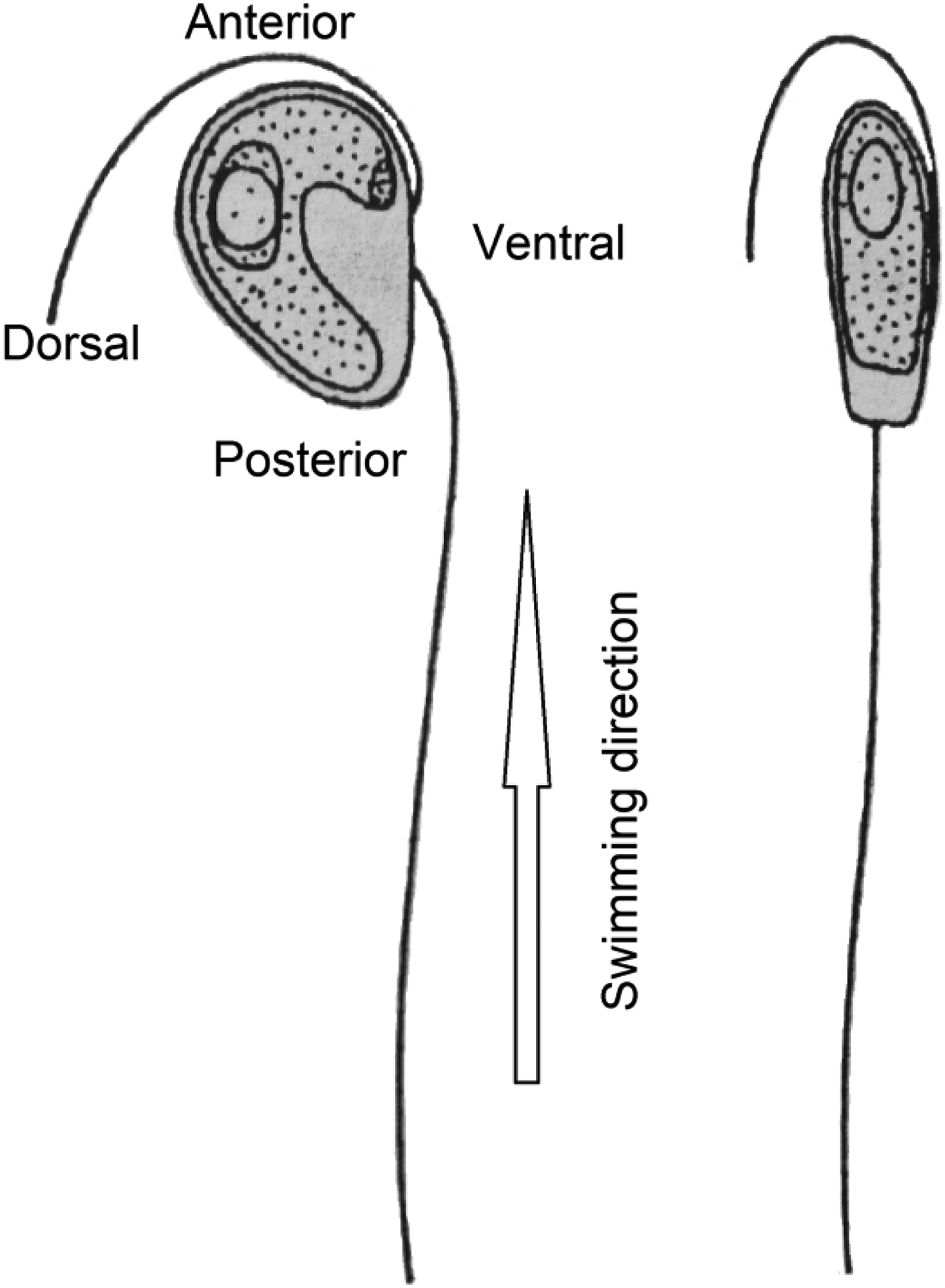
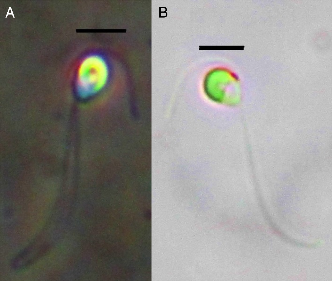
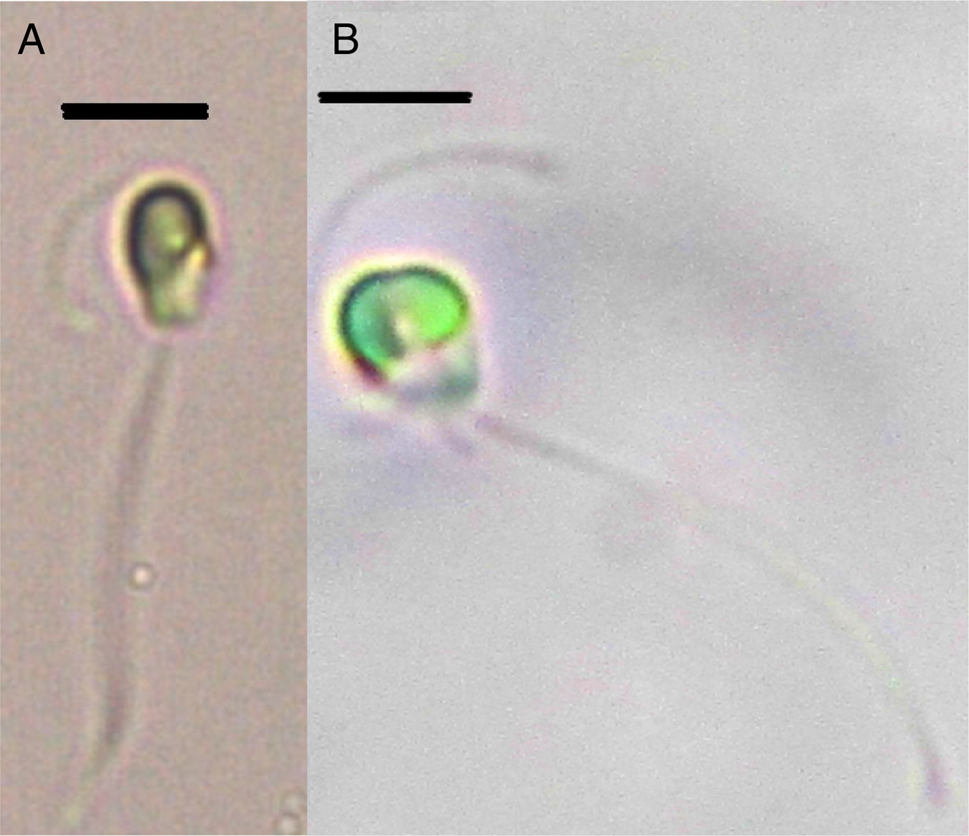
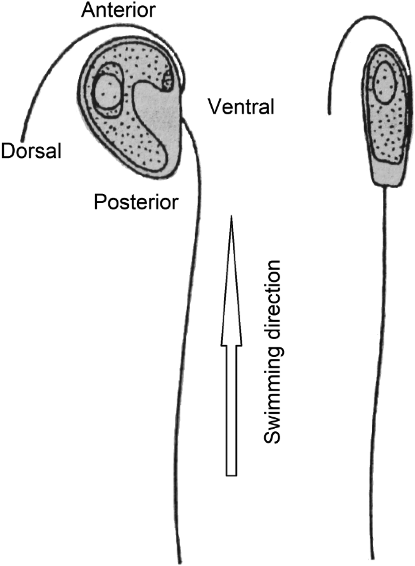
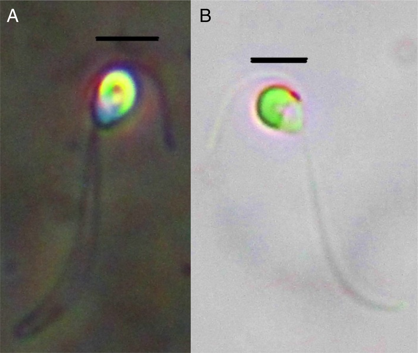
Cells are rounded, drop-shaped and laterally compressed (Throndsen et al., Reference Throndsen, Hasle and Tangen2007) (Figure 2). Cell length 5–7 μm, cell width 5–6 μm, cell thickness 2 μm. Two heterodynamic and unequal flagella, 20 μm and 7–8 μm (Throndsen, Reference Throndsen and Tomas1996; Throndsen et al., Reference Throndsen, Hasle and Tangen2007). One flagellum is typically wrapped around the cell while the other is bent away from the cell during resting (Throndsen et al., Reference Throndsen, Hasle and Tangen2007). Heterodynamic mode of flagellar action may also be observed at rest. The cup-shaped chloroplast extends anteriorly and posteriorly from the ventral surface to the dorsal surface (Figures 2 & 3; Bell, Reference Bell2008). Chloroplast with pyrenoid is yellowish green (Figure 3A) with an eyespot under the short flagellum on the anterior and ventral side of the body (Figure 3B; Throndsen et al., Reference Throndsen, Hasle and Tangen2007; Bell, Reference Bell2008; Yamaguchi et al., Reference Yamaguchi, Nakayama and Inouye2013). Three flagellar scale types (two flagellar scale layers, which are pentagonal and rod-shaped and flagellar hair scales) are present. Only two layers of body scales are present. The swimming direction of the cells are towards the anterior side where the short flagellum is present (Figures 2 & 4A, see also video images 1, 2 and 3 in the Supplementary Materials).
Nephroselmis pyriformis reproduced from Throndsen et al. (Reference Throndsen, Hasle and Tangen2007). Left cell in lateral view. Right cell in dorsal view with short flagellum curved around dorsal portion of the cell.
(A) Cup-shaped chloroplast and starch grains with pyrenoid are shown in the dorsal portion of the cell; (B) Eyespot under the short flagellum is displayed. Scale bar shows 5 μm.
(A) A cell from the dorsal view; (B) A dividing cell. Scale bar shows 5 μm.
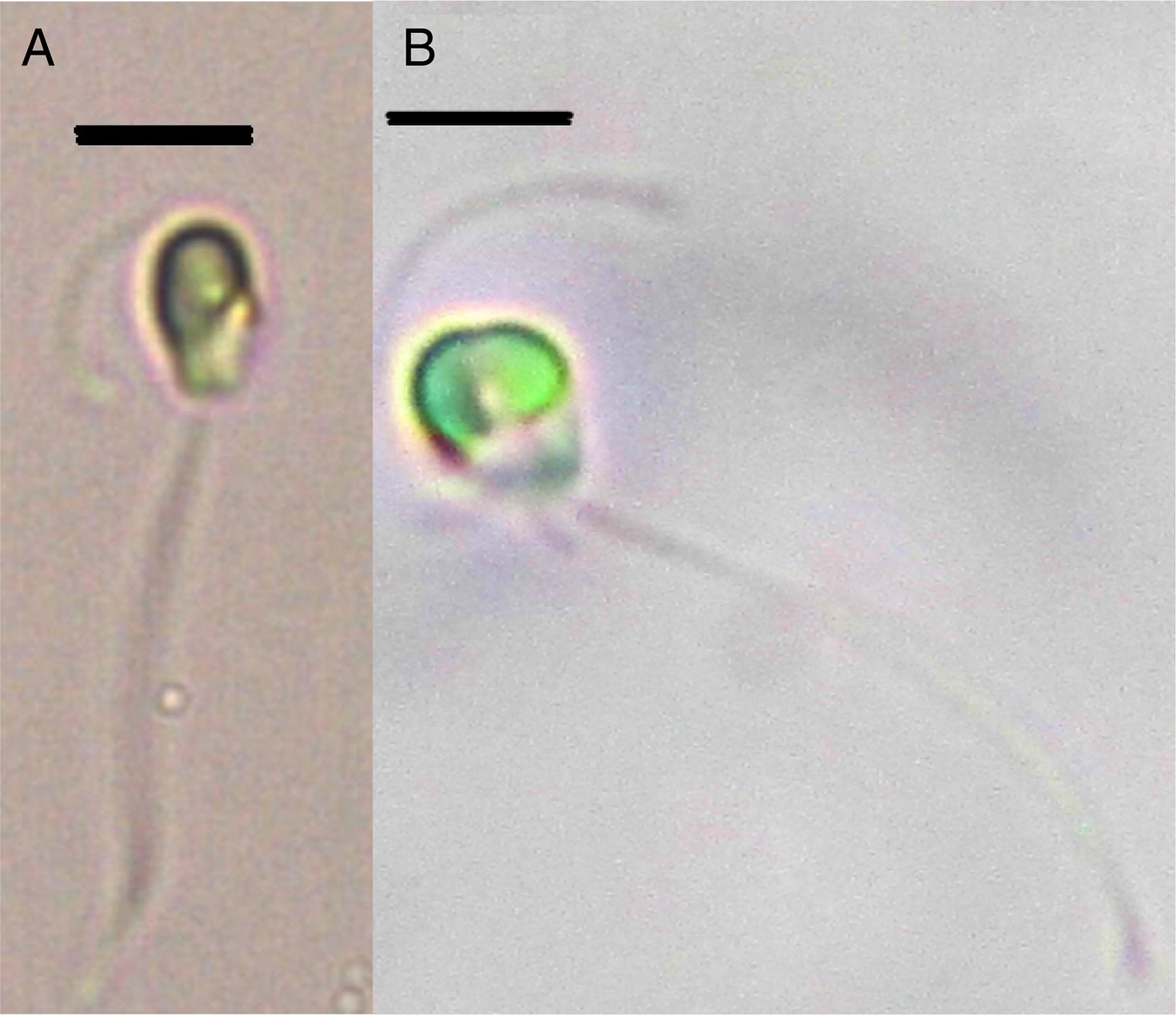
A dividing cell is observed in Figure 4B. Among the members of Prasinophytes, sexual reproduction was only observed in Nephroselmis olivacea (a freshwater species) (Suda et al., Reference Suda, Watanabe and Inouye2004). It is unknown if other species of prasinophytes have this sexuality (Suda et al., Reference Suda, Watanabe and Inouye2004; Bell, Reference Bell2008). Since the phylogenetic position of Nephroselmis is suggested to be an ancestral branch of the Chlorophyta (Suda et al., Reference Suda, Watanabe and Inouye2004; Turmel et al., Reference Turmel, Gagnon, O'Kelly, Otis and Lemieux2009), the Nephroselmis genus is proposed to retain the ancestral features of sexual reproduction.
There are numerous studies related to the composition of diatom and dinoflagellate species along the Turkish Sea coasts (Koray, Reference Koray1995; Eker & Kıdeyş, Reference Eker and Kıdeyş2000; Polat et al., Reference Polat, Sarıhan and Koray2000; Polat & Işık, Reference Polat and Işık2002; Türkoğlu & Koray, Reference Türkoğlu and Koray2002; Polat & Koray, Reference Polat and Koray2007; Özman-Say & Balkıs, Reference Özman-Say and Balkıs2012). However, there is a lack of data related to the species composition of chlorophytes in the same region. Among 11 genera and 28 species belonging to Nephrophyceae class, only Nephroselmis marina was reported previously in the Adriatic Sea. Furthermore, there are only a few studies related to the composition of nanoflagellates in the whole of the Mediterranean Sea, such as studies on coccolithophores (Cros & Fortuno, Reference Cros and Fortuno2002), cryptomonads (Novarino, Reference Novarino2005), prymnesiophytes (Leadbeater, Reference Leadbeater1974; Puigserver et al., Reference Puigserver, Chrétiennot-Dinet and Nezan2003) and chrysophytes (Leadbeater, Reference Leadbeater1974; Novarino et al., Reference Novarino, Oliva and Pérez-Uz2002). This study will facilitate the recognition of the chlorophyte Nephroselmis pyriformis along the Turkish coastal waters and possibly provide additional records for the Mediterranean Sea.
Supplementary material
To view supplementary material for this article, please visit http://dx.doi.org/10.1017/S1755267215000469
ACKNOWLEDGEMENT
This study was supported by the Bilimsel Araştırma Projeleri, Mersin University, Project No. BAP-EF FBE (EED) 2009–7.