Junctional tachycardia is a rare arrhythmia in the paediatric populationReference Garson and Gillette 1 and its mechanism is thought to be due to abnormal automaticity in the region of the atrioventricular junction.Reference Amrousy, Elshehaby, Feky and Elshmaa 2 The arrhythmia has been described most commonly after cardiac surgery in children.Reference Zampi, Hirsch and Gurney 3 , Reference Makhoul, Oster, Fischbach, Das and Deshpande 4 The congenital form of junctional tachycardia often presents ⩽6 months of age and is associated with high morbidity and mortality.Reference Sarubbi, Musto and Ducceschi 5 , Reference Collins, Van Hare and Kertesz 6 There are, however, only a few case reports of isolated junctional tachycardia in children.Reference Ward 13 – Reference Sanyal, Thapar, Sharma and Shrivastava 18 Although junctional tachycardia has been associated with acute rheumatic fever and is even hypothesised to be disease specific,Reference Clarke and Keith 7 – Reference Karacan, Isikay, Olgun and Ceviz 9 it has not been previously described in isolated non-rheumatic streptococcal infection. We report a 9-year-old girl with streptococcal pharyngitis who did not meet revised Jones criteria for rheumatic fever and presented with asymptomatic junctional tachycardia during the acute infectious period.
Case report
A previously healthy, 9-year-old girl presented to the emergency department of our tertiary-care centre with complaints of sore throat, low-grade fever (Tmax 37.8°C), and intermittent chest pain lasting for the previous 4 days. There was no previous history of recurrent sore throat. She did not receive any previous outpatient medical treatment. She was afebrile during the physical examination without signs or symptoms suggestive of arthralgia/arthritis, skin rash, subcutaneous nodules, abnormal movements of her extremities, or any other neurological involvement. Her cardiovascular examination revealed an irregular heart rate of 130–140 beats per minute (bpm) without abnormal praecordial pulsations, third or fourth heart sounds, cardiac murmurs, or rubs. She was haemodynamically stable, breathing comfortably in room air with normal oxygen saturation, and her lung fields were clear to auscultation. In addition, there was no hepatosplenomegaly or peripheral oedema, and her neurological examination was normal.
Her pharyngeal swab was positive for the rapid streptococcal antigen test, and acute-phase reactants were elevated with a C-reactive protein (CRP) of 82 mg/L and an erythrocyte sedimentation rate (ESR) of 42 mm/hour. Her anti-streptolysin O (ASO) titres were not elevated. Complete blood count and basic metabolic panel were within the normal range. Autoimmune panel including C3 and C4 complement levels, rheumatoid factor, myeloperoxidase, and anti-proteinase 3 antibodies were within the normal range.
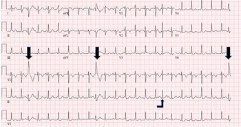
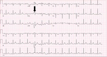
A cardiology consultation was requested after performing a 12-lead electrocardiogram. The initial electrocardiogram showed atrioventricular dissociation due to junctional tachycardia with a narrow QRS (130 bpm) (Fig 1). In addition, there were intermittently conducted sinus impulses, demonstrating both normal and abnormal QRS (right bundle branch) patterns as well a first-degree atrioventricular block (P-R interval 200 ms) (Fig 1). The initial transthoracic echocardiogram revealed a structurally normal four-chamber anatomy with normal biventricular size and function. The atrioventricular and semilunar valves were normal without any stenosis or regurgitation. No pericardial effusion, vegetation, tumour, or thrombi were visible. She was started on oral amoxicillin and was admitted for evaluation of any progression of her rhythm abnormalities. She remained afebrile and haemodynamically stable throughout her stay of 4 days as an inpatient without any new symptoms. Bedside telemetry and follow-up, electrocardiograms showed sinus rhythm with continued episodic junctional tachycardia but no sustained arrhythmias. There was no progression of any atrioventricular block. A repeat echocardiogram performed after 4 days of admission showed continued normal biventricular size and function with no changes from the initial study. She was discharged home on oral amoxicillin to complete a 10-day course and remained clinically asymptomatic. During an outpatient clinic visit 4 weeks later, she had a normal physical examination and a normal electrocardiogram (Fig 2).
Initial electrocardiogram of the patient showing junctional tachycardia with atrioventricular dissociation at a heart rate of ~140 beats per minute (bpm) in deference to the sinus P wave rate of 120 bpm. There are intermittently conducted sinus complexes resulting in variable QRS complex intervals, some with a right bundle branch pattern (large arrows) as well as prolonged P-R intervals (elbow arrow)

Electrocardiogram showing normal sinus rhythm with normal atrioventricular conduction, at 4 weeks after hospital discharge. Compared with Figure 1, each QRS is preceded by a P wave with a normal interval (140 ms) and there are no abnormal QRS complexes. The arrow indicates a recording artefact.

Discussion
Junctional tachycardia has been reported from 9.4 to 20% of the electrocardiograms or Holter recordings among children in the setting of acute rheumatic fever;Reference Clarke and Keith 7 – Reference Ceviz, Celik, Olgun and Karacan 10 however, we describe a hitherto unreported case of isolated junctional tachycardia in a child with streptococcal pharyngitis who did not meet the revised Jones criteria for rheumatic fever.Reference Gewitz, Baltimore and Tani 11 She did not manifest any major criteria such as carditis, arthritis, erythema marginatum, or subcutaneous nodules. Chorea is another major criterion, but this is a late finding, and was not noted in the acute setting in our patient.
The P-R interval was prolonged (200 ms) for the occasional sinus beats that were conducted to the ventricle and this may represent a minor criterion. The presence of junctional tachycardia and intermittently conducted sinus beats with prolonged P-R interval and right bundle branch aberrancy (Fig 1) may constitute evidence of possible acute inflammation to the atrioventricular conduction system.Reference Clarke and Keith 7 , Reference Caraco, Arnon and Raz 12 An alternative explanation can be speculated for the conducted beats with prolonged P-R interval and right bundle branch. This may be because of the premature timing of the sinus beats relative to the atrioventricular conduction system that had not fully recovered following its depolarisation from the preceding junctional beat.Reference Ward 13 Our patient also had elevated acute-phase reactants, which is another minor criterion, but did not have arthralgia or fever (>38.5°C).
Streptococcal infection is quite common in paediatric practice; however, in the current era of antibiotic usage, rheumatic fever has, fortunately, become a rare complication in the developed world.Reference Gewitz, Baltimore and Tani 11 Electrocardiogram changes such as prolonged P-R interval and inverted T waves are not specific for group A haemolytic streptococcal infection. These changes have also been associated with other infections such as influenza, mumps, and gonorrhoea.Reference Rosenberg 14 – Reference Bang 16 .
In recent studies, accelerated junctional rhythm was hypothesised to be quite specific to acute rheumatic fever. It was noted in 10/64 (15.6%) patients with rheumatic fever, but in none with streptococcal upper respiratory tract infection who did not develop rheumatic fever (0/60 patients) or other rheumatological diseases (0/37 patients). In these studies, clinical carditis was associated with the presence of premature contractions. Junctional tachycardia was associated with absence of carditis, and specifically seen in conjunction with polyarthritis.Reference Karacan, Isikay, Olgun and Ceviz 9 , Reference Ceviz, Celik, Olgun and Karacan 10 In two other studies involving patients with rheumatic fever, junctional tachycardia was also associated with the absence of carditis. No relationship was found between this rhythm and inflammatory markers such as ASO titres as well as clinical evidence of carditis, suggesting that it is not related to the severity of myocardial inflammation.Reference Clarke and Keith 7 , Reference Cristal, Stern and Gueron 8
In another case–control study of children following a streptococcal infection, however, junctional tachycardia was only noted in the setting of carditis associated with rheumatic fever (1/28 patients). It was not present in rheumatic fever without carditis (0/5 patients) nor in patients in the “post-streptococcus state” (0/15 patients). The diagnosis of “post-streptococcus state” was made when revised Jones criteria were not met, but there was evidence of streptococcal infection with a positive throat culture, elevated ESR or CRP, or elevated ASO antibodies;Reference Izumi, Yonesaka and Saito 17 one study concluded that, in the absence of other conditions prolonging P-R interval, any abnormal atrioventricular conduction in a child with recent streptococcal infection is specific for acute rheumatic fever.Reference Sanyal, Thapar, Sharma and Shrivastava 18
Contrary to the above reports, in our patient with isolated streptococcal pharyngitis and junctional tachycardia, the clinical criteria for rendering a diagnosis of acute rheumatic fever could not be validated. She presented during the acute phase of streptococcal pharyngitis without any elevated ASO titres and not a few weeks after the infection. Serial echocardiograms documented normal biventricular size and function without any evidence of valvulitis or pericardial effusion; moreover, she remained asymptomatic during the ensuing weeks, and her arrhythmia completely resolved.
Isolated junctional tachycardia has been reported in children in settings such as non-cardiac surgery,Reference Sriram, White, Madhavan and Cannon 19 Lyme disease,Reference Frank, Patel, Sanchez, Shah and Bonney 20 and viral myocarditis,Reference Maiers and Ebenroth 21 but not in association with isolated streptococcal pharyngitis; however, there are reports of group A streptococcal tonsillitis followed acutely by non-rheumatic myopericarditis and associated chest pain.Reference Karjalainen 22 – Reference Said and Severin 24 It is rarely recognised and is thought to be due to a bacterial toxin-induced myocardial inflammation. It is considered a benign condition in the absence of arrhythmias. Selective damage to the cardiac conduction system without other myocardial involvement can occur in the setting of inflammation. This may be due to the higher glycogen content of the conduction system.Reference Clarke and Keith 7 , Reference Caraco, Arnon and Raz 12 , Reference Kirmser, Umbach, Rowett and Ross 25 A co-existent sub-clinical myocarditis cannot be entirely excluded in our patient, despite the benign clinical course and normal echocardiograms. A cardiac MRI scan was not obtained, which is a limitation of this case report.
In summary, we report a unique case of junctional tachycardia in the setting of an acute streptococcal upper respiratory tract infection without evidence of associated rheumatic fever. The mechanism of the junctional rhythm is speculative; however, because of the presence of associated first-degree atrioventricular block as well as a right bundle branch seen on the electrocardiogram, it may be hypothesised as streptococcal toxin-induced acute inflammation/irritation of the atrioventricular conduction system, which resolved without clinical sequelae.
Acknowledgement
None.
Conflicts of Interest
None.