Introduction
Besnoitia besnoiti is a cyst-forming apicomplexan parasite (Marotel, Reference Marotel1912) responsible for bovine besnoitiosis, a re-emerging disease in Europe characterized by both local and systemic clinical signs (Álvarez-García et al., Reference Álvarez-García, Frey, Mora and Schares2013; Cortes et al., Reference Cortes, Leitao, Gottstein and Hemphill2014). Besnoitia besnoiti, together with the closely related parasites Toxoplasma gondii and Neospora caninum, belongs to subfamily Toxoplasmatinae (Tenter et al., Reference Tenter, Barta, Beveridge, Duszynski, Mehlhorn, Morrison, Thompson and Conrad2002). Members of this subfamily are characterized by the presence of two sequential asexual parasite stages that develop in the intermediate hosts. First, rapidly dividing tachyzoites are responsible for the acute stage of the disease, and subsequently tachyzoites undergo differentiation to bradyzoites that form tissue cyst during the chronic stage (Álvarez-García et al., Reference Álvarez-García, García-Lunar, Gutiérrez-Expósito, Shkap and Ortega-Mora2014; Gutiérrez-Expósito et al., Reference Gutiérrez-Expósito, Ferre, Ortega-Mora and Álvarez-García2017).
Morphologically, B. besnoiti tachyzoites closely resemble the tachyzoite stage of N. caninum and T. gondii. The B. besnoiti invasive stages possess the typical apicomplexan secretory organelles of apicomplexan parasites such as rhoptries, micronemes and dense granules, which may most likely, similar to T. gondii and N. caninum, play an important role in various aspects of the host–parasite relationship (Dubey et al., Reference Dubey, Shkap, Pipano, Fish and Fritz2003; Langenmayer et al., Reference Langenmayer, Gollnick, Scharr, Schares, Herrmann, Majzoub-Altweck and Hermanns2015).
To date, only a few Besnoitia proteins have been studied. Protein disulfide isomerase (BbPDI), a protein that acts as a chaperone in the endoplasmatic reticulum, is thought to be secreted by B. besnoiti micronemes during host cell invasion (Naguleswaran et al., Reference Naguleswaran, Alaeddine, Guionaud, Vonlaufen, Sonda, Jenoe, Mevissen and Hemphill2005; Marcelino et al., Reference Marcelino, Martins, Morais, Nolasco, Cortes, Hemphill, Leitao and Novo2011). On the other hand, calcium-dependent protein kinase 1 (BbCDPK1), which is expressed exclusively in plants, fungi algae and apicomplexan parasites, represents a promising target for a new generation of drugs named bumped kinase inhibitors, and is expected to play a role in tachyzoite invasion and proliferation (Jiménez-Meléndez et al., Reference Jiménez-Meléndez, Ojo, Wallace, Smith, Hemphill, Balmer, Regidor-Cerrillo, Ortega-Mora, Hehl, Fan, Maly, Van Voorhis and Álvarez-García2017). Moreover, a few proteomic studies have been carried out in order to investigate relevant biological processes as well as the host immune response associated with B. besnoiti infection that identified several proteins involved in conservative pathways among Toxoplasmatinae parasites (Fernández-García et al., Reference Fernández-García, Álvarez-García, Marugán-Hernández, García-Lunar, Aguado-Martínez, Risco-Castillo and Ortega-Mora2013; García-Lunar et al., Reference García-Lunar, Regidor-Cerrillo, Gutiérrez-Expósito, Ortega-Mora and Álvarez-García2013b). However, the contribution of these studies has been hampered by the absence of B. besnoiti genome sequence which became available only very recently (Schares et al., Reference Schares, Venepally and Lorenzi2017). Surface antigens and proteins from secretory organelles, which have been described to be highly immunogenic in N. caninum and T. gondii, and which are therefore considered to be valuable and specific diagnostic targets (Schares et al., Reference Schares, Rauser, Söndgen, Rehberg, Bärwald, Dubey, Edelhofer and Conraths2000), could not be identified in B. besnoiti proteome (García-Lunar et al., Reference García-Lunar, Regidor-Cerrillo, Gutiérrez-Expósito, Ortega-Mora and Álvarez-García2013b).
We have shown earlier that at least 25 B. besnoiti antigens cross-reacted with specific anti-N. caninum antibodies, and these may be responsible for false-positive reactors (García-Lunar et al., Reference García-Lunar, Regidor-Cerrillo, Gutiérrez-Expósito, Ortega-Mora and Álvarez-García2013b; García-Lunar et al., Reference García-Lunar, More, Campero, Ortega-Mora and Álvarez-García2015). Thus, the development of monoclonal antibodies (MABs) was regarded as a suitable approach to develop novel markers for diagnostic purposes and for studies on B. besnoitia cell biology. Eight MABs directed against different compartments of B. besnoiti tachyzoite antigens were identified and respective epitopes were localized during the lytic cycle in tachyzoites by immunofluorescence and immunogold transmission electron microscopy (TEM). In addition, the genus, species and stage specificity was investigated in the closely related protozoans B. tarandi, N. caninum, T. gondii and Sarcocystis spp. as well as in the bradyzoite stage of B. besnoiti.
Materials and methods
Parasites
Tachyzoites from the BbSpain-1 isolate of B. besnoiti (Fernández-García et al., Reference Fernández-García, Álvarez-García, Risco-Castillo, Aguado-Martínez, Marugán-Hernández and Ortega-Mora2009b), B. tarandi (Dubey et al., Reference Dubey, Sreekumar, Rosenthal, Vianna, Nylund, Nikander and Oksanen2004), the Nc-1 isolate of N. caninum (Dubey et al., Reference Dubey, Hattel, Lindsay and Topper1988) and TgME49 isolate of T. gondii (Lunde and Jacobs, Reference Lunde and Jacobs1963) were grown in Marc-145 cell monolayers with DMEM supplemented with 5% fetal calf serum. Tachyzoites were separated from host cells as previously described (Fernández-García et al., Reference Fernández-García, Risco-Castillo, Pedraza-Díaz, Aguado-Martínez, Álvarez-García, Gómez-Bautista, Collantes-Fernández and Ortega-Mora2009a), centrifuged at 1350 × g for 10 min, and stored at −80 °C until use for Western blot assays (Fernández-García et al., Reference Fernández-García, Álvarez-García, Risco-Castillo, Aguado-Martínez, Marugán-Hernández and Ortega-Mora2009b).
Besnoitia besnoiti bradyzoites were released by a trypsin treatment of a skin biopsy sample from a chronically naturally infected cow, following a previously described method (Fernández-García et al., Reference Fernández-García, Risco-Castillo, Pedraza-Díaz, Aguado-Martínez, Álvarez-García, Gómez-Bautista, Collantes-Fernández and Ortega-Mora2009a). Sarcocystis spp. cystozoites were purified from naturally infected bovine hearts as previously described by More et al. (Reference More, Basso, Bacigalupe, Venturini and Venturini2008) and were pelleted by centrifugation and stored at −80 °C until use for Western blot experiments.
Production of hybridomas
MABs were developed against a whole B. besnoiti tachyzoite extract and an enriched membrane extract that were prepared following previous procedures (Wouda et al., Reference Wouda, Brinkhof, van Maanen, de Gee and Moen1998; Schares et al., Reference Schares, Langenmayer, Scharr, Minke, Maksimov, Maksimov, Schares, Barwald, Basso, Dubey, Conraths and Gollnick2013). Briefly, whole B. besnoiti tachyzoite extract was obtained from detergent lysate of purified B. besnoiti tachyzoites that were pelleted through a 20% sucrose cushion in PBS for 1 h at 13 000 × g at 4 °C (Wouda et al., Reference Wouda, Brinkhof, van Maanen, de Gee and Moen1998). Next, tachyzoite pellets were suspended in PBS containing 1% (vol/vol) Triton X-100. Enriched membrane extract was prepared by surface biotinylation and subsequent immunoprecipitation (Schares et al., Reference Schares, Langenmayer, Scharr, Minke, Maksimov, Maksimov, Schares, Barwald, Basso, Dubey, Conraths and Gollnick2013).
For the production of MABs against intact whole purified B. besnoiti tachyzoites, BALB/C mice were immunized with 50 µg of the respective extract four times with 15 days intervals. Three days prior to the fusion, mice were boosted by an intraperitoneal injection of 50 µg of whole B. besnoiti tachyzoite extract. After euthanasia, splenocytes were fused with P3 × 63 Ag8653 myeloma cells. The supernatants of the hybridomas were screened for antibodies by ELISA. Positive hybridomas were cloned by limiting dilution and recloned at least four times.
MABs against the enriched membrane extract of B. besnoiti tachyzoites were obtained following a previously reported procedure (Schares et al., Reference Schares, Conraths and Reichel1999a,Reference Schares, Dubremetz, Dubey, Barwald, Loyens and Conrathsb; Aguado-Martínez et al., Reference Aguado-Martínez, Álvarez-García, Schares, Risco-Castillo, Fernández-García, Marugán-Hernández and Ortega-Mora2010). Briefly, mice were immunized by an intraperitoneal injection of 50 µg of B. besnoiti membrane-enriched extract on day 0. Three days prior the fusion, mice were boosted by a subcutaneous injection of 50 µg of the respective extract. Then, mice were euthanized and splenocytes were fused with SP2/0 myeloma cells. The supernatants of the resulting hybridomas were screened for antibodies against B. besnoiti tachyzoites by Western blot and positive hybridomas were cloned by limiting dilution and recloned at least twice.
Isotyping of MABs was performed with a commercially available kit (IsoQuick™ Kit for Mouse Monoclonal Isotyping, Sigma-Aldrich, Madrid, Spain).
SDS-PAGE and Western blot
SDS-PAGE was carried out using 15% polyacrylamide gels (Fernández-García et al., Reference Fernández-García, Risco-Castillo, Pedraza-Díaz, Aguado-Martínez, Álvarez-García, Gómez-Bautista, Collantes-Fernández and Ortega-Mora2009a; Schares et al., Reference Schares, Basso, Majzoub, Rostaher, Scharr, Langenmayer, Selmair, Dubey, Cortes, Conraths and Gollnick2010; García-Lunar et al., Reference García-Lunar, Ortega-Mora, Schares, Gollnick, Jacquiet, Grisez, Prevot, Frey, Gottstein and Álvarez-García2013a; Schares et al., Reference Schares, Langenmayer, Scharr, Minke, Maksimov, Maksimov, Schares, Barwald, Basso, Dubey, Conraths and Gollnick2013). A total of 4 × 10⁷ tachyzoites of B. besnoiti and B. tarandi, 2 × 10⁷ tachyzoites of N. caninum and T. gondii and 107 Sarcocystis spp cystozoites were used as antigens. Both reducing and non-reducing conditions were employed for each antigen. For reducing conditions, dithiothreitol or β-mercapto-ethanol was added to the five times concentrated loading buffer. Parasite antigens and marker proteins separated in SDS-PAGE were electrophoretically transferred to polyvinylidene fluoride membranes (Immobilon-P, Millipore, Germany). After blocking with PBS-TG [PBS, 0.05% (v/v) Tween 20, 2% (v/v) fish gelatine liquid (Serva, Heidelberg, Germany)], the antigen-coated membrane was cut into strips and stored frozen at −20 °C until used (Schares et al., Reference Schares, Basso, Majzoub, Rostaher, Scharr, Langenmayer, Selmair, Dubey, Cortes, Conraths and Gollnick2010). To detect antibodies against parasite antigens, the incubation of the strips with serum was performed as previously described by Schares et al. (Reference Schares, Conraths and Reichel1999a) with few modifications. Strips were probed with undiluted hybridoma supernatants and reactions visualized using peroxidase conjugates [anti-mouse IgG (H + L) or anti-mouse IgG (Fc), MinX Hu,Bo,Ho, Dianova, Hamburg, Germany] and 4-chloro-1-naphthol as a substrate.
Protein localization dynamics throughout the lytic cycle by immunofluorescence
Protein localization dynamics alongside the lytic cycle of B. besnoiti tachyzoites were studied using infected Marc-145 cells cultured on coverslips, and six out of the eight MABs, namely MAB 2.F.3, 2G.4, 2.G.A, 8.9.2, 5.5.11 and 3.10.8. For this, 5 × 10⁴ Marc-145 cells were placed on sterile 13 mm coverslips in 24-well plates and cultured overnight at 37 °C/5% CO2. Subsequently, B. besnoiti BbSpain-1 tachyzoites were collected by scrapping an infected culture flask, passing the suspension through a 25-gauge needle, and the number of viable parasites was determined in a Neubauer chamber by trypan blue exclusion. Subsequently, the Marc-145 monolayers were exposed to B. besnoiti tachyzoites during 30 min, 1, 2, 6, 24, 48 and 72 h. After these time points, extracellular/non-adherent parasites were removed by three PBS washes, and the specimens were fixed in either 2% paraformaldehyde in PBS (2% PFA-PBS) or in 2% paraformaldehyde-0.05% glutaraldehyde in PBS (2% PFA + 0.05%GA-PBS) for 10–30 min at room temperature (RT). After fixation, the infected monolayers were permeabilized in PBS containing 0.2% Triton X-100 (Merck Chemicals) for 20 min at RT, and blocking of unspecific binding sites was carried out using PBS/0.1% Triton X-100/3% bovine serum albumin (BSA; Roche) for 15 min at RT. Then, cultures were labelled with a polyclonal rabbit antiserum against B. besnoiti tachyzoites (Gutiérrez-Expósito et al., Reference Gutiérrez-Expósito, Ortega-Mora, Gajadhar, García-Lunar, Dubey and Álvarez-García2012) (1:1000 dilution) to delineate the tachyzoite surface, and with non-diluted hybridoma, supernatants (MABs 2.F.3, 2G.4, 2.G.A, 8.9.2, 5.5.11 and 3.10.8) for 30 min at RT. Following three washes with PBS, coverslips were incubated with Alexa Fluor 488-conjugated goat anti-mouse IgG (1:1000) and Alexa Fluor 594-conjugated goat anti-rabbit IgG (1:1000) (Invitrogen, Thermo Fisher Scientific, Waltham, MA, USA) for 30 min at RT. Parasite and host cell nuclei were stained with 40, 6-diamidino-2-phenylindole (DAPI), and finally, coverslips were mounted on glass slides with ProLong® Gold antifade reagent (Molecular Probes, Thermo Fisher Scientific, Waltham, MA, USA) and sealed with clear nail polisher. Single stacks of immunofluorescence staining were captured with a Leica TCS-SP5 confocal laser-scanning microscope (Leica Microsystems) in the Spanish National Centre for Biotechnology (CNB, Madrid). Image processing was performed using the LAS AF (Leica Microsystems) and the ImageJ software (NCBI, http://rsb.info.nih.gov/ij/).
Transmission electron microscopy
Besnoitia besnoiti tachyzoite-infected Vero cells were grown in T25 tissue culture flasks. After a 72 h culture period, the medium was removed and cells were washed with 100 mm sodium cacodylate buffer, pH 7.2, and were fixed in cacodylate buffer containing 3% paraformaldehyde and 0.05% glutaraldehyde for 1 h at 20 °C. The cell layer was removed with a cell scraper, washed in cacodylate buffer, centrifuged, and the pellet was resuspended in cacodylate buffer. Cells were then dehydrated by sequential washing in progressively increasing concentrations of pre-cooled (−20 °C) ethanol (30–50–70–90–100%), and finally embedded in LR-White resin as previously described (Hemphill et al., Reference Hemphill, Vonlaufen, Naguleswaran, Keller, Riesen, Guetg, Srinivasan and Alaeddine2004; Risco-Castillo et al., Reference Risco-Castillo, Fernández-García, Zaballos, Aguado-Martínez, Hemphill, Rodríguez-Bertos, Álvarez-García and Ortega-Mora2007). Infiltration of the resin was carried out over 3 days at −20 °C, with one resin change/day. Finally, the specimen blocks were polymerized at 60 °C over night. Sections of 80 nm thickness were prepared using a Reichert and Jung ultramicrotome, placed onto formvar-carbon-coated nickel grids (Plano GmbH, Wetzlar, Germany), and air-dried.
For immunogold labelling, sections were on-grid-labelled in a moist chamber with MAB culture supernatants that were used undiluted for 1 h at 20 °C, followed by three washes in PBS, 10 min each. Goat anti-rabbit conjugated to 10 nm diameter gold particles (Aurion, Wageningen, The Netherlands) was applied at a dilution of 1:5 in PBS-0.3% BSA as secondary antibody. After another three washes, 10 min each, grids were air-dried and contrasted with uranyle acetate and lead citrate (Hemphill et al., Reference Hemphill, Vonlaufen, Naguleswaran, Keller, Riesen, Guetg, Srinivasan and Alaeddine2004). Specimens were viewed on a CM12 TEM operating at 80 kV.
Results and discussion
MABs labelled the surface, the apical tip and the granular content of B. besnoiti tachyzoites
We have developed eight MABs that clearly label different subcellular compartments of B. besnoiti tachyzoites (Fig. 1). The characterization of these antibodies is timely, since tools for cell biological studies on B. besnoiti are essentially missing. Only five MABs against B. besnoiti tachyzoites have been developed in the past. Njagi et al. (Reference Njagi, Entzeroth, Nyaga and Musoke2004) developed three MABs that were shown to label the apical cone and one MAB that stained cytoplasmic granules of the tachyzoites. Based on immunofluorescence results and the in vitro neutralizing properties of these MABs, it was suspected that these antibodies recognized proteins localized in secretory organelles, but the precise subcellular location was not confirmed. Similarly, Shkap et al. (Reference Shkap, Pipano and Zwernemann1995) characterized one MAB that showed different recognition patterns, labelling a 70 kDa polypeptide in the soluble cytosol and the membrane fraction of B. besnoiti tachyzoites. Thus, the MABs we have generated can be added to this limited panel of biological reagents on B. besnoiti cellular biology and, potentially, also diagnosis.
Confocal laser scanning microscopy of MABs labelling the surface (A and B), the apical tip (C–E) and granular content (F–H) of Besnoitia besnoiti tachyzoites. Infected cultures were fixed with either PFA or PFA + GA and double labelled with anti-B. besnoiti polyclonal antibody (green) and mouse MABs (red). Nuclei were stained with DAPI (blue). All the images show a single-stack 1 µm slice. Scale bars represent 5 µm.

In the present study, MABs 2.A.12, 2.F.3 and 2.G.4 labelled cytoplasmic granular content compatible with what has been observed for dense granule proteins (Leineweber et al., Reference Leineweber, Spekker-Bosker, Ince, Schares, Hemphill, Eller and Däubener2017), MABs 2.G.A, 1.17.8 and 8.9.2 labelled the apical tip of the tachyzoites, which resembles microneme staining (Li et al., Reference Li, Liu, Wang, Fu, Nan and Liu2015), whereas MABs 3.10.8 and 5.5.11 appeared to bind to structures on the tachyzoite surface (Schares et al., Reference Schares, Dubremetz, Dubey, Barwald, Loyens and Conraths1999b). Interestingly, the later ones were produced using an enriched membrane antigen extract of tachyzoites, prepared by surface biotinylation and subsequent immunoprecipitation (Schares et al., Reference Schares, Langenmayer, Scharr, Minke, Maksimov, Maksimov, Schares, Barwald, Basso, Dubey, Conraths and Gollnick2013). In the past, Schares et al. (Reference Schares, Dubremetz, Dubey, Barwald, Loyens and Conraths1999b) obtained four MABs directed against biotinylated N. caninum tachyzoites and, similarly, three of them labelled the outer membrane surface of the parasite. TEM analysis was carried out with all MABs. However, only MABs 2.A.12 and 2.F.3 gave conclusive results, confirming dense granule staining as shown in Fig. 2.
Immunogold-TEM of Besnoitia besnoiti tachyzoites stained with MABs 2.A.12 (A) and 2.F.3 (B) and anti-mouse-10 nm gold particles. Arrows pint towards gold particles indicating dense granule staining. Bars = 1.8 µm.

Epitope co-localization dynamics showed distinctive pattern for each MAB along the lytic cycle
The lytic cycle in Toxoplasmatinae parasites is a conserved process, and involves key events that occur sequentially, during which specialized organelles secrete their contents in an orchestrated manner. First, apical micronemes release their contents during the early attachment–invasion process, mediating attachment and also re-orientation of tachyzoites. At the onset, and also during the invasion process, rhoptry proteins are released, and the dense granules discharge their contents shortly prior, or upon completion of host cell invasion, and they are involved in parasitophorous vacuole (PV) formation and maturation, and eventually parasite egress to ensure parasite dissemination and survival (Black and Boothroyd, Reference Black and Boothroyd2000; Blader et al., Reference Blader, Coleman, Chen and Gubbels2015). Initially, two MABs representative of each subcellular localization were selected for the studies of localization dynamics during the lytic cycle of B. besnoiti tachyzoites: 2.F.3, 2G.4 (dense granules), 2.G.A, 8.9.2 (micronemes), and 5.5.11 and 3.10.8 (tachyzoite surface). Moreover, different fixatives should be employed to characterize proteins in closely related parasites in order to obtain a concise picture of their localization, particularly for those proteins located in subcellular compartments. Thus, we selected two fixation methods for crosslinking proteins, one comprised only of paraformaldehyde, and the other one employing combined paraformaldehyde/glutaraldehyde fixation. In addition, a precipitation fixation protocol using methanol was also employed, but this resulted in either absence of labelling or lower staining intensity (data not shown). In general, paraformaldehyde fixation was more efficient in retaining the reservoirs of intracellular epitopes, whilst combined paraformaldehyde/glutaraldehyde fixation resulted in improved preservation of proteins after secretion. This is in agreement with the previous studies (Sohn et al., Reference Sohn, Cheng, Drummond, Peng, Vermont, Xia, Cheng, Wastling and Bradley2011; Pastor-Fernández et al., Reference Pastor-Fernández, Regidor-Cerrillo, Álvarez-García, Marugán-Hernández, García-Lunar, Hemphill and Ortega-Mora2016a, Reference Pastor-Fernández, Regidor-Cerrillo, Jiménez-Ruiz, Álvarez-García, Marugán-Hernández, Hemphill and Ortega-Mora2016b). Membrane staining, most likely on the surface of tachyzoites, was visualized regardless the fixation protocol.
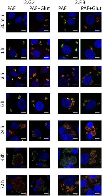
Immunofluorescence staining with MABs 2.F.3 and 2.G.4 confirmed the punctated, granular-dense granules-like staining (Fig. 3). Secretion of respective proteins recognized by these antibodies was observed with both fixatives, and with MAB 2.F.3 most pronounced after 1–2 h post infection (hpi), extending up to PV maturation at 24 hpi. Interestingly protein secretion in evacuoles was visible in samples fixed with paraformaldehyde and glutaraldehyde at 6 hpi. In T. gondii, numerous studies have demonstrated that the parasite modifies the PV and its host cell by secreting numerous rhoptry and dense granule proteins. In particular, Dunn et al. (Reference Dunn, Ravindran, Kim and Boothroyd2008) reported that the dense granule protein TgGRA7 was present in strand-like structures in the host cytosol together with other dense granule proteins. Similar findings were reported for the Neospora rhoptry protein NcROP2 (Pastor-Fernández et al., Reference Pastor-Fernández, Regidor-Cerrillo, Jiménez-Ruiz, Álvarez-García, Marugán-Hernández, Hemphill and Ortega-Mora2016b). Accordingly, the proteins labelled with our MABs are likely to participate in invasion, PV formation and/or PV maturation. However, B. besnoiti Sp-1 tachyzoites are able to survive in the extracellular environment for extended periods of time, and thus exhibit a more asynchronous invasion process compared with Neospora or Toxoplasma (Frey et al., Reference Frey, Regidor-Cerrillo, Marreros, García-Lunar, Gutiérrez-Expósito, Schares, Dubey, Gentile, Jacquiet and Shkap2016). From 24 hpi onwards, the granular staining pattern observed with MAB 2.F.3 changed to a more pronounced staining of the membrane, and protein secretion was again detected during parasite egress at 72 hpi. The labelling pattern of MAB 2.G.4 was similar to the one displayed by 2.F.3, but a few remarkable differences were noted (Fig. 3). Protein secretion into evacuoles was detected earlier at 1 hpi, in close proximity to the host cell nucleus, and secretion extended up to 6 hpi in parallel with a punctated staining. Once the PV was formed, a slight labelling seemed to be associated with the membrane of the tachyzoites, and a granular staining was again observed during egress. Similar findings regarding dense granules staining were observed in closely related parasites. In Neospora, NcNTPase and NcGRA7 protein secretion was also evident during early invasion, PV maturation and egress. However, NcNTPase was scattered throughout the tachyzoite cytoplasm, whereas NcGRA7 labelling was observed closer to the parasite surface, suggestive of differential protein trafficking (Pastor-Fernández et al., Reference Pastor-Fernández, Regidor-Cerrillo, Álvarez-García, Marugán-Hernández, García-Lunar, Hemphill and Ortega-Mora2016a).
Confocal laser scanning microscopy at different stages of the lytic cycle of Besnoitia besnoiti tachyzoites, stained with MABs displaying a granular staining pattern compatible with dense granule localization (2.F.3 and 2.G.4). Infected cultures were fixed with paraformaldehyde (PFA) or paraformaldehyde combined with glutaraldehyde (PFA + GA), and infected cells were double labelled with a polyclonal antibody against B. besnoiti tachyzoites (green) and monoclonal antibodies (red). Nuclei were stained with DAPI (blue). All the images show a single 1 µm slice. Scale bars represent 5 µm.

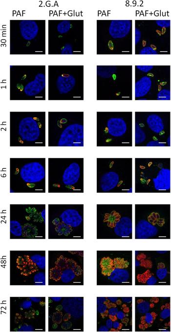
MABs 2.G.A and 8.9.2 showed a distinct apical staining upon paraformaldehyde fixation (Fig. 4). Interestingly, at 6 hpi, both MABs labelled the anterior as well as the posterior end of intracellular parasites, and staining was also found on the surface membrane. This clearly indicates that the proteins recognized by these MABs are secreted, and most likely micronemal components. During Toxoplasma gliding motility and invasion, microneme proteins have been reported to migrate across the tachyzoite surface and redistribute towards the posterior pole of the parasite (Soldati et al., Reference Soldati, Dubremetz and Lebrun2001) as shown, e.g. by the apical membrane antigen AMA1 during moving junction formation (Alexander et al., Reference Alexander, Mital, Ward, Bradley and Boothroyd2005). Similar findings were reported for the Neospora microneme antigen MIC3, which was shown to be secreted at the anterior end, and then redistributed along the tachyzoite surface, finally ending up at the posterior pole (Naguleswaran et al., Reference Naguleswaran, Cannas, Keller, Vonlaufen, Schares, Conraths, Bjorkman and Hemphill2001). The expected microneme protein discharge induced upon host cell contact was not observed. However, Lodoen et al. (Reference Lodoen, Gerke and Boothroyd2010) postulated that secretion of low-abundance proteins may be undetectable by immunofluorescence approaches, so that further secretion assays could be more suitable to address this issue.
Confocal laser scanning microscopy at different stages of the lytic cycle of Besnoitia besnoiti tachyzoites, stained with MABs that exhibit a staining pattern compatible with microneme localization (2.G.A and 8.9.2). Infected cultures were fixed with paraformaldehyde (PFA) or paraformaldehyde combined with glutaraldehyde (PFA + GA), and were double labelled with a polyclonal antibody against B. besnoiti tachyzoites (green) and monoclonal antibodies (red). Nuclei were stained with DAPI (blue). All the images show a single 1 µm slice. Scale bars represent 5 µm.

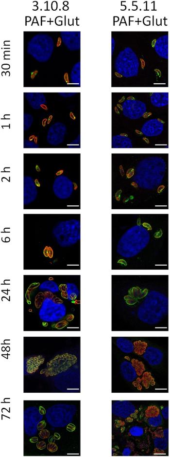
Finally, MABs 3.10.8 and 5.5.11 labelled epitopes that are associated with, or in close proximity to the surface membrane and staining did not vary along the lytic cycle (Fig. 5). However, it is not clear whether the epitopes recognized by these two MABs are oriented towards the extracellular space. If so, these MABs could be of special interest for the prevention of infection by immunological means.
Confocal laser scanning microscopy at different stages of the lytic cycle of Besnoitia besnoiti tachyzoites, stained with MABs labelling the tachyzoite surface membrane (3.10.8 and 5.5.11). Infected cultures were fixed with paraformaldehyde (PFA) or paraformaldehyde combined with glutaraldehyde (PFA + GA). Specimens were double labelled with a polyclonal antibody against B. besnoiti tachyzoites (green) and monoclonal antibodies (red). Nuclei were stained with DAPI (blue). All the images show a single 1 µm slice. Scale bars represent 5 µm.

MABs 2.G.A, 2.A.12, 2.G.4 and 1.17.8 represent promising diagnostic markers
The development of MABs against specific B. besnoiti proteins is an optimal approach to develop new diagnostic markers for Besnoitia spp. infection. Similar approaches were successfully employed in related parasites. For instance, Baszler et al. (Reference Baszler, Knowles, Dubey, Gay, Mathison and McElwain1996, Reference Baszler, Adams, Vander-Schalie, Mathison and Kostovic2001) generated a MAB that recognized the surface protein NcSRS2, and applied this antibody for the development of a competitive ELISA that has been widely used to detect anti-Neospora antibodies in cattle. Another MAB was developed by Ghalmi et al. (Reference Ghalmi, China, Kaidi and Losson2009) to detect N. caninum infection in wildlife species. Uzeda et al. (Reference Uzeda, Schares, Ortega-Mora, Madruga, Aguado-Martinez, Corbellini, Driemeier and Gondim2013) employed a combination of MABs targeting the N. caninum tachyzoite surface protein NcSRS2 and the dense granule protein NcGRA7 to improve the immunohistochemical detection of N. caninum in tissue sections from naturally infected cattle.
In order to verify the genus, species and stage specificity of our MABs, potential cross-reactions with antigens of the closely related apicomplexans N. caninum, T. gondii, and Sarcocystis spp and B. tarandi, and with B. besnoiti bradyzoites, were investigated by Western blot. Table 1 summarizes the results, and also provides information on the Ig isotypes of the MABs. As can be seen, the results of the present study revealed that most MABs developed herein were genus specific. None of the MAB showed cross-reactions with T. gondii antigens, neither under reducing nor non-reducing conditions. None of the MABs cross-reacted with N. caninum antigens. Only MAB 2.F.3 exhibited cross-reaction with Sarcocystis spp. cystozoites when proteins were separated under non-reducing conditions (Table 1). Previously, Shkap et al. (Reference Shkap, Pipano and Zwernemann1995) investigated the specificity of the MAB against a soluble fraction of T. gondii tachyzoites, and a negative result was observed. However, cross-reactions against N. caninum and Sarcocystis spp. were not studied, although it is widely known that N. caninum and Sarcocystis spp. infections are highly prevalent in cattle worldwide (Bartels et al., Reference Bartels, Arnaiz-Seco, Ruiz-Santa-Quitera, Bjorkman, Frossling, von, Conraths, Schares, van, Wouda and Ortega-Mora2006; Moré et al., Reference More, Basso, Bacigalupe, Venturini and Venturini2008; Eiras et al., Reference Eiras, Arnaiz, Álvarez-García, Ortega-Mora, Sanjuanl, Yus and Dieguez2011). Moreover, antisera obtained from N. caninum- and Sarcocystis spp.-infected animals cross-reacted with B. besnoiti antigens, causing false-positive results (García-Lunar et al., Reference García-Lunar, More, Campero, Ortega-Mora and Álvarez-García2015) and at least 25 B. besnoiti antigens are recognized by anti-N. caninum antibodies (García-Lunar et al., Reference García-Lunar, Regidor-Cerrillo, Gutiérrez-Expósito, Ortega-Mora and Álvarez-García2013b). Previous MABs developed against other Toxoplasmatinae members also showed cross-reactions with closely related parasites. A MAB directed against a marker of T. gondii bradyzoites – TgBAG-1 – cross-reacted with N. caninum bradyzoites (Weiss et al., Reference Weiss, Ma, Halonen, McAllister and Zhang1999). Similarly, the MAB CC2, which reacts with T. gondii bradyzoites (Gross et al., Reference Gross, Bohne, Soete and Dubremetz1996) was shown to also cross-react with N. caninum tissue cyst-like structures generated in vitro (Vonlaufen et al., Reference Vonlaufen, Muller, Keller, Naguleswaran, Bohne, McAllister, Bjorkman, Muller, Caldelari and Hemphill2002; Risco-Castillo et al., Reference Risco-Castillo, Fernández-García and Ortega-Mora2004).
Characterization of the MABs obtained against Besnoitia besnoiti tachyzoites: IFAT co-localization, immunoglobulin isotyping and protein bands detected by Western blot under reducing and non-reducing conditions

T, tachyzoite; B, bradyzoite; C, cystozoite; nr, non-reducing conditions; r, reducing conditions.
a Localization of the protein band recognized by the MABs established by IFAT.
b Values in parenthesis indicate the molecular size of additional faint reactivity.
–No reactivity observed.
In contrast to the non-reactivity with T. gondii, N. caninum and Sarcocystis antigens, most MABs, with the exception of MABs 1.17.8 and 2.G.A, recognized B. tarandi antigens under both, reducing and non-reducing, conditions. These results agree with the previous findings where B. besnoiti- and B. tarandi-infected animals exhibited the same pattern of immunodominant antigens and no specific antigenic spots for B. besnoiti and B. tarandi were recognized by 2-DE immunoblotting (Gutiérrez-Expósito et al., Reference Gutiérrez-Expósito, Ortega-Mora, Gajadhar, García-Lunar, Dubey and Álvarez-García2012; García-Lunar et al., Reference García-Lunar, Regidor-Cerrillo, Ortega-Mora, Gutiérrez-Expósito and Álvarez-García2014). Thus, we corroborate the difficulty in developing specific tools to differentiate infections by these two species. The species specificity of MABs 1.17.8 and 2.G.A should be further assessed with other Besnoitia species affecting ungulates such as B. bennetti and B. caprae.
None of the MABs recognized B. besnoiti bradyzoite antigens, despite the similarity in the proteome profiles of these two stages (Fernandez-García et al., Reference Fernández-García, Álvarez-García, Marugán-Hernández, García-Lunar, Aguado-Martínez, Risco-Castillo and Ortega-Mora2013). However, differences in the immunodominant antigenic pattern between B. besnoiti tachyzoites and bradyzoites has been observed by others (Fernández-García et al., Reference Fernández-García, Risco-Castillo, Pedraza-Díaz, Aguado-Martínez, Álvarez-García, Gómez-Bautista, Collantes-Fernández and Ortega-Mora2009a; Schares et al., Reference Schares, Basso, Majzoub, Rostaher, Scharr, Langenmayer, Selmair, Dubey, Cortes, Conraths and Gollnick2010).
In conclusion, we have generated a set of MABs that will be invaluable tools as markers for studying the localization and the role of proteins originating from secretory organelles of these parasites. Two MABs bind to the tachyzoite surface membrane, three MABs label the apical part and show a localization pattern during the lytic cycle that indicates that they react with micronemal proteins, and three show a staining pattern that clearly suggest a dense granule localization, with secretion observed during early invasion in evacuoles, PV formation and egress. The molecular identity of the antigens recognized by these MABs is currently under investigation, and these MABs will be useful tools to elucidate the role of the respective antigens in the invasion process. Ideally, MABs of diagnostic value (e.g. to develop a competitive ELISA) should recognize B. besnoiti-specific antigens. Thus, the MABs 2.G.A, 2.A.12 and 2.G.4, together with those recognizing the apical tip of the tachyzoite (1.17.8 and 8.9.2), and the surface of the tachyzoites (3.10.8 and 5.5.11) are promising candidates to be applied as diagnostic tools. The absence of cross-reactions between MABs 1.17.8 and 2.G.A with B. tarandi antigens supports their additional value as diagnostic markers to differentiate between these two closely related species. Moreover, tachyzoite-specific MABs could be employed as markers for monitoring tachyzoite–bradyzoite conversion; however, bradyzoite-specific markers in B. besnoiti remain to be discovered.
Acknowledgements
We gratefully acknowledge Andrea Bärwald and Vanesa Navarro for their excellent technical assistance. We also acknowledge Professor Dr Mª Teresa Miras Portugal's research group from the Department of Biochemistry and Molecular Biology IV of the Complutense University of Madrid and people from the Spanish National Centre for Biotechnology (CNB, Madrid) for their kind help with the confocal laser microscopy system.
Financial support
This work was financed by the Spanish Ministry of Economy and Competitiveness (AGL 2010-20561) and CYTED (Thematic Network 113RT0469 Protozoovac) and by the Community of Madrid (PLATESA S20137ABI-2906). Paula García Lunar was supported by a fellowship from Complutense University of Madrid. Alejandro Jiménez-Meléndez and Iván Pastor-Fernández were supported by two fellowships from the Spanish Ministry of Education, Culture, and Sports (M.E.C.D.), as part of the Program of Training of University Staff (F.P.U., grant numbers FPU13/05481 and AP2009-0354, respectively). Andrew Hemphill was supported by the Swiss National Science Foundation (grant number 310030_165782).
Conflict of interest
None.
Ethical standards
All mice-handling procedures complied with the EU legislation and were approved by the Ethics Committee of the Complutense University of Madrid, Spain and by the Landesamt für Landwirtschaft, Lebensmittelsicherheit und Fischerei, Mecklenburg-Vorpommern, Germany.