Introduction
Controlled ovarian stimulation (COS) is an important step for the optimization of in vitro fertilization (IVF) success, as it leads to the retrieval of multiple mature oocytes, which in turn increases the odds of having embryos available for transfer. However, the ovarian response to COS may be suboptimal or poor, having negative consequences for the patients (La Marca and Sunkara Reference La Marca and Sunkara2014, Conforti et al. Reference Conforti, Cariati, Vallone, Alviggi and de Placido2017).
Poor ovarian responders (POR) represent 9–24% of patients undergoing ovarian stimulation and the list of its aetiologies include metabolic, autoimmune and infectious diseases, pelvic adhesions, advanced endometriosis, ovarian surgery, reduction of ovarian follicles due to ageing and chromosomal and genetic alterations (Drakopoulos et al. Reference Drakopoulos, Bardhi, Boudry, Vaiarelli, Makrigiannakis, Esteves, Tournaye and Blockeel2020).
The clinical management of these patients remains a challenge in everyday practice. To standardize the diagnosis of POR, the medical community developed the Bologna Criteria and then shifted to the Patient-Oriented Strategies Encompassing IndividualizeD Oocyte Number (POSEIDON), which was launched to identify and stratify women undergoing IVF with ‘expected’ or ‘unexpected’ compromised ovarian response to COS with exogenous gonadotropins. Four groups of low prognosis patients were formed based on female age, ovarian biomarkers and patient’s ovarian response in a previous stimulation, if any (Alviggi et al. Reference Alviggi, Andersen, Buehler, Conforti, De Placido, Esteves, Fischer, Galliano, Polyzos and Sunkara2016).
Recent studies have shown that patients stratified in POSEIDON groups have distinct reproductive outcomes (Leijdekkers et al. Reference Leijdekkers, Eijkemans, van Tilborg, Oudshoorn, van Golde, Hoek, Lambalk, de Bruin, Fleischer, Mochtar, Kuchenbecker, Laven, Mol, Torrance and Broekmans2019, Chinta et al. Reference Chinta, Antonisamy, Mangalaraj, Kunjummen and Kamath2021). Therefore, we hypothesized that patients categorized in different POSEIDON groups not only respond distinctly to COS, but also present with different oocyte and embryo qualities.
Embryo monitoring with timelapse imaging (TLI) may allow the identification of morphokinetic events potentially affected by ovarian response. Consequently, it may be a valuable approach to evaluate the relationship between POSEIDON classification and the competences of developing embryos, considering morphokinetic parameters. The aim of this study was to compare embryo quality, and speed and pattern of cell divisions in the four groups of POR as stratified by the POSEIDON criteria.
Materials and methods
Patients and experimental design
This historical cohort-study was performed in a private university-affiliated IVF centre, between March 2019 and April 2022. Kinetic data were analysed in 3326 embryos, individually cultured in a TLI incubator (EmbryoScope+, Unisense Fertilitech, Aarhus, Denmark) until day five of development, deriving from 846 patients undergoing their ICSI cycles. Timing of specific events from the point of insemination was determined using TLI. Patients were split into four groups according to the POSEIDON criteria: POSEIDON group 1 (n = 144), consisting of patients < 35 years old and AFC ≥ 5 and an unexpected poor or suboptimal response (up to nine oocytes retrieved); POSEIDON group 2 (n = 570) consisting of patients ≥ 35 years old with AFC ≥ 5 and an unexpected poor or suboptimal ovarian response; POSEIDON group 3 (n = 24), consisting of patients < 35 years old and AFC < 5, and POSEIDON group 4 (n = 108), consisting of patients ≥ 35 years and AFC < 5. Morphokinetic events and ICSI clinical outcomes were compared among the groups.
All patients signed a written informed consent form in which they agreed to share the outcomes of their cycles for research purposes, and the study was approved by the local Institutional Review Board.
Controlled ovarian stimulation and laboratory procedures
On the third day of the cycle, COS was started by the administration r-FSH (300IU follitropin alpha, Gonal-F, Serono, Geneva, Switzerland or 16 mcg follitropin delta, Rekovelle®, Ferring, Saint-Prex, Switzerland) daily doses. r-FSH dose was adjusted according to follicular development, which was monitored by ultrasound scan.
When at least one follicle ≥ 14 mm was visualized, pituitary blockage was performed using gonadotropin-releasing hormone (GnRH) antagonist (GnRHa, Cetrotide®; Merck KGaA, Darmstadt, Germany). When three or more follicles attained a mean diameter of ≥ 17 mm and adequate serum estradiol levels were observed, r-FSH and GnRH antagonist administrations were stopped, and final follicular maturation was triggered by the administration of recombinant human chorionic gonadotropin (r-hCG, 250 µg, Ovidrel®, Merck KGaA, Geneva, Switzerland). Oocyte retrieval was performed 37 h later. Oocytes in metaphase II were selected for ICSI.
Semen analysis and preparation
Semen samples were collected in the laboratory by masturbation and were prepared using a two-layered density gradient centrifugation technique (50% and 90% Isolate, Irvine Scientific, Santa Ana, CA, USA).
Intracytoplasmic sperm injection
Intracytoplasmic sperm injection was performed according to Palermo et al. (Reference Palermo, Joris, Devroey and Van Steirteghem1992). Sperm was selected at 400x magnification using an inverted Nikon Eclipse TE 300 microscope and injected into the oocytes in a micro-injection dish prepared with buffered medium (Global w/HEPES, LifeGlobal, Guilford, USA) covered with paraffin oil (Paraffin oil P.G., LifeGlobal), on an inverted microscope heated stage (37.0°C ± 0.5°C).
Embryo culture
Injected oocytes were individually cultured in a 16-well culture dish (Embryoslide, Unisense Fertilitech, Aarhus, Denmark), in 360 μl of continuous single-culture media (Global® total®, LifeGlobal), overlaid with 1.8 ml of mineral oil (Paraffin oil P.G., LifeGlobal) in a TL-monitored incubator (EmbryoScope+, Unisense Fertilitech, Aarhus, Denmark) set at 37°C with an atmosphere of 6% O2 and 7.2% CO2 until day five of embryo development. The incubator high-definition camera was set up to record embryos’ images, in eleven focal planes, every 10 min. Recorded kinetic markers were timing to pronuclei appearance (tPNa) and fading (tPNf), timing to two (t2), three (t3), four (t4), five (t5), six (t6), seven (t7), and eight cells (t8), and timing to start blastulation (tSB) and to blastulation (tB). Durations of the second (cc2, t3-t2) and third cell cycles (cc3, t5-t3) and timing to complete synchronous divisions t2-tPNf (s1), t4-t3 (s2) and t8-t5 (s3) were calculated. Data generated from EmbryoScope+ was analysed using the EmbryoViewer software (Vitrolife, Denmark). The incidences of multinucleation at 2- and 4-cell stages and of abnormal cleavage patterns (direct or reverse cleavage) were recorded for each embryo.
Clinical follow-up
Embryo transfer was performed on day 5 of embryo development and one or two embryos were transferred per patient.
Women with a positive pregnancy test, performed 10 days post embryo transfer, had a transvaginal ultrasound scan 2 weeks later. The clinical pregnancy was diagnosed upon detection of foetal heartbeat. The pregnancy rate was calculated per embryo transfer. The implantation rate is the number of gestational sacs with foetal heartbeats divided by the number of transferred embryos. Miscarriage was defined as clinical pregnancy loss before 20 weeks.
Data analysis and statistics
The primary outcome measure was tB since it is the most advanced key-stage of embryonic development recorded in our centre. Post hoc power analysis was calculated, given α of 5%, sample size of 1328 embryos that reached blastocyst stage at day 5 of development, and effect size for tB. The achieved power was superior to 80%. The calculation was performed using Hotelling Lawley Trace test in GLIMMPSE App for multilevel data (15), which accounted for correlation between embryos from the same cycle.
Generalized mixed models (GMM), adjusted for potential confounders, were used to study the relationship between POSEIDON groups and embryo morphokinetics. The models were generated using POSEIDON groups as independent variables and kinetic markers as dependent variables. Maternal and paternal ages were included as covariates in all models to control for their influence.
A random effect was added to account for the correlation between the embryos within the same cycle, with linear distribution for morphokinetic data in hours (h) and known implantation diagnosis score (KIDScore) ranking, and binomial distribution for the incidences of abnormal cleavage patterns and multinucleation.
For clinical outcomes, which were based on a single observation per couple, regression models were used without random effects, with linear distribution for implantation rate and binomial distribution for clinical pregnancy and miscarriage rates.
The results are expressed as the mean ± standard error (SD), odds ratio (OR) or beta coefficient (B) with 95% confidence interval (CI) and p-values. P < 0.05 was considered statistically significant. Data analysis was conducted using the Statistical Package for the Social Sciences (SPSS) 21 (IBM, New York, NY, USA).
Results
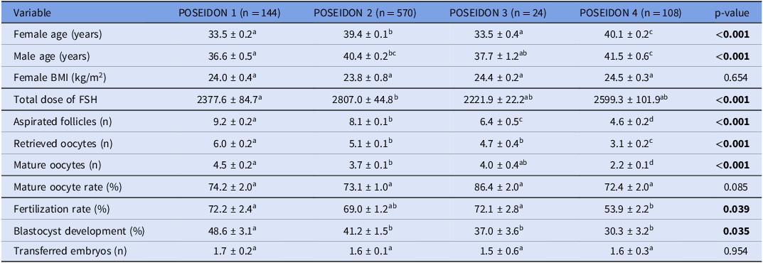
Demographic data concerning female and male partners, and the outcomes of ICSI cycles are shown in Table 1.
Comparison of demographic data and cycles’ characteristics among POSEIDON groups (n = 846 cycles)

Note: Values are means ± standard error, unless otherwise noted. Different letters within the same line represent statistically significant differences.
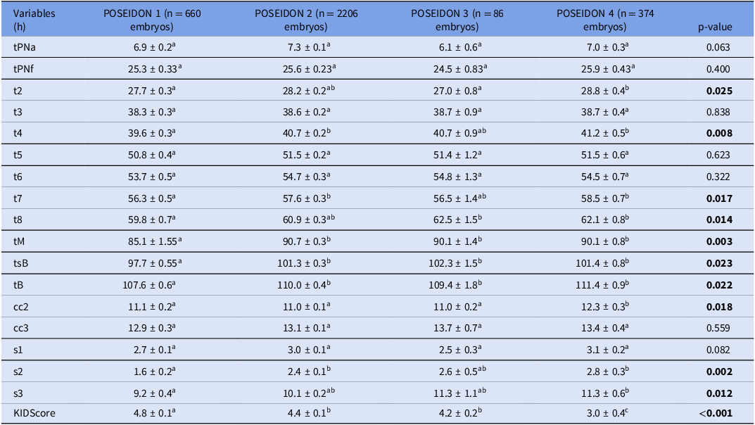
The results from the generalized mixed models followed by Bonferroni post hoc for the comparison of means among groups showed that embryos derived from patients in the POSEIDON groups 2, 3 and 4 showed significantly slower divisions compared to those from POSEIDON 1 group (Table 2). Considering POSEIDON 1 as the reference group, it was observed that (i) group POSEIDON 4 took longer to achieve t2, t4, t7, t8, tM, tSB, tB, cc2, s2 and s3; (ii) group POSEIDON 2 needed more time to achieve a t4, t7, tM, tSB, tB and s2; and (iii) group POSEIDON 3 took longer to achieve t8, tM, tSB and tB (Table 2).
Comparison of morphokinetic parameters among the POSEIDON groups (n = 846 cycles and 3326 injected oocytes)

Note: Values are means ± standard error, unless otherwise noted. tPNa – timing to pronuclei appearance, tPNf – timing to pronuclei fading, t2 – timing to two cells, t3 – timing to three cells, t4 – timing to four cells, t5 – timing to five cells, t6 – timing to six cells, t7 – timing to seven cells, t8 – timing to eight cells, tSB – timing to start blastulation, tB – timing to blastulation, s1 – timing to complete t2-tPNf synchronous divisions, s2 – timing to complete t4-t3 synchronous divisions, s3 – timing to complete t8-t5 synchronous divisions, cc2 – duration of the second cell cycle (t3-t2), cc3 – duration of third cell cycle (t5-t3). Different letters within the same line represent statistically significant differences.
The KIDScore rank was significantly lower for embryos derived from POSEIDON groups 2, 3 and 4 compared to those derived from POSEIDON 1 group (Table 2).
Group POSEIDON 1 showed higher implantation rate compared to groups POSEIDON 2, 3 and 4 (26.9% vs. 2: 22.4% vs. 3: 20.0% vs. 4: 14.0, p < 0.001), and improved clinical pregnancy (34.6% vs. 2: 20.1 vs. 4: 18.0%, p = 0.043), and miscarriage rates (5.6% vs. 2: 31.2% vs. 4: 50.0%, p = 0.013) compared to groups POSEIDON 2 and 4 (Table 3).
Comparison of intracytoplasmic sperm injection (ICSI) clinical outcomes among POSEIDON groups (n = 846 cycles)

Note: Values are means ± standard error, unless otherwise noted. Different letters within the same line represent statistically significant differences.
Discussion
The Poseidon criteria was launched to identify and stratify women undergoing IVF with ‘expected’ or ‘unexpected’ compromised ovarian response to COS with exogenous gonadotropins. Four groups of low prognosis patients were formed based on (i) female age and the expected euploidy rate, (ii) ovarian biomarkers, named antral follicle count (AFC) and/or anti-Müllerian hormone (AMH),and (iii) patient’s ovarian response in terms of number of retrieved oocytes in a previous stimulation, if any (Alviggi et al. Reference Alviggi, Andersen, Buehler, Conforti, De Placido, Esteves, Fischer, Galliano, Polyzos and Sunkara2016).
The number of retrieved oocytes after COS has been proposed as an independent predictor of pregnancy chance (Sunkara et al. Reference Sunkara, Rittenberg, Raine-Fenning, Bhattacharya, Zamora and Coomarasamy2011, De Geyter et al. Reference De Geyter, Fehr, Moffat, Gruber and von Wolff2015, Drakopoulos et al. Reference Drakopoulos, Blockeel, Stoop, Camus, de Vos, Tournaye and Polyzos2016). The definition of a typical ovarian response is the retrieval of 10–15 oocytes following conventional COS (Sunkara et al. Reference Sunkara, Rittenberg, Raine-Fenning, Bhattacharya, Zamora and Coomarasamy2011). Nevertheless, poor (< 4 oocytes) or suboptimal (4–9 oocytes) number of retrieved oocytes is often observed in patients undergoing COS (3–9), leading to a reduced number of embryos available for utilization (transfer of freezing), reduced treatment clinical outcomes, repeated treatment cycles and higher IVF treatment cost, causing emotional and financial burdens for the patient and her partner (Haahr et al. Reference Haahr, Esteves and Humaidan2018a, Reference Haahr, Esteves and Humaidan2018b).
In the present study we hypothesized that patients categorized in different POSEIDON groups would not only respond distinctly to COS, but also present with different oocyte and embryo qualities. Our results show that embryos derived from patients sorted in group POSEIDON 1 have faster embryo development than their counterparts sorted into groups POSEIDON 2, 3 and 4, demonstrated by shorter time to achieve several kinetic events. Group POSEIDON 1 showed significantly higher KIDScore ranking and blastocyst development rate contrasted to groups POSEIDON 2, 3 and 4. Finally, improved pregnancy and miscarriage rates were observed for group POSEIDON 1 compared to groups POSEIDON 2 and 4.
Low ovarian response is generally mediated by a reduced number of follicles responsive to FSH. Most often observed in women of advanced maternal age, this event occurs mainly because of faster follicular loss leading to reduced ovarian reserve (Wallace and Kelsey Reference Wallace and Kelsey2010). In some cases, patients with good ovarian reserve may also present with low prognosis, which can be related to an insufficient dose of gonadotropin used for COS (Sampo et al. Reference Sampo, Palena, Ganzer, Maccari, Estofán and Hernández2017), or to genetic polymorphisms in FSH and LH and their receptors (La Marca et al. Reference La Marca, Papaleo, Alviggi, Ruvolo, De Placido, Candiani, Cittadini, De Michele, Moriondo, Catellani, Volpe and Simoni2013a). In both cases the response of recruitable follicles to exogenous gonadotrophins is compromised (Alviggi et al. Reference Alviggi, Pettersson, Longobardi, Andersen, Conforti, De Rosa, Clarizia, Strina, Mollo, De Placido and Humaidan2013, La Marca et al. Reference La Marca, Sighinolfi, Argento, Grisendi, Casarini, Volpe and Simoni2013b).
A higher number of women are postponing motherhood to their late thirties and early forties, motivated by socioeconomic and other issues. Women of advanced maternal age are prone to have a reduced ovarian response due to ovarian ageing which causes not only a progressive reduction in the number of antral follicle but also an increased oocyte aneuploidy rate and cytoplasmic abnormalities (Wallace and Kelsey Reference Wallace and Kelsey2010). These phenomena are responsible for reducing both oocyte quantity and quality, which can explain the inferior clinical outcomes observed in older women compared to younger women (Oudendijk et al. Reference Oudendijk, Yarde, Eijkemans, Broekmans and Broer2012), and corroborates the findings here presented. In fact, the impact of a maternal age on embryo morphokinetics has yet to be completely understood. Given that a higher percentage of low-quality embryos is observed in mothers of advanced age (Cimadomo et al. Reference Cimadomo, Fabozzi, Vaiarelli, Ubaldi, Ubaldi and Rienzi2018), it would be natural to expect slower embryonic kinetics in these patients. Despite some studies found no significant association between maternal age and embryo morphokinetics (Gryshchenko et al. Reference Gryshchenko, Pravdyuk and Parashchyuk2014, Warshaviak et al. Reference Warshaviak, Kalma, Carmon, Samara, Dviri, Azem and Ben-Yosef2019), there is also evidence reporting faster embryo morphokinetics in patients aged 20–30 years compared to those aged 30–40 years (Akarsu et al. Reference Akarsu, Gode, Isik, Celenk, Tamer and Erkilinc2017). Another study indicated that embryos capable of leading to a live birth generally exhibit faster early developmental kinetics compared to embryos that do not result in a live birth, however, this difference tended to diminish as the maternal age increased (Dal Canto et al. Reference Dal Canto, Bartolacci, Turchi, Pignataro, Lain, De Ponti, Brigante, Mignini Renzini and Buratini2021). On the other hand, Lebovitz et al. (Reference Lebovitz, Michaeli, Aslih, Poltov, Estrada, Atzmon and Shalom-Paz2021) demonstrated that advanced maternal age is associated with delayed embryonic development, and that embryos that yielded pregnancy cleaved faster compared to those which did not, irrespective of maternal age. Variations in maternal age across the studies could possibly explain these inconsistent results.
On the other hand, in patients with good ovarian reserve and low prognosis, other mechanisms can be suggested. Polymorphic variants of gonadotropins and their receptors’ genes modulate the reproductive functions and diseases (Casarini et al. Reference Casarini, Moriondo, Marino, Adversi, Capodanno, Grisolia, La Marca, La Sala and Simoni2014, Casarini et al. Reference Casarini, Santi and Marino2015, Alviggi et al. Reference Alviggi, Conforti, Santi, Esteves, Andersen, Humaidan, Chiodini, De Placido and Simoni2018). The mechanisms by which these genetic variations may be associated with reduced embryo competence is not fully understood. One could argue that follicles achieving a diameter that allows their aspiration may contain oocytes developed under gonadotropin deprivation, which may affect their further development, demonstrated by the slower cell divisions, lower KIDScore, blastocyst formation and clinical outcomes.
Proliferation, growth and differentiation of granulosa cells is driven by FSH. It also regulates the expression of oocyte-derived BMP-15 and GDF-9 (Roy et al. Reference Roy, Gandra, Seger, Biswas, Kushnir, Gleicher, Kumar and Sen2018), which are involved in regulating the intrafollicular glycolysis and synthesis of cholesterol, playing an important role in oocyte development, ovulation, and fertilization, and embryonic competence (Turathum et al. Reference Turathum, Gao and Chian2021).
Oxidative stress affects folliculogenesis (Agarwal et al. Reference Agarwal, Gupta and Sharma2005, Mailloux and Harper Reference Mailloux and Harper2012, Velthut et al. Reference Velthut, Zilmer, Zilmer, Kaart, Karro and Salumets2013, Dumesic et al. Reference Dumesic, Meldrum, Katz-Jaffe, Krisher and Schoolcraft2015, Lin and Wang Reference Lin and Wang2021) and the quality of oocytes and embryos (Gardner et al. Reference Gardner, Lane and Lane2000) and may also negatively impact the ovarian response to COS, which was confirmed by increased number of retrieved and mature oocytes when antioxidants were administered prior to COS (Alviggi et al. Reference Alviggi, Andersen, Buehler, Conforti, De Placido, Esteves, Fischer, Galliano, Polyzos and Sunkara2016).
In addition, mechanisms related with the endocrine changes during ovarian ageing and across women with different ovarian reserves that could impair embryo cytoskeleton dynamics can also be proposed. In both scenarios, ovarian reserve markers progressively deteriorate, indicated as increased levels of FSH and LH, and decreased AMH, inhibin B, AFC, estradiol, and testosterone levels (Ubaldi et al. Reference Ubaldi, Cimadomo, Vaiarelli, Fabozzi, Venturella, Maggiulli, Mazzilli, Ferrero, Palagiano and Rienzi2019, Jiao et al. Reference Jiao, Meng, Zhai, Zhao, Luo, Liu and Qin2021), which may lead to molecular and cellular impairment such as mitochondrial dysfunction (May-Panloup et al. Reference May-Panloup, Boucret, Chao de la Barca, Desquiret-Dumas, Ferré-L’Hotellier, Morinière, Descamps, Procaccio and Reynier2016), telomere shortening, cohesion dysfunction, spindle instability, alteration of gene expression patterns and epigenetic modifications (Cimadomo et al. Reference Cimadomo, Fabozzi, Vaiarelli, Ubaldi, Ubaldi and Rienzi2018).
In the present study, the POSEIDON groups showed significantly different times to reach several milestones that was seen as early as at t2 and progressed until tB. It has been previously showed that embryos cleaving earlier from the 2- to 8-cell stage present with higher blastulation and implantation potential than slower embryos (Meseguer et al. Reference Meseguer, Herrero, Tejera, Hilligsoe, Ramsing and Remohi2011, Dal Canto et al. Reference Dal Canto, Coticchio, Mignini Renzini, De Ponti, Novara, Brambillasca, Comi and Fadini2012, Kirkegaard et al. Reference Kirkegaard, Kesmodel, Hindkjær and Ingerslev2013). Shorter t4 was specifically correlated with euploidy (Minasi et al. Reference Minasi, Colasante, Riccio, Ruberti, Casciani, Scarselli, Spinella, Fiorentino, Varricchio and Greco2016). From the results of this study, it can be suggested that the laboratory and clinical outcomes in POSEIDON patients is influenced not only by maternal age but also by the number and quality of oocytes retrieved.
It was expected that groups POSEIDON 1 and 3 would present with similar embryo development, since women in both groups are under 35 years old, and the only difference between these groups is the AFC cut-off value. However, this was not the result obtained, which suggests that POSEIDON stratification may be not only a quantitative evaluation but also a qualitative assessment of the ovarian response to COS. Our findings agree with previous studies showing that quantitative markers of ovarian reserve are correlated with oocyte quality in stimulated cycles (Borges et al. Reference Borges, Braga, Setti, de Cássia Figueira and Iaconelli2017). A direct correlation between the ability of granulosa cells to produce anti-mullerian hormone (AMH) and oocyte function has been previously suggested (Fanchin et al. Reference Fanchin, Mendez Lozano, Frydman, Gougeon, di Clemente, Frydman and Taieb2007), and increasing AMH levels have been associated with improved embryo quality and morphokinetics, KIDScore ranking and implantation rate (Braga et al. Reference Braga, Setti, Guilherme, Vingris, Provenza, Iaconelli and Borges2021).
The use of historical cohort groups is a limitation of this study. Despite the eligibility criteria for inclusion in the analysis, potential differences in the baseline characteristics cannot be ruled out. Therefore, a cautious interpretation is due.
In conclusion, the results here presented contribute to the understating of POSEIDON stratification. Embryo quality and morphokinetic behaviour differed across the POSEIDON groups, being more favourable in POSEIDON group 1, as well as implantation, clinical pregnancy and miscarriage rates. Embryo development was more favourable in POSEIDON group 1 (young age and adequate ovarian reserve), suggesting that oocyte quality is determinant of embryo developmental potential. These findings show the reasonability of stratifying POR by the POSEIDON criteria and provide information for the appropriate counselling of POR regarding their possible prognosis.
Funding
This research did not receive any specific grant from any funding agency in the public, commercial or not-for-profit sector.
Competing interests
All authors declare that there is no conflict of interest that could be perceived as prejudicing the impartiality of the research reported.