Introduction
Cholesterol is a fat-like substance in the body. Despite cholesterol’s bad reputation, the body needs cholesterol to maintain cell membrane functionality, synthesis of steroid hormones and production of bile acids, which aid in fat absorption in the intestines. Moreover, cholesterol is a major component of the myelin sheaths and therefore is essential for optimal neuronal health (Tabas, Reference Tabas2002). As a result, the body needs cholesterol, which can be derived from dietary sources or synthesized by the liver. Myant (Reference Myant1990) has previously reported that precise control of plasma cholesterol levels is critical because excessive cholesterol is known to predispose to the development of atherosclerosis and cardiovascular disease.
3-Hydroxy-3-methylglutaryl-CoA reductase (HMGR) catalyzes the conversion of HMG-CoA to mevalonate and is considered as the rate-limiting enzyme in the overall pathway of cholesterol biosynthesis (Istvan and Deisenhofer, Reference Istvan and Deisenhofer2000; Jiang et al., Reference Jiang, Kai, Cao, Chen, He and Liu2006). Leveille et al. (Reference Leveille, Romsos, Yeh and O’Hea1975) showed that liver is an important site of cholesterol synthesis and is controlled by gene of HMGR. Moreover, liver is also the only site in which substantial amounts of cholesterol are removed from the body by excretion into bile acids. However, Sato et al. (Reference Sato, Ohuchi, Sook, Toyomizu and Akiba2003) found that lipogenic activity in chicken and human liver is much greater than that in adipose tissue, whereas in rat and swine, adipose tissue is the major site of cholesterol synthesis.
The objective of the present study was to evaluate the effects of breed and tissue on cholesterol content and HMGR expression, and analyzed the reason of cholesterol content difference between Chinese local breed of pig (Huai pig) and exotic breed (Landrace).
Material and methods
Animals and tissue collection
Six Huai and Landrace pigs each were individually housed at the Fujian Shanghang Huai pig breeding farm under same feeding and pen conditions. Pigs were slaughtered at saleable body weight (Landrace: 100 kg; Huai pig: 70 kg) by commercially acceptable practices. Within 20 min of exsanguination, approximately 50 to 100 mg samples were collected from the liver, muscle (Longissimus dorsi), and adipose tissues (back-fat). Samples were snap-frozen in liquid nitrogen, and stored at −80°C for later mRNA extraction.
Molecular cloning of pig HMGR
General methods
Our research groups had obtained the complete pig HMGR cDNA (GenBank accession No. DQ432054) by reverse transcriptase-PCR (RT-PCR) and 3-and 5-RACE (rapid amplification of cDNA ends) (Wang, Reference Wang2006).
RNA extraction
Total RNA was extracted by TRIzol Reagent (Life Technologies, Inc., Gaithersburg, MD, USA) in accordance with manufacturer’s instructions, and then stored at −80°C. The purity and integrity of RNA was evaluated using electrophoresis and ethidium bromide staining and by optical density (OD) absorption ratio OD260/OD280 and rRNA (28S/18S) ratios. Total RNA (2 μg) was reverse-transcribed in the presence of polythymidine oligonucleotide primers (Oligo-dT18) and Moloney Murine Leukemia Virus Reverse Transcriptase (M-MLVRT; Promega Corporation, Madison, WI, USA) in 20 μl of reaction volume.
Quantification of genes mRNA using real-time quantitative RT-PCR
PCR amplification
The primers of HMGR and glycerolaldehyd-3-phosphat-dehydrogenase (GAPDH) genes were designed through using the Beacon Designer 2.1 software. Primers were synthesized in Beijing Ubiogene Science and Technology Corporation (Beijing, China). Sequences of primers and annealing temperature are shown in Table 1.
Summary of oligonucleotide primer pairs

To validate primers, PCR was performed using 1.5 μl cDNA template, 2.5 μl 10× PCR buffer (containing 100 mM Tris-HCl (pH 8.0), 500 mM KCl, 10 mM of MgCl2 and 0.1% glutin), 2.0 μl 10 mM dNTPs mix, 0.6 μl forward and reverse primers (10 pmol/μl), 0.5 μl AmpliTaq DNA polymerase (5 U/μl) and 17.3 μl double-distilled water (the reagents all came from the National Laboratories for Agro-biotechnology, China Agricultural University, Beijing, China). Amplifying conditions of PCR were 94°C for 4 min, followed by 35 cycles of 94°C for 30 s, annealing for 30 s, 72°C for 40 s, then 72°C for 7 min. The PCR products were observed on 1.5% agarose gels, visualized by ethidium bromide staining and analyzed using an Alpha Innotech (San Leandro, CA, USA) imaging system.
Once validated, multiplex PCR was carried out in 96-well plates through using 1 μl cDNA template. A 15 μl reaction sample contained 7.5 μl SYBR Green PCR Master Mix (Applied Biosystems Incorporation, Foster City, CA, USA), 0.5 μl forward and reverse primers (10 pmol/μl) and 5.5 μl double-distilled water. Thermal cycling conditions were 2 min at 50°C and 10 min at 95°C followed with 40 cycles at 95°C for 30 s and 60°C for 1 min. Data were collected using the ABI 7900HT Sequence Detection System (Applied Biosystems). Samples were normalized using the housekeeping gene GAPDH (Table 1). All real-time PCR reactions were established in duplicate for target gene and standard curve were established in duplicate for each gene. Pooled liver cDNA was used to create a standard curve for quantification of the transcript using the relative standard curve method as described by Applied Biosystems (Use Bulletin, 1997). Standard curve arbitrary units were set at one for pooled samples and six gradient dilutions were then performed. For each experimental sample, the amount of target gene mRNA relative to GAPDH was determined from their respective standard curves. Relative quantity ratios were obtained by dividing the relative quantity units of target genes by those of GAPDH. Mean values from duplicates were then used to perform statistical analyses.
Relative quantification of genes expression
Quantification of transcripts was performed by comparative CT method and standard curve method (Fink et al., Reference Fink, Seeger, Ermert, Hanze, Stahl, Grimminger, Kummer and Bohle1998; Favy et al., Reference Favy, Lafarge, Rio, Vissac, Bignon and Bernard-Gallon2000; Lord et al., Reference Lord, Murphy, Desmarais, Ledoux, Beaudry and Palin2006). Standard curve values were used for statistical analyses.
The CT method consists of the normalization of the number of target gene copies to an endogenous reference gene (i.e. GAPDH) and is designated as the calibrator, in order to normalize quantity and quality of the cDNA samples.
Relative quantification is given by the following equation:
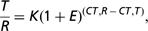
where T is the initial number of target gene copies, R, initial number of standard gene copies, E, efficiency of amplification, CT,T, threshold cycle of target gene, CT,R, threshold cycle of standard gene and K, constant.
In general, the efficiency of PCR reaction was close to one (Use Bulletin, 1997). Although the exact value of K does not be equal to one, the affecting factors will vary only negligibly among single samples so that K is assumed to be equal. So the following equation is given:

The second method was performed according to the standard curve described by Applied Biosystems.
Cholesterol level detection
High performance liquid chromatography (HPLC) method was applied to detect the cholesterol concentration in tissues. Liver (0.15 g), adipose (0.3 g) and muscle (1 g) tissues were respectively put in Pyrex test tube; internal standard liquid (1 ml) and saponified liquid (7.3 ml) were added into it. Air above the liquor was removed by overflowing nitrogen gas and placed the test tube in 80°C water-bath until the solution clarified completely. Then the tubes were placed on ice to chill and directly extracted with petroleum ether. The supernatant liquor was taken out and put in a clean test tube. The appropriate volume liquor was collected by drying and concentrating and then analyzed in capillary chromatography. The recovery of this method was between 92.58% and 104.68%. The satisfied results can be obtained under these chromatographic conditions, and cholesterol level determination can be finished within 12 min for each sample (Fang Wenning, unpublished data).
Statistical analysis
The data when comparing two breeds were determined using t-test. The other data were analyzed by ANOVA. Significant differences were detected by Duncan’s multiple range tests using GLM procedure of SAS software (version 8.02; SAS Institute, Inc., Cary, NC, USA). Results were presented as means of mRNA relative abundance and cholesterol content ± s.e. Statistical significance was set at P < 0.05 and P < 0.01.
Results
Cloning and sequence analysis of porcine HMGR cDNA
The pig HMGR gene contained 20 exons and 19 introns. This gene was assigned to pig chromosome 2q21-22 by RH (radiation hybrid) mapping. The length of its cDNA is 2864 bp (GenBank accession No. DQ432054), including an open reading frame covering 2658 bp. The homology analysis by BLAST software (Basic Local Alignment Search Tool) showed that the identity of pig HMGR and human HMGR was very high (91%; Wang, Reference Wang2006).
Real-time quantitative PCR
The housekeeping gene, GAPDH, was used as the control to show the normalization of the templates in reactions, and standard curves were prepared through using highly concentrated cDNA and gradient dilution, and was used when R was above 0.997.
Cholesterol content analysis
Breed effects
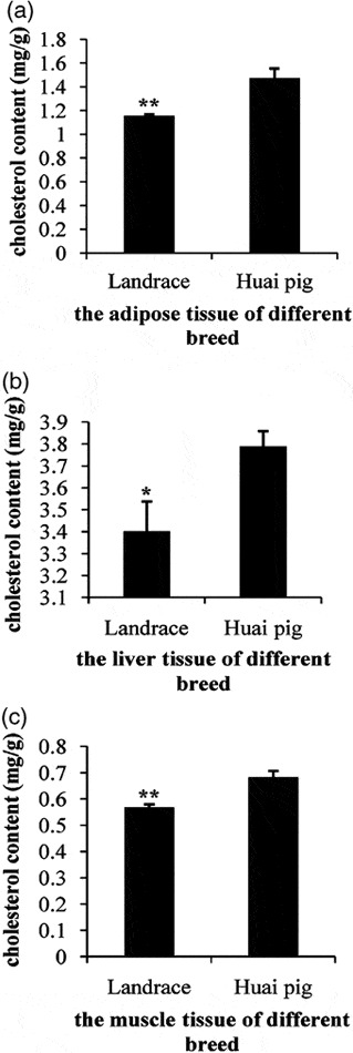
Cholesterol concentrations were higher in Huai pigs as compared to Landrace in all tissues (Figure 1): adipose tissue (P < 0.01), liver (P < 0.05) and muscle (P < 0.01).
The cholesterol concentration difference in three tissues adipose (a), liver (b), muscle (c) of two breeds (Landrace and Huai pig). Each bar represents mean ± s.e. *P < 0.05; **P < 0.01.

Tissues effects
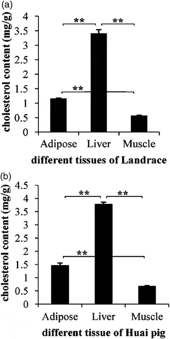
Within both breeds, cholesterol concentrations were higher in liver, intermediate in adipose tissue and lowest in muscle (Figure 2). Landrace’s result is showed in Figure 2a and Huai pig’s result is showed in Figure 2b.
The difference of the cholesterol concentration in tissues of adipose, liver, muscle. (a) Landrace; (b) Huai pig. Each bar represents least-square mean ± s.e. **P < 0.01.

Gene expression analysis
Differences in mRNA abundance of HMGR in three tissues across breed
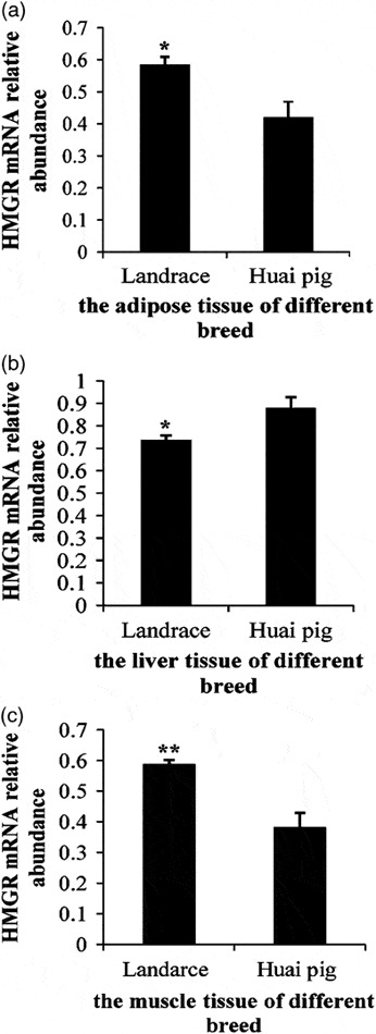
Using real-time PCR, porcine HMGR mRNA levels were quantified in three tissues between two different kinds of breeds. Results showed that there were differences in HMGR expression in adipose tissue (Figure 3a; P < 0.05), liver tissue (Figure 3b; P < 0.05) and muscle tissue (Figure 3c; P < 0.01) between two breeds.
Three tissues: adipose (a), liver (b), muscle (c) effects on relative expression of HMGR mRNA in two breeds (landrace and Huai pig). Data are ratios of HMGR genes relative mRNA levels normalized to GAPDH (housekeeping gene) mRNA levels. Each bar represents mean ± s.e. *P < 0.05; **P < 0.01.

Differences of mRNA abundance of HMGR in three tissues of each breed
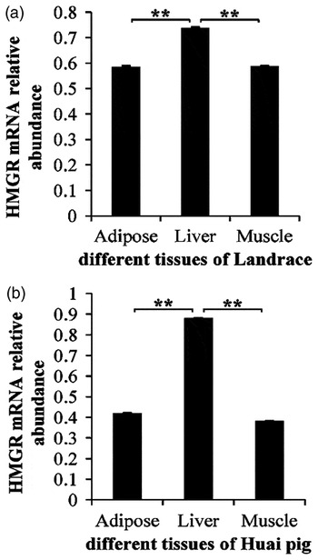
Porcine HMGR mRNA levels were quantified in three tissues of each breed. The results showed that the HMGR mRNA level in liver in Landrace was higher than in adipose (Figure 4a; P < 0.01) and muscle (Figure 4a; P < 0.01), and the difference between the adipose and muscle was not significant (Figure 4a; P > 0.05). The trend in Huai pig was the same as in Landrace (see Figure 4b).
Three tissues: adipose, liver, muscle effects on relative expression of HMGR mRNA in two breeds, (a) landrace and (b) Huai pig. Data are ratios of selected genes relative mRNA levels normalized to GAPDH (housekeeping gene) mRNA levels. Each bar represents least-square mean ± s.e. **P < 0.01.

Pearson Correlation Coefficients between HMGR gene and cholesterol
We compared the correlation between cholesterol concentration and gene expression of HMGR. The result showed that the Pearson Correlation Coefficients between HMGR gene and cholesterol was 0.46 (P < 0.01).
Discussion
Huai pig is considered a highly desirable Chinese local breed because it is a miniature breed and produces high quality pork. However, Huai pig is a lard-type breed and the fat content of Huai pigs is considerably higher than that of Landrace pigs. The present research showed that cholesterol content was higher in liver, adipose and muscle from Huai pig than in the corresponding tissues from Landrace pigs. Harris et al. (Reference Harris, Pond, Mersmann, Smith, Cross and Savell2004) reported genetic selection for high or low plasma cholesterol is not a good selection indicator of cholesterol or fat accretion, yet argued that the source of dietary fat may influence total fat content in muscle tissues. According to Dorado et al. (Reference Dorado, Martin Gomez, Jimenez-Colmenero and Masoud1999), there is a high correlation between cholesterol content and fat content (r = 0.88, P < 0.05). Thus, the difference in cholesterol content between the two breeds may be related to overall fat content.
The HMGR gene has been widely cloned in many species, i.e. the human HMG-CoA reductase gene has been localized to human chromosome 5q12 by in situ hybridization and contains 20 exons and 19 introns (Humphries et al., Reference Humphries, Tata, Henry, Barichard, Holm, Junien and Williamson1985; Luskey and Stevens, Reference Luskey and Stevens1985), chicken (Sato et al., Reference Sato, Ohuchi, Sook, Toyomizu and Akiba2003), rat (Simonet and Ness, Reference Simonet and Ness1988; Sundaresan et al., Reference Sundaresan, Yang-Feng and Francke1989; Duckworth et al., Reference Duckworth, Vlahcevic, Studer, Gurley, Heuman, Beg and Hylemon1991) and some plants (Ha et al., Reference Ha, Kim, Hwang and Lee2003; Jiang et al., Reference Jiang, Kai, Cao, Chen, He and Liu2006). Information regarding this gene in pigs, however, is limiting. In recent years, our group reported firstly, the HMGR gene and the sequence information of HMGR in pig. The research found that nucleotide acid (91%) and amino acid sequence (95%) of pig HMGR were very similar to human HMGR gene (Wang, Reference Wang2006).
In this study, Huai pig HMGR mRNA level was lower than the corresponding tissues of Landrace in muscle and adipose tissues. However, the cholesterol content in adipose and muscle tissues of Huai pig was higher than the corresponding tissues of Landrace. Cholesterol content as a biological trait was affected by many factors. Besides the level of HMGR mRNA expression, the cholesterol content was affected by other levels such as the processing and modifying of transcription and translation levels. In addition, the cholesterol 7 alpha-hydroxylase (CYP7A1) is an essential enzyme of cholesterol excretion. The substantial amounts of cholesterol are removed from the body by this enzyme. Because the role of this enzyme was different in different tissues, it led to different cholesterol content in different tissues. These factors may affect the amount of cholesterol in tissues. At present, there was no research of CYP7A1 in pig, so a further research of this enzyme would be done.
In pigs, HMGR mRNA level was high in most tissues, as measured by RT-PCR and Northern blot analysis, however, the highest levels were found in liver, brain and ileum (Wang, Reference Wang2006). The tissue expression pattern showed that HMGR had strongly expressed differences, which suggested that HMGR might be a constitutively expressing gene. The similar results were reported by Jiang et al. (Reference Jiang, Kai, Cao, Chen, He and Liu2006). In addition, the expression pattern in different tissues suggested that this gene is expressed in a tissue-dependent manner in pigs. Our results also show that HMGR expression in liver is higher than that in the adipose and muscle. This suggested that liver is the major site of the cholesterol synthesis and it is possible to decrease the cholesterol content by controlling HMGR expression.
HMGR is the essential enzyme in cholesterol synthesis pathway. Initially, physicians were skeptical of a causal link between cholesterol and coronary heart disease because most patients with the disease had plasma cholesterol only slightly higher than the general population average (The Lipid Research Clinics Coronary Primary Prevention Trial results (LRC-CPPT), 1984; Baum et al., Reference Baum, Teng, Erdman, Weigel, Klein, Persky, Freels, Surya, Bakhit, Ramos, Shay and Potter1998; Tobert, Reference Tobert2003). Furthermore, Ness (Reference Ness2003) reported that supplementing animal diets with cholesterol can lower hepatic HMGR activity. The efficacy of clinically inhibiting HMGR by various statins and reducing cardiovascular morbidity and mortality has been convincingly demonstrated in primary and secondary prevention trials (Scandinavian Simvastatin Survival Study, 1994; Shepherd et al., Reference Shepherd, Cobbe, Ford, Isles, Lorimer, MacFarlane, McKillop and Packard1995; Rasmusseen et al., Reference Rasmusseen, Hanzen, Nabipour, Olesen, Kristiansen and Ledet2001). Recently, HMGR inhibitors have been applied as therapy for the diseases caused by the high level of cholesterol in human; the clinical effect has won widely approval.
Because the similarities between humans and pigs also exist in renal function, vascular structure and respiratory rates, pigs are used as model organism in many areas of medical research including obesity, cardiovascular disease, diabetes, nephropathy and organ transplantation (Dodds, Reference Dodds1982; Lee et al., Reference Lee, Kim and Thomas1986). The evidence points out that the pig is accepted as an appropriate animal model to access the relationship between plasma cholesterol concentration and coronary artery disease, because of its similarities to human (Pond et al., Reference Pond, Mersmann and Young1986; Stanton and Mersmann, Reference Stanton and Mersmann1986; Van et al., Reference Van, Van, Scheek, Groener, Sassen, Lamers and Verdouw1991). Due to the pressing research for human disease, the small-size pigs have been deemed to ideal subscriber. Thus, the Huai pig may serve as an ideal research model for studying this malady, as it is small and easy to handle as well.
Acknowledgements
The authors greatly appreciate Fujian Shanghang Huai pig breeding farm for collecting samples. This study was supported by National Scientific and Technical Supporting Programs of China (No. 2006BAD01A08 & 2006BAD13B08).