Bovine mastitis is an economic burden for dairy farmers and control measures to prevent mastitis are crucial for dairy farm sustainability. The identification of aetiological agents is necessary to control the disease in herds, reduce the risk of chronic infections and target antimicrobial therapy. Staphylococcus aureus is considered a major mastitis pathogen due to its impact on udder health (Bradley, Reference Bradley2002) and coagulase-negative staphylococci are considered minor mastitis pathogens, but they are the most common agents isolated from milk samples in several large scale surveys worldwide (Tenhagen et al. Reference Tenhagen, Koster, Wallmann and Heuwieser2006). Measures to control mastitis due to S. aureus include the segregation of infected animals, to reduce exposure of healthy animals. That segregation is commonly based on results of conventional bacteriological identification, which is still the most widely used technique in mastitis diagnostic laboratories. This methodology presents some pitfalls (Duarte et al. Reference Duarte, Freitas and Bexiga2015b), including a relatively long time to obtain results, a high proportion of negative results that may correspond to false negatives, and an improbable accuracy in identification of certain species. Phenotypic identification is, therefore, frequently supplemented with genotypic methods, such as Polymerase Chain Reaction (PCR) (Taponen et al. Reference Taponen, Salmikivi, Simojoki, Koskinen and Pyörälä2009) for a more accurate identification of bacteria associated with intramammary infections. The high sensitivity of PCR may, however, present a problem in itself, as this method leads to the amplification of genetic sequences from microorganisms present in small amounts, some with debatable clinical relevance (Hiitiö et al. Reference Hiitiö, Riva, Autio, Pohjanvirta, Holopainen, Pyörälä and Pelkonen2015).
Development prospects for new bovine mastitis diagnosis methodologies point to new biomarkers and technological advances for highly sensitive and specific, fast and efficient devices that can offer a ‘cow-side’ use (Duarte et al. Reference Duarte, Freitas and Bexiga2015b). This would allow for timely segregation of animals, or for decisions to be made on duration of treatment, since cure rates for mastitis due to S. aureus have been shown to increase with longer treatments (Truchetti et al. Reference Truchetti, Bouchard, Descôteaux, Scholl and Roy2014). Biosensors are fast becoming the next generation of tools in analysing areas such as environmental research, medicine, biodefense, agriculture, and food control (Lazcka et al. Reference Lazcka, Del Campo and Muñoz2007). Biosensors use biological receptor molecules (e.g., antibody, enzyme, and nucleic acid) combined with a transducer to produce a signal that shows a specific biological event (e.g., an antibody–antigen interaction). Our previous work (Fernandes et al. Reference Fernandes, Duarte, Cardoso, Bexiga, Cardoso and Freitas2014; Duarte et al. Reference Duarte, Costa, Carneiro, Soares, Jitariu, Cardoso, Piedade, Bexiga and Freitas2016) describes magnetic detection of bovine mastitis pathogens based on immunological recognition of its immunogenic proteins by specific antibodies.
The aim of this study was to develop and validate a sensitive method for magnetic detection of S. aureus and of the genus Staphylococcus in raw milk samples. For both magnetic detection and conventional bacterial culture methods, sensitivity, specificity and positive predictive value (PPV) were calculated in comparison with the PCR reference method as gold standard.
Material and methods
Milk samples
Raw milk was collected aseptically from two healthy cows for biosensor calibration measurements. Conventional bacterial culture was performed according to NMC (1999) protocols, to confirm no bacterial growth. Briefly, 10 µl of milk were plated on Columbia agar supplemented with 5% sheep blood (43021, bioMérieux) and on MacConkey agar plate (CM0007, Oxoid) and both were incubated at 37 °C for 48 h. The absence of growth on both plates was considered to be equivalent to the presence of no viable bacteria in the milk.
Mastitic milk samples were needed to validate biosensor detection. For this, milk samples with known bacteriological results were used. The milk samples tested were collected aseptically from cows (n = 47) of 12 Portuguese dairy farms. Animal selection was based on a somatic cell count higher than 1 000 000 cells/ml and infected quarter screening was based on California Mastitis Test results with score 3. A total of 49 quarter samples were submitted to conventional bacteriology as previously described. These 49 quarter samples yielded Staphylococcus aureus (n = 9), Streptococcus agalactiae (n = 3), Streptococcus uberis (n = 1), Streptococcus spp. (n = 1), CNS (n = 11), Enterococcus spp. (n = 4), Escherichia coli (n = 7), yeasts (n = 6) and Prototheca spp. (n = 7). These samples were submitted to biosensor analysis with the following distribution: 18 samples with the pAb anti-S. aureus, 18 samples with the mAb anti-Staphylococcus spp. and 13 samples with both antibodies, amounting to 31 samples per antibody being screened by the biosensor.
PCR analysis
This genotypic method was used to validate the new methodology based on immunological magnetic detection. Mastitic milk samples with a preservative (6·65 µl of azidiol per 2 ml of milk) were sent to an external laboratory (VACUNEK, SL) and were submitted to real time PCR analysis with the PathoProof Mastitis Complete-16® assay kit (Thermo Scientific), a development of the PCR assay initially described in Koskinen et al. (Reference Koskinen, Holopainen, Pyörälä, Bredbacka, Pitkälä, Barkema, Bexiga, Roberson, Sølverød, Piccinini, Kelton, Lehmusto, Niskala and Salmikivi2009), allowing for the simultaneous detection of 15 mastitis pathogens.
Bacterial cells
Suspensions
In order to distinguish biosensor signal outputs from different bacterial concentrations in raw milk samples, calibration curves were developed for the pairs: Staphylococcus aureus/pAb anti-S. aureus ScpA; Staphylococcus aureus/mAb anti-Staphylococcus spp. and Staphylococcus epidermidis/mAb anti-Staphylococcus spp. The target bacteria used were ATCC 29213 for Staphylococcus aureus and a clinical bovine mastitis isolate of Staphylococcus epidermidis which identification had been previously confirmed genotipically (Bexiga et al. Reference Bexiga, Rato, Lemsaddek, Semedo-Lemsaddek, Carneiro, Pereira, Mellor, Ellis and Vilela2014).
Bacterial cells were grown separately on Columbia agar supplemented with 5% sheep blood (43021, bioMérieux) and incubated at 37 °C, overnight. A single colony of each isolate was selected and re-suspended on 4 ml of tripticase soy broth overnight at 37 °C. Subsequently, the bacterial cells were collected through centrifugation (15 min, 17 °C, 2700 rpm) and re-suspended in PBS 1X (pH 7·2) to allow for optical density measurement (at 600 nm) (BECKMAN DU-68 Spectrophotometer) and for colony-forming unit estimation. A bacterial suspension with a known concentration of 104 cfu/μl was the starting point to obtain seven different bacterial concentrations for each species, in sterile raw milk samples: 0·1; 0·3; 0·5; 1; 2; 10 and 20 cfu/μl.
Magnetic labelling
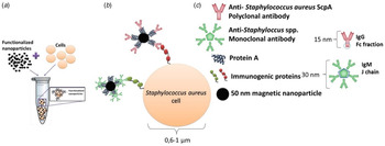
The immunological recognition of Staphylococci was achieved by biological functionalisation with antibodies of iron oxide nanoparticles, which could be detected by the biosensor (Duarte et al. Reference Duarte, Fernandes, Cardoso, Bexiga, Cardoso and Freitas2015a). The antibodies were expected to attach to protein A of those nanoparticles (79-20-501, Micromod Partikeltechnologie GmbH) by the Fc fraction in immunoglobulins G and by the joining chain in immunoglobulins M (Fig. 1). The antibodies used separately in this work were a rabbit polyclonal IgG to ScpA protein (ab92983, Abcam) and a mouse monoclonal IgM anti-S. aureus ATCC 29740 (MCA 5793, AbDSerotec). The polyclonal antibody used for S. aureus identification recognises the extracellular protease cysteine proteinase staphopain A (ScpA) which is considered a putative virulence factor (Ohbayashi et al. Reference Ohbayashi, Irie, Murakami, Nowak, Potempa, Nishimura, Shinohara and Imamura2011). Regarding S. epidermidis, the monoclonal antibody used recognises its cell wall peptidoglycan as well as S. aureus and protein A – negative S. aureus, herein designated as ‘anti-Staphylococcus spp.’. Antibodies (Walstra et al. Reference Walstra, Wouters and Geurts2006) and bacterial cell dimensions are shown in Fig. 1.
Schematics of immuno-magnetic detection of cells. (a) Incubation of functionalised nanoparticles with bacterial cells; (b) biological affinities between different functionalised nanoparticles with bacterial cell wall immunogenic proteins; (c) predictable protein A binding site to each antibody.

As explained in our previous work (Duarte et al. Reference Duarte, Fernandes, Cardoso, Bexiga, Cardoso and Freitas2015a), nanoparticles (NP's) (7·27 µl from an original vial with 5·5 × 1013 nanoparticles per ml) were incubated with 1·08 µl of pAb anti- S. aureus (0·5 mg/ml) (or with 2·65 µl of mAb anti-Staphylococcus spp. (1 mg/ml)) in 492·2 µl of PBS (or in 490·08 µl for mAb), during 1 h at room temperature (RT), under agitation. Final functionalised nanoparticles were magnetically isolated by magnetic separation (MS) column (130-042-201, Miltenyi) and eluted with buffer (PBS + 0·5% BSA + 2 mM EDTA) after removal of the MS column from the magnet. A volume of 2 µl of this final suspension with 8 × 10⁶ functionalised nanoparticles was diluted in 98 µl of PBST and added to each milk sample with bacteria.
Biosensor detection
This method is based on the detection of the fringe field created by magnetic particles attached to the bacterial cells. It is a dynamic detection where milk that is potentially contaminated with bacterial cells flows within a microchannel. By selecting the suitable antibodies, it is possible to perform immunological recognition of bacteria in milk samples. As described in our previous work (Fernandes et al. Reference Fernandes, Duarte, Cardoso, Bexiga, Cardoso and Freitas2014; Duarte et al. Reference Duarte, Fernandes, Cardoso, Bexiga, Cardoso and Freitas2015a), an integrated cytometer platform was used, consisting of magnetoresistive sensors and readout/acquisition electronics and a microfluidic channel where the milk was injected.
Calibration trials
A blank sample (only sterile raw milk) and a control sample (sterile raw milk with functionalised nanoparticles) were always measured before samples with known bacterial concentrations, to give the background signal of the system. All calibration points resulted from three different days’ measurements leading to three independent trials for each bacterial concentration sample.
Each milk sample with bacteria for biosensor analysis had a 500 µl volume consisting of 2 µl of functionalised nanoparticles suspension, 98 µl of PBST, and 400 µl of a bacterial suspension volume corresponding to one of seven pre-defined bacterial concentrations and the remaining volume of sterile raw milk. The incubation of these samples was performed at RT for 3 h, under agitation.
All raw milk samples with bacteria and PBST were submitted to a pre-treatment of 15 min at 60 °C in a dry bath incubator (model QBD2, Grant) and 15 min of continuous centrifugation in a vortex mixer (Labnet). Only then, the functionalised nanoparticles suspension volume was added for the final incubation step.
The calibration range between 0·1 and 20 cfu/μl was established taking into account the detection limit for conventional bacteriological culture of 500 cfu/ml (0·5 cfu/μl) (NMC, 1999).
Mastitic milk sample evaluation
Mastitic milk samples for evaluation were obtained by mixing 400 µl of mastitic milk and 98 µl of PBST.
Mastitic milk samples (n = 31) were distributed so that each trial with a different antibody (anti-S. aureus ScpA or anti-Staphylococcus spp.) could be performed independently, amounting to 62 trials for magnetic method validation.
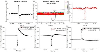
Samples with functionalised NP's on PBS or sterile milk (negative controls) evidenced magnetic signal lower than 50 µV (Fig. 2a). Mastitic milk samples without the targeted bacteria (proved by PCR) and tested with NP's functionalised with chosen antibodies, also evidenced magnetic signal lower than 50 µV (Fig. 2b). Samples used for calibration assays spiked with bacterial cells on sterile milk showed magnetic signal higher than 50 µV (Fig. 2c–e).
Sensor output for (a) negative control with the higher amplitude of 23 µV, (b) mastitic milk (9077 T sample code without S. aureus accordingly with PCR) with NP's functionalised with pAb anti-S. aureus (Ab 92983) which presents an amplitude peak of 10·7 µV. The higher amplitude peaks found for each pair of bacteria-antibody were, (c) 1703·4 µV in raw milk with pAb anti-S. aureus and 10 cfu/μl of S. aureus, (d) 964·4 µV in raw milk with mAb anti-Staphylococcus spp. (MCA 5793) and 10 cfu/μl of S. aureus and (e) 1030·5 µV in raw milk with mAb anti-Staphylococcus spp. and 10 cfu/μl of S. epidermidis.

Biosensor validation was based on mastitic milk sample classification as having or not having the targeted bacteria present. Consequently, the ‘Positive’ samples were those with at least one magnetic peak above 50 µV, therefore higher than the magnetic signal found in negative control samples and in mastitic milk samples without targeted bacteria. Next, this ‘Positive’ sample magnetic peak should evidence a bipolar or unipolar shape similar to the ones found in samples used for calibration trials as shown in Fig. 2c–e.
Biosensor analysis was a dynamic detection where a heterogeneous milk sample flowed inside the microchannel. Magnetically labelled bacterial cells were mixed randomly in milk leading to the impossibility of predicting its position above the sensor over time. Consequently, the magnetic peaks’ shape and time resulting from biosensor analysis were expected to be different between samples (Fig. 2).
Data analysis
PCR identification was considered the gold standard against which both magnetic identification and conventional bacterial culture were compared. Isolates were considered to be correctly identified by magnetic detection if the same species was found with the PCR, or if the magnetic detection did not identify as the agents it was targeting, one of the other bacteria identified as such by the PCR. For example, a correct identification referred to a S. aureus being identified magnetically in a sample that PCR had identified as S. aureus, but also when not identifying as S. aureus a sample that through PCR was identified as Streptococcus agalactiae for example. Misidentification was considered when the magnetic detection identified a different species than the PCR. For example, a sample that was identified as Streptococcus agalactiae by PCR and that was identified as S. aureus by magnetic detection or a sample that was identified as S. aureus by PCR and not identified as such by magnetic detection. The same principles were used for conventional bacterial culture. For both magnetic detection and conventional bacteriological culture, sensitivity, specificity and positive predictive value were calculated in comparison with PCR species identification. Sensitivity was calculated as the proportion of true positive isolates that were correctly identified with magnetic detection. Specificity was calculated as the proportion of true negatives that were correctly identified with magnetic detection. Finally, PPV was calculated as the proportion of isolates identified as a specific species based on magnetic detection that truly represented that particular species when compared with PCR results. The same principles were used to compare conventional bacterial culture with PCR.
Results
Evaluation of biosensor's quantification
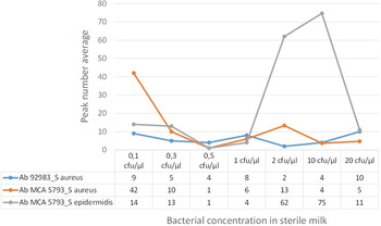
The calibration trials’ outcome is shown in Fig. 3. The number of peaks per signal amplitude were calculated evidencing no linear correlation with increasing bacterial concentration. The milk samples with functionalised nanoparticles with the anti-Staphylococcus spp. antibody, evidenced the highest peak number with S. epidermidis when compared with the other two bacteria-antibody pairs (Fig. 3). Overall, the biosensor could detect S. aureus and S. epidermidis in milk samples from 100 cfu/ml.
Calibration trial results for milk samples with seven bacterial concentrations (S. aureus or S. epidermidis) and functionalised NP's with pAb anti-S. aureus (Ab 92983) or mAb anti-Staphylococcus spp. (Ab MCA 5793). Peak number average for each bacteria-antibody pair are counted.

Validation of magnetic detection
Forty nine mastitic milk samples with known bacteriology results, obtained through conventional bacteriological culture were analysed by PCR, leading to 123 agent identifications. As a result of the high sensitivity of the PCR methodology, an average of 2·5 different pathogens were detected per mastitic milk sample, not allowing for the true causative agent of mastitis to be determined. Therefore, it was decided to use the conventional bacteriology results as the basis for sensitivity, specificity and PPV calculation confirmed by the PCR (Table 1).
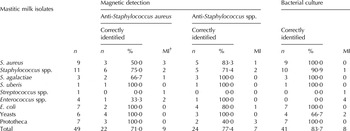
Identification of isolates in mastitic milk samples with both magnetic detection (pAb anti- Staphylococcus aureus; mAb anti-Staphylococcus spp.) and with conventional bacterial culture, compared to PCR analysis as the reference method

Correctly identified = True positives + True negatives.
† MI (misidentified) = False Negatives + False positives.
Magnetic detection with the anti-S. aureus antibody tested 31 mastitic milk samples, of which 6 were identified as S. aureus by conventional bacteriological culture (Table 1). The magnetic detection with the anti-S. aureus antibody identified 4 S. aureus isolates correctly out of 7 (57·1%) milk samples with this species according to PCR results. Eighteen mastitic milk samples did not lead to any identification by this polyclonal antibody as they did not present S. aureus according to the PCR, thus being true negatives (Table 2). Misidentification was observed for 9 of 31 (29%) isolates in all mastitic milk samples tested with the anti-S. aureus antibody (Table 1). Only 3 misidentified S. aureus isolates were found in milk samples with this species analysed by PCR, evidencing a failure of recognition by this monoclonal antibody (Table 1). Adding to that, the remaining 6 mastitic milk samples were misidentified as having S. aureus (Table 1), while really presenting S. uberis, yeasts, S. agalactiae and other staphylococcal species according to the PCR. Overall 57·1% sensitivity, 75% specificity and 40% PPV were found for magnetic detection with the anti-S. aureus ScpA antibody (Table 2).
Sensitivity, specificity and positive predictive value of conventional bacterial culture and the magnetic detection with the polyclonal antibody anti-Staphylococcus aureus and with the monoclonal antibody anti-Staphylococcus spp., using PCR analysis as the reference method

† PPV, Positive predictive value.
Using the monoclonal anti-Staphylococcus spp. antibody in magnetic detection, the 31 mastitic samples tested included 6 S. aureus and 7 Staphylococcus spp. identified by conventional bacteriological culture (Table 1). However, PCR analysis identified 29 as Staphylococcus spp. of which 8 samples also evidenced S. aureus, showing incomplete identifications by microbiological analysis. The magnetic detection with the anti-Staphylococcus spp. antibody identified correctly 23 staphylococci present in 29 (79·3%) milk samples with mostly staphylococci other than S. aureus, or also with S. aureus according to PCR analysis. Only one mastitic milk sample was not detected by this monoclonal antibody (true negative) because it did not present any Staphylococcus spp. according to the PCR (Table 2). Misidentification was observed for 7 isolates out of the 31 (22·6%) mastitic samples tested. Six of them were found in milk samples with staphylococcal species analysed by PCR, evidencing a failure of recognition by this anti-Staphylococcus spp. antibody (Table 1). Moreover, only one false positive was found in a mastitic sample without any staphylococci evidenced by PCR. Overall a sensitivity of 79·3%, a specificity of 50% and a PPV of 95·8% were found for magnetic detection with the anti-Staphylococcus spp. antibody (Table 2).
Regarding bacteriological testing for all samples considered, 100% of S. aureus, S. agalactiae, S. uberis, E. coli and Prototheca were correctly identified when comparing with PCR (Table 1). Incomplete microbiological identifications of 85·7% and a misidentification of 14·3% were observed. Microbiological tests evidenced a PPV value of 85·4% and a sensitivity of 97·6% to identify mastitis pathogens in milk samples, showing that conventional microbiology identified correctly true positives (Table 2).
Discussion
Sensitivities of 57·1 and 79·3% and specificity values of 75 and 50% were obtained for magnetic identification of staphylococci species in mastitic milk samples with an anti-S. aureus ScpA antibody and an anti-Staphylococcus spp. antibody, respectively. The higher PPV value (95·8%) evidenced for magnetic detection with the anti-Staphylococcus spp. antibody may reinforce its bonding avidity for each immunogenic cell wall protein of S. aureus and of the genus Staphylococcus comparing to the polyclonal antibody. These specificities were confirmed by our Western Blotting assays (data not shown) which evidenced three to six stained immunogenic proteins in S. aureus cell wall proteins’ pattern and one immunogenic protein in both S. aureus and genus Staphylococcus, recognised by the selected polyclonal and monoclonal antibodies, respectively.
The knowledge that S. aureus is an important cause of udder infections in dairy herds sustains the interest in identification for treatment and prevention of S. aureus mastitis (Fabres-Klein et al. Reference Fabres-Klein, Aguilar, Silva, Silva and Ribon2014). Comparing the sensitivity (57·1, 79·3%) and specificity (75, 50%) values of this magnetic detection with another study based on immuno-agglutination, which compared 6 commercially available slide agglutination tests for S. aureus identification in milk samples (Zschöck et al. Reference Zschöck, Nesseler and Sudarwanto2005), the highest sensitivity (86·7%) and specificity (90·1%) values were obtained for a test consisting of latex particles coated with human fibrinogen and immunoglobulin G. Strain typing methods that are DNA sequence-based have also been used to improve S. aureus detection. Bittar et al. (Reference Bittar, Ouchenane, Smati, Raoult and Rolain2009) attempted to differentiate between positive and negative S. aureus strains for Panton–Valentine leucocidin, used a MALDI-TOF MS (matrix-assisted laser desorption ionisation time-of-flight mass spectrometry) analysis and obtained higher sensitivity (100%) and specificity (90·6%), compared with our magnetic detection method. On the other hand, considering that the minimum bacterial presence detected by the present immunological recognition was 100 cfu/ml, independently of which antibody and targeted bacteria were considered, when compared to a sandwich ELISA test recently patented (Libing et al. Reference Libing, Chuanlai, Qianqian, Xiaofang, Shanshan and Xun2012) to detect S. aureus in artificially contaminated milk, a lower detection limit of 105 cfu/ml was found. However, a competitive immunoassay performed by an amperometric magnetoimmunosensor (de Ávila et al. Reference de Ávila, Pedrero, Campuzano, Escamilla-Gómez and Pingarrón2012) for the specific detection and quantification of staphylococcal protein A and S. aureus cells, evidenced a better detection limit of 1 cfu/ml, also in artificially contaminated milk samples.
The conventional bacterial culture misidentification ratio of 14·3% observed in our study was due to 7 erroneous identifications compared with PCR analysis. The use of PCR for the identification of mastitis pathogens may have the advantage (Taponen et al. Reference Taponen, Salmikivi, Simojoki, Koskinen and Pyörälä2009) of leading to decreased false negative results, but it may also be clinically challenging. PCR's higher sensitivity leads to the identification of all milk sample pathogens and contaminants alike (Hiitiö et al. Reference Hiitiö, Riva, Autio, Pohjanvirta, Holopainen, Pyörälä and Pelkonen2015), being difficult to assign mastitis causality to a particular microorganism.
Regarding the validation of the magnetic detection method, some false positive results (6 for pAb anti-S. aureus and 1 for mAb anti-Staphylococcus spp.) could be explained by NP's agglomeration by the mastitic milk matrix heterogeneity, sporadic low cleaning efficiency of the channel's inlet chamber and also the electrical conductivity of mastitic milk samples. Bovine mastitis leads to changes in ion concentration of milk, leading to modifications in electrical conductivity of milk (Hovinen et al. Reference Hovinen, Aisla and Pyörälä2006). The conductance in milk causes sensor's resistivity variation, translated by higher background noise. Despite a detergent (Tween 20) being added to milk samples to reduce fat globule dimensions, to distribute the bacterial cells in the milk, to improve nanoparticle mobilisation and to allow a more homogeneous milk matrix, the optimisation trials (not described in this manuscript) showed the need for a compromise between Tween 20 quantity and magnetic peak discrimination to avoid bubbling and signal misunderstanding. Different volumes of PBST in 500 µl of milk samples were used. The higher volumes (>100 µl) created bubbles inside the microchannel which hampered milk flow, caused nanoparticle agglomeration and did not help magnetic peaks discrimination between control samples (milk with only NP's) and samples with bacteria. On the other hand, false negatives (3 for pAb anti-S. aureus and 6 for mAb anti-Staphylococcus spp.) may have occurred because of three circumstances. Firstly, the binding yield variations between antibodies and NP's and/or failure in bacterial cells magnetic labelling could narrow bacterial cell identification. This fact should be recognised as possible because IgM and nanoparticle's dimensions are close, so it would be more difficult to have the same number of attached IgM when comparing with NP's functionalisation with smaller IgG. Secondly, according to Henriksen's study (Reference Henriksen, Wang and Hansen2015), it is possible that the rotating nature of the magnetic dipole field of NP's magnetised by an external magnetic field, may induce signal cancelation. Therefore, the fields from two differently placed NP's can partially cancel each other. Finally, microchannel height (50 µm) could be reduced to improve detection, but mastitic milk trials showed that milk clots hampered sample flow and height decrease led to microchannel obstruction, pointing to the need for a compromise between sensor's detection and sample fluidity.
With regards to calibration trials, bacterial cells group together randomly depending on growth conditions (Quinn et al. Reference Quinn, Carter, Markey and Carter1994). Each bacteria may express a different number of cell wall proteins, including the immunogenic ones (van der Woude & Baümler, Reference van der Woude and Baümler2004). Together, these facts limit the knowledge of how many immunogenic proteins there are per cell and consequently, how many proteins will be recognised by each specific antibody used. On the other hand, the chemical and colloidal changes of milk components in a state of inflammatory response (Walstra et al. Reference Walstra, Wouters and Geurts2006) as occurs with mastitis, prevent and reduce bacterial magnetic labelling efficacy (Duarte et al. Reference Duarte, Costa, Carneiro, Soares, Jitariu, Cardoso, Piedade, Bexiga and Freitas2016). Consequently, it was not possible to predict peaks profile (number, shape) for each bacterial concentration.
Although the sensitivity of the immuno-magnetic detection method was important to determine the potential future use of such technology, many additional factors must be considered, including speed and ease of use, flexibility, portability and costs (Mortari & Lorenzelli, Reference Mortari and Lorenzelli2014). This methodology showed it was possible for a mastitic milk sample to be processed until a final result was obtained in 5 h, but was not suitable for processing a large number of samples (maximum of 10–12 per day). It also showed technical simplicity when established, and ease of scoring and interpreting the results. Despite the lower sensitivities obtained, both antibodies used were capable of detecting bacterial cells in real milk samples. However, other antibodies could be used for further identification of different bovine mastitis pathogens, reinforcing this method's flexibility.
This biosensor can be submitted to further improvements which may include milk pre-treatment step incorporated into the microfluidic platform and also further studies on electronics to allow multiplex analysis of several samples at the same time. Currently, this biosensor requires an external computer for system operation and displaying test results, but a system fully integrated into a single device could also be made.
Work partially supported by the project FCT-EXCL/CTM-NAN/0441/2012 and by the CIISA-46 project UID/CVT/00276/2013. INESC-MN acknowledges FCT funding through the IN-Associated laboratory (|Pest-OE/CTM/LA0024/ 2011).