
I. INTRODUCTION
Despite many efforts in the field of clinical hygiene research, healthcare-associated infections are still a serious problem and frequently lead to revision surgeries. According to recent studies, joint replacements require still almost 7% revision, and infections due to surfaces harboring bacteria, such as gram positive Staphylococci, Reference Pauksch, Rohnke, Schnettler and Lips1 are amongst the most prominent post-operative incidents. Reference Pauksch, Rohnke, Schnettler and Lips1,Reference Grandfield2 As the aging population and hence the need for implants grows in Western countries, this serious problem will presumably even increase in the future. Novel and innovative scientific approaches thus, amongst others, aim toward an appropriate biofunctionalization of implant surfaces. Especially for articulated joint surfaces, the most important demands are biocompatibility, time-dependent antibacterial activity, and profound corrosion as well as wear resistance. In terms of antibacterial performance, modern implant surfaces should ideally provide a tailored antibacterial behavior with a large initial antibacterial activity during the first 24 h after implantation. This way, implant-associated infections could be avoided and the implant surface would be protected against bacterial adhesion and subsequent biofilm formation. Reference Monteiro, Gorup, Takamiya, Ruvollo-Filho, de Camargo and Barbosa3,Reference McCoy, Craig, McGlinchey, Carson, Jones and Gorman4 The initial fast action should be followed by a continuously decreasing antibacterial activity within the subsequent few days.
To create such intelligent implant surfaces with optimized hardness, wear and corrosion resistance and at the same time exhibiting the desired antibacterial properties, we used an energetic ion treatment of a functionalized polymeric surface layer. This polymer layer is containing metallic nanoparticles (NP) and then is transformed into a very hard and wear resistant DLC layer, still containing the metal NPs.
The transformation method of our choice is the plasma immersion ion implantation (PIII), a surface modification process that improves the wear resistance by forming a very hard layer on the material surface. Reference Mohammed, Khan and Siddiquee5 During the PIII process, the polymeric surface is surrounded by noncondensing plasma, whereupon a pulsed bias (–20 kV) is applied to an electrode coupled to the polymer surface to be modified. Due to their acceleration in the plasma, the impinging ions are implanted below the surface leading to hydrogen effusion. Subsequent rebonding of the displaced atoms then results in a well-defined DLC layer, having the desired low friction coefficient, high density and perfect hardness. DLC has been proven in the past to represent an outstanding coating material and has been in the focus of research for medical materials for several years. Reference Mohammed, Khan and Siddiquee5–Reference Schwarz and Stritzker8 By our transformation process of functional polymer layers containing metal NP, we combine these outstanding properties with additional antibacterial ones.
In terms of antibacterial applications, Ag, Cu, and ZnO nanoparticles (NPs), of order 100 nm in size, are receiving more and more attention. Several mechanisms have been reported for the antibacterial activity of metal NPs. Their antibacterial activity is related to the generation and release of ions from the particle surface into the biological system, where smaller particles result in faster ion release than larger ones. Reference Elkhawass, Mohallal and Soliman9–Reference Ma, Williams and Diamond11 The antibacterial effectiveness of NPs is assumed to result from a combination of their small size, fast dissolution in a given volume, and high surface-to-volume ratio providing close interactions with microbial membranes. Reference Karlsson, Cronholm, Hedberg, Tornberg, De Battice, Svedhem and Wallinder12 Especially silver NPs have been proven to ensure high antimicrobial efficacy while showing low cytotoxicity, Reference Pauksch, Rohnke, Schnettler and Lips1 but antimicrobial activity has also been demonstrated for nano-sized Cu particles. Reference Karlsson, Cronholm, Hedberg, Tornberg, De Battice, Svedhem and Wallinder12 Because of its selective toxicity mainly targeting prokaryotic systems, also ZnO NPs can be used to reduce microbe attachment and viability on medical surfaces. Reference Salem, Leitner, Zingl, Schratter, Prassl, Goessler, Reidl and Schild13,Reference Ma, Williams and Diamond14 According to Ref. Reference Sowa-Söhle, Schwenke, Wagener, Weiss, Wiegel, Sajti, Haverich, Barcikowski and Loos15, AgNO3, CuCl2 and ZnCl2 metal salts display a concentration-dependent cytotoxicity with inhibitory concentration (IC50) values of 3.23, 5.08, and 4.58 mg/L, respectively. Hence, the cytotoxic effect of the NPs mentioned above seems to decrease following the sequence Ag > Cu > ZnO. Various biomedical applications using antibacterial NPs have emerged on the market over the last years: Prominent examples include venous catheters coated with Ag NPs or Cu and ZnO NPs within dental materials. Reference Chaloupka, Malam and Seifalian16–Reference Aydin Sevinç and Hanley18 Regarding medical DLC applications, DLC coated stents were also investigated. Reference Gutensohn, Beythien, Bau, Fenner, Grewe, Koester, Padmanaban and Kuehnl6 Moreover, DLC was found to be a versatile coating material for endoprosthesis and dental implants and other applications. Reference Schwarz, Hauser-Gerspach, Waltimo and Stritzker19 While there are publications on the copper ion release kinetics of plasma-polymer coatings, Reference Zhang, Zhang, Ji, Yan, Huang and Chu20,Reference Chu21 there are no publications on quantitative metal ion release from DLC. Some reports, however, on doping DLC with Ag, Cu, and V to create antibacterial properties, Reference Schwarz, Hauser-Gerspach, Waltimo and Stritzker19,Reference Francz, Schröder and Hauert22,Reference Hauert23 exist in the literature. These publications report in detail about the material properties and the biological effect in vitro but not on quantitative release kinetics.
However, there is still a lack of promising implant materials being wear resistant and antimicrobial due to tailorable Ag, Cu, or Zn release at the same time. Here, we demonstrate a novel approach to synthesize antibacterial implant materials by the combination of a dip-coating polymer deposition process and subsequent DLC transformation employing our PIII technique. We thus obtain materials fulfilling the above mentioned essential requirements for articulated joint implants like mechanical stability, biocompatibility, and time-dependent antibacterial metal ion release. These modified DLC surfaces are supposed to have low abrasive properties. In this study, we report for the first time on the ion release kinetics of Ag, Cu, and Zn from DLC. Knowledge about these kinetics is crucial for tailoring desired ion release from medical implants.
II. MATERIALS AND METHODS
A. Materials
Cylindrical titanium alloy (Ti6Al4V) samples (Ø: 10 mm, thickness: 2 mm), provided by Aesculap AG Tuttlingen, were used as substrate material. The sample surfaces exhibit a roughness of R q = 5 µm, which is due to a corundum blasting step. For the coating, we prepared a colloidal poly(vinylpyrrolidone) (PVP) solution, containing the Ag, Cu, or ZnO NPs. Ag NPs were directly formed in the polymer solution while the Cu or ZnO NPs were synthesized in a separate step beforehand.
To prepare the Ag containing polymer dip-coating solution, stock solutions of PVP (MW 55,000, Sigma–Aldrich, 66.7 g/L) in ethanol, silver nitrate (AgNO3, 17.0 g/L) in ethanol and benzoin (10.6 g/L) in acetone were prepared. Here benzoin acts as photoinitiator. These stock solutions were mixed in the volume ratio 1:3:3 (‘Ag 1’), 1:0.6:0.6 (‘Ag 1/5’), and 1:0.3:0.3 (‘Ag 1/10’) starting with 75 mL of PVP solution. After the complete dissolution of all reactants, the solution was UV-irradiated with a mercury lamp (36 W) from a distance of 25 cm for 12 h leading to a light-induced reduction of the AgNO3 and subsequent formation of Ag NPs. These NP dispersions were used for the dip-coating process without further treatment for the samples Ag 1/5 and Ag 1/10, whereas the total volume of the highest Ag concentration was reduced to 150 mL by evaporation.
The ZnO NPs were prepared separately from the PVP solution as described elsewhere Reference Demir, Munoz-Espi, Lieberwirth and Wegner24,Reference Herrmann, García-García and Reller25 and stored in dry ethanol (Emsure quality, Merck). For the according coating, we prepared a dispersion containing 500 mg of ZnO NPs and 5.0 g of PVP in 145 mL dry ethanol.
Similarly, the Cu NPs were synthesized employing a modification of a standard protocol. Reference Chowdury, Shaik and Chakraborty26 Here, a copper salt (CuCl2 ≥98%, Merck, 0.4 g), PVP (6.48 g) and poly(ethylene glycol) (PEG, MW 8000, Sigma Aldrich, 8.52 g), were dissolved under a slow N2 stream in 120 mL deionized water and heated to 70 °C (oil bath temperature). Aqueous NH3 solution (25%, 3.0 mL) was added and the mixture was stirred for 45 min. Hydrazine sulfate (≥99%, Sigma–Aldrich, 2.22 g) was added and stirred for another 100 min at 70 °C, followed by 16 h stirring at room temperature. The NPs were finally separated by centrifugation (6700 g, 15 min), re-dispersed in 70 mL of deionized water, centrifuged again and re-dispersed in 70 mL of ethanol (last step repeated). The particles were stored in ethanol (16 mL). For the Cu NP containing samples, dispersions containing 500 mg of NPs and 5.0 g of PVP in 145 mL of ethanol were used.
B. Sample preparation and characterization
To create a well-defined polymeric film on top of our implant Ti6Al4V samples, we used a dip coating technique. The samples were mounted on a steel plate using adhesive carbon tape and then dipped into the colloidal polymer solution containing either the Ag, Cu or ZnO NPs. The film thickness was adjusted by a variation of the dipping velocity (here 0.75 mm/s) and the viscosity of the solution. After dipping, the complete sample was covered with the polymer film except for the backside which stuck to the holder.
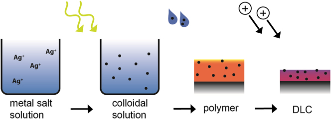
To obtain a polymer-to-DLC surface transformation, the coated samples were dried in air and then treated by a PIII process. The samples were immersed into a microwave plasma (f = 2.45 GHz) consisting of Ne (47.7 %), Ar (32.7 %), and CH4 (19.6 %) for t = 73 min at P = 3 × 10⁻³ mbar, while a pulsed bias U = −20 kV was applied to the sample holder at a frequency of 200 Hz and a pulse length of 5 µs, leading to a DLC layer of 40–50 nm thickness. To further elucidate the sample preparation procedure, we schematically presented it in Fig. 1. To characterize the final functional DLC layers, Raman spectroscopy (sp3 content, see Fig. 2) as well as nano-hardness measurements were performed. We used an ASMEC UNAT nanoindenter (Asmec GmbH, Rossendorf, Germany) in QCSM mode. Here, an additional force vibration is modulated on top of the force ramp which allows to measure depth-dependent hardness. We applied a maximum force of 0.6 mN using a diamond Berkovich tip.
Sample preparation procedure. The figure schematically shows the dip-coating process used to cover the samples' surface by a polymer film for Ag containing samples, reproduced from Ref. Reference Schwarz27. Accelerated ions were implanted into the surface creating a modified DLC layer with a thickness of tens of nanometers. The first two steps were omitted for Cu and ZnO samples.

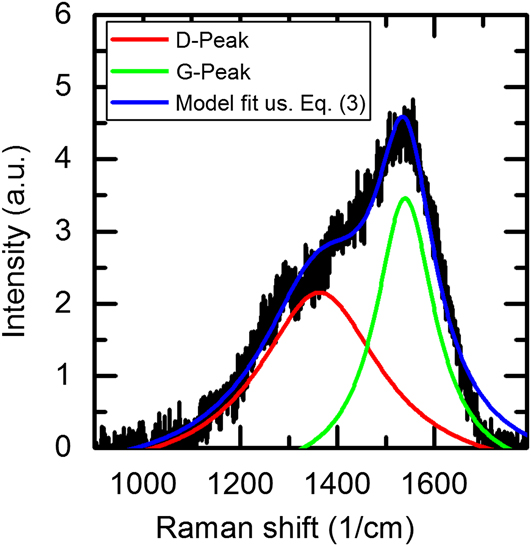
Raman spectrum of the DLC layer. The green and red lines show the Lorentzian of D and G peaks, respectively. The cumulative fit is represented by the blue line. The I(D)/I(G) ratio is a measure for the sp3 fraction of the carbon atoms.

C. Nanoparticle characterization
To obtain information about the Ag, Cu, and ZnO NPs, their size and morphology, we performed transmission electron microscopy (TEM) investigations with a JEM 2100 F instrument (JEOL Ltd., Tokyo, Japan) (see Fig. 3). Here, small droplets of the colloidal solutions were deposited and dried on standard carbon-coated copper grids (Plano GmbH).
Microscopical investigation of NPs. TEM micrographs of (a and b) Cu NPs, (c) Ag NPs and (d) ZnO NPs dispersed in polymer solution. Cu NPs formed large agglomerates (a) consisting of nano-sized nearly spherical particles as depicted in micrograph (b) Ag NPs and ZnO NPs (c and d) show a greater dispersion while ZnO NPs forms smaller aggregates (d) and have a less spherical shape than Ag NPs and Cu NPs.

D. Metal ion release measurements
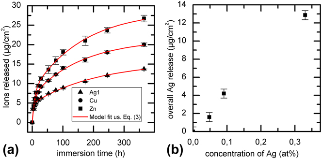
To investigate the metal ion release for the functionalized DLC samples, two identical samples from each batch (Ag, Cu, ZnO) were embedded in paraffin wax, leaving a total surface area of 1.57 cm2 exposed. To prevent ion release from the sample edges, they were covered by a metal protective paint (AkzoNobel). Afterward, all samples were immersed into 10 mL calf serum solution (Biochrom GmbH, protein concentration of 30 g/L, containing EDTA as stabilizer and Amphotericin B from Calbiochem to prohibit fungal infection) and kept in a standard incubator at 37 °C. After defined periods of time, the calf serum solution was completely exchanged and the total concentrations were measured employing inductively coupled plasma-optical emission spectroscopy (Varian ICP-OES) equipped with an autosampler. The presented data in Fig. 4 represent the mean values from three independent samples.
Metal ion release. (a) Cumulative ion release from DLC films into calf serum as a function of immersion time. The red lines are model fits [eq. (3)]. In most cases, error bars were smaller than respective data points and (b) linear relationship between Ag concentration in the DLC sample and cumulative Ag ion release.

III. RESULTS AND DISCUSSION
A. DLC characterization
To characterize the hybridization type in the DLC-layer, we performed Raman spectroscopy measurements (see Fig. 2). The so-called D and G peaks in the spectra were fitted by Lorentzians and the intensity ratio of both modes, I(D)/I(G), was calculated. This ratio as well as the shift of the G-Peak was compared with literature values published by Robertson et al. Reference Robertson28 to determine the sp3 fraction of the carbon atoms. All Raman spectra resulted in sp3 fractions of approximately 35% in the material, which is a typical value for a-C:H networks. Also, nano-hardness testing was done to prove the success of the DLC-transformation. Hardness measurements resulted in typical DLC hardness values of about 14 GPa for the samples produced. Due to the low thickness of the DLC layer compared to the indentation depth, however, some influence from the substrate might inflict the measurements. However, in presently ongoing studies, we also measured the hardness of nominally identical but thicker coatings (multi-layer DLC, produced by repeated dip-coating and ion implantation process, to be published elsewhere) with thicknesses d > 500 nm. For those samples, the indentation depth was below 10% of the film thickness. The hardness of such multi-layer DLC samples is expected to be lower than a single layer DLC coating as the range in the repeated ion bombardment process is greater than the layer thickness. This leads to partial graphitization of the lower layer, which in turn leads to an overall hardness decrease. Nevertheless, hardness values of 12 GPa, typical for a-C:H networks, were measured. As the focus of this work, however, lies on the tailored metal ion release from DLC layers, we did not intensively study wear resistance. Nevertheless, preliminary studies using a standardized tribological setup, where a DLC coated sphere was moved with a defined load on a flat DLC surface in calf serum solution, showed no significant debris according to ISO 17853 after 2 million cycles. Using a steel tip, it is not possible to scratch the coating by hand. Thus, we feel safe to claim typical DLC properties for the samples used here.
B. Nanoparticle characterization
Morphology and size of the synthesized nanoparticles were characterized using TEM. The Cu NPs formed large agglomerates [Fig. 3(a)] consisting of nearly spherical nanoparticles as depicted in Fig. 3(b). The ZnO NPs are less spherical and tend to form small aggregates [Fig. 3(d)] whereas the spherical Ag NPs were more dispersed [Figs. 3(c) and 3(d)] but show a wider size distribution than the Cu and ZnO NPs.
C. Metal ion release
As depicted in Fig. 4, all samples tested kept releasing metal ions into the calf serum solution for several days. The absolute amount of ions released from the DLC layer differed strongly for different metals, with highest ion release for Zn, followed by Cu and Ag.
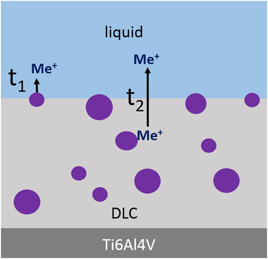
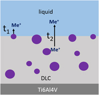
Moreover, the ion release was found to be a time-dependent two-stage process, with fast increase during the first days followed by a decrease within the next few days. We attribute this behavior to a purely geometrical effect: NPs at or partially sticking out of the surface and NPs completely “buried” in the DLC contribute differently to the ion release. Whereas NPs in contact to the aqueous solution immediately release free ions, metal ions from those in the bulk need to diffuse to the surface before entering the fluid. Hence, the ion release during the first decay time t1 predominantly depends on the fast oxidation by oxygen (of Ag and Cu) and the hydration of the ions, as depicted in Fig. 5.
Assumed ion release process. Schematic representation of the proposed ion release process with ion release from NPs at the surface during the initial decay time t 1 and diffusion of ions toward the DLC surface occurring during the second decay time t 2.

The kinetics of nanoparticle dissolution was considered before Reference Zhang, Yao, Sullivan and Chen29–Reference Ho, Yau, Lok, So and Che31 by looking at the oxidation reaction stoichiometry of Ag NPs being (partially) exposed to the aqueous environment (scenario 1 in Fig. 5):
 $$2{\rm{A}}{{\rm{g}}_{\left( {\rm{s}} \right)}} + {1 \mathord{\left/ {\vphantom {1 2}} \right. \kern-\nulldelimiterspace} 2}{{\rm{O}}_{2\left( {{\rm{aq}}} \right)}} + 2{{\rm{H}}^ + }_{\left( {{\rm{aq}}} \right)} \leftrightarrow 2{\rm{A}}{{\rm{g}}^ + }_{\left( {{\rm{aq}}} \right)} + {{\rm{H}}_2}{\rm{O}}\quad {\rm{.}}$$
Using an Arrhenius ansatz, the Ag+ release rate
 ${{\rm{\gamma }}_{{\rm{A}}{{\rm{g}}^ + }}}$
was derived as
Reference Zhang, Yao, Sullivan and Chen29
:
 $${\gamma _{{\rm{A}}{{\rm{g}}^ + }}} \sim {r^2}{T^{{1 \mathord{\left/ {\vphantom {1 2}} \right. \kern-\nulldelimiterspace} 2}}}\exp \left( {{{ - {E_{\rm{a}}}} \over {{k_{\rm{B}}}T}}} \right)\left[ {{\rm{AgNPs}}} \right]{\left[ {{{\rm{O}}_2}} \right]^{0.5}}{\left[ {{{\rm{H}}^ + }} \right]^2}\quad .$$
Here, r is the radius of Ag NPs, k B is the Boltzmann constant, E a is the activation energy, and [Ag NPs], [O2] and [H+] are respective molar concentrations. Further adaptations of Eq. (1) lead to the total amount of released ions which was used before to describe the experimental data with a and [Ag+]0 as fitting parameters.
 $${\left[ {{\rm{A}}{{\rm{g}}^ + }} \right]_{{\rm{released}}}} = {\left[ {{\rm{Ag}}}^+ \right]_0}\left[ {1 - \exp \left( { - at} \right)} \right]\quad .$$
However, the microscopic origin and mechanism leading to the second time constant in the release measurements is not yet clear, to date. Theoretically, this mechanism could be diffusion of either bare or hydrated metal ions through the DLC. In the latter case, water could either diffuse into the DLC or be formed by hydrogen from the DLC and oxygen diffusing into the DLC. To the knowledge of the authors, however, there are no reports on these mechanisms for amorphous DLC, yet. In any case, the fact that ions are released and detected at two different time scales indicates that there must be one escape mechanism available which seems not to be present for dry samples, which remain stable for weeks after preparation. Figure 5 illustrates the assumed ion release process described above. Independent of the actual transport mechanism, a diffusive behavior and hence a kinetic of the same type as Eq. (3) is obvious. Consequently, the total amount A of released metal ions was modeled as a combination of ion release contributing from the surface (*, scenario 1 in Fig. 5) and from the bulk (**, scenario 2 in Fig. 5).
 $$A\left( t \right) = \underbrace {{A_1}\left[ {1 - \exp \left( { - {t \over {{t_1}}}} \right)} \right]}_{\left( * \right)} + \underbrace {{A_2}\left[ {1 - \exp \left( { - {t \over {{t_2}}}} \right)} \right]}_{\left( {**} \right)}$$
Equation (4) was used to fit the experimental results with A 1, A 2, t 1, and t 2 being fitting parameters using the nonlinear Levenberg Marquardt algorithm (see Fig. 4). The release in the first few days is dominated by the amount A 1 released from or near the surface.
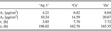
A summary of all fitting parameters is shown in Table I. The fitted values of A 1, t 1 and t 2 for Cu and Zn are quite similar, whereas the values of A 2 were slightly different, but still comparable in the order of magnitude. This finding suggests that the release and subsequent transport mechanism of Cu and Zn ions through the DLC is rather similar. This might be due to the similar ion radii of Cu and Zn and charge of the released ions (Cu2+ and Zn2+ versus Ag+). The differences in total ion release A tot = A 1 + A 2 of Zn2+ and Cu2+ ions can be explained by deviations of the precursor film thickness and used NP concentration. The mass ratio of embedded ZnO and Cu determined by RBS measurements (data not shown) displays qualitatively the ratio of A tot,Zn:A tot,Cu.
Calculated fitting parameters. Parameters A 1, A 2, t 1, and t 2, of Ag NPs, Cu NPs, or ZnO NPs containing DLC samples calculated from the developed model fit.

Figure 4(b) depicts a nearly linear ion release as a function of Ag concentration for three initial concentrations in the colloidal solution (‘Ag 1’ → ‘Ag 1/5’ → ‘Ag 1/10’), Hence, we deduce from our data that a tailorable Ag ion release seems to be adjustable through the proper choice of initial AgNO3 concentration.
IV. CONCLUSION
The presented study was undertaken to synthesize novel implant materials fulfilling the basic desired requirements for artificial joint applications including appropriate wear resistance and time-dependent antibacterial activity. As a matter of fact, all fabricated samples show DLC characteristics in terms of hardness as well as sp3 content and all samples in fact released the desired Ag, Cu and Zn ions from the DLC surface layer thus providing antibacterial functionality. From our findings, we deduct a model to further fine tune various ion release kinetic aspects by a variation of the ratio of near-surface and ‘buried’ NPs.
To give a rough estimate for the relevance of an antimicrobial effect, we here exemplarily consider the Ag ion release within 24 h for an ‘Ag 1’ sample: If we assume the knee joint's surface area to be 50 cm2, our model reveals an amount of 0.3 mg Ag ions released into the knee joint. For an average volume of a male adult's knee joint of V = 103.6 mL, Reference Visuri and Kiviluoto32 a global Ag concentration of 2.9 mg/L in the knee is obtained. This value is higher than the minimum inhibitory concentration against S. aureus (2.5 mg/L) but lower than IC50 (3.23 mg/L) for the inhibition of fibroblasts' cell metabolism. Reference Chernousova and Epple33 This ensures the potential of the material for an effective antibacterial treatment, especially as we may assume that locally the Ag concentration might be even higher.
In summary, we have demonstrated a method for tailored ion release from Ag NPs containing DLC surfaces exhibiting a linear relationship between the initial AgNO3 concentration in the sample and the amount of released Ag ions. This relationship in fact allows for the precise adaption of the desired antibacterial activity of wear and corrosion resistant DLC coatings on medical implants. Our experimental findings and results have the evident potential to provide a platform for the design and development of various novel wear resist and antibacterial implant materials, being highly relevant for future clinical applications.
DISCLOSURES
The authors have no financial conflicts of interest.
ACKNOWLEDGMENTS
The authors like to thank the “Deutsche Forschungsgemeinschaft (DFG)” for the financial support of this research work in form of the “Erkenntnistransferprojekt Antibakterielle und abriebarme Beschichtung von Gleitflächen in orthopädischen Implantaten” and Aesculap AG Tuttlingen for providing required materials. CW likes to acknowledge funding by Nanosystems Initiative Munich (NIM).