Background
Takayasu arteritis is a chronic, idiopathic, granulomatous vasculitis involving the aorta, its major branches, and occasionally the pulmonary arteries. Although rare in children, it is the third most common vasculitis occurring in the paediatric population.Reference Lindsley and Laxer 1 Presenting symptoms are typically non-specific and include headache, nausea/vomiting, palpitations, arthralgia, weight loss, and fever. Stenosis is the most common type of vascular involvement in Takayasu arteritis, affecting the majority of patients.Reference Zhu, Shen and Neubauer 2 Although aneurysmal disease has been reported in adults with Takayasu arteritis, it is a rare entity in children. We present the case of a 10-year-old boy with a giant ascending and arch aneurysm that necessitated follow-up surgery for a new aneurysm and occlusive disease.
Clinical summary
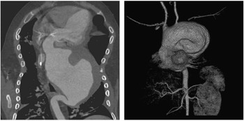
A 10-year-old Caucasian male was referred to our centre for the management of ascending and arch aneurysms. He had been diagnosed with Takayasu arteritis at the age of 3 years with poor response to anti-inflammatory medications in the past, including dual therapy with glucocorticoids and infliximab. CT showed a significant increase in his ascending aorta, arch, and proximal descending aortic aneurysm, measuring 8×8 cm at the level of the left subclavian (Fig 1). He had a 4- to 5-L oxygen requirement due to compression of the pulmonary arteries by the aneurysm. Given the failure of medical management, and after extensive discussion with the parents of the patient, he was taken to the operating room for total ascending aorta, aortic arch, and proximal descending aorta replacement with a 20-mm Vascutek graft via clamshell incision with re-implantation of the innominate artery and the left common carotid arteries under hypothermic circulatory arrest. His postoperative course was unremarkable and he was discharged after 2 weeks. Histological analysis of the resection specimen confirmed the diagnosis of Takayasu arteritis.
Three-dimensional and coronal CT angiogram images showing a giant ascending, arch, and transverse aortic aneurysm at the time of presentation.

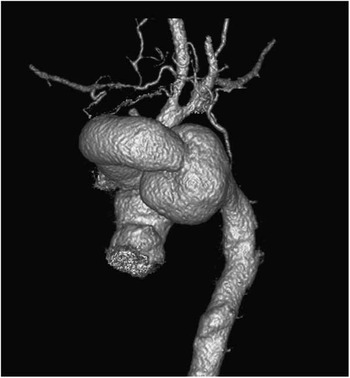
The patient presented 11 months later with an anastomotic aneurysm between the graft and the proximal descending aorta (Fig 2). He was felt to be at high risk for a redo-left thoracotomy and replacement of his distal aorta, and therefore underwent attempted thoracic aortic endovascular repair; however, this failed due to complete disruption of his right femoral artery that was repaired with a bovine pericardial patch; 2 months later, he ultimately underwent thoracic aortic endovascular repair via open exposure of the infrarenal aorta and creation of a conduit for delivery of a Medtronic Valiant thoracic endograft with coverage of the origin of the left subclavian artery.
Three-dimensional CT angiogram showing a distal anastomotic aneurysm.

On follow-up CT 1 month later, he was noted to have focal vasculitis of the left carotid artery with complete occlusion, and only the innominate artery supplying the head vessels with a high-grade stenosis noted. A covered stent was delivered through the right brachial artery for dilation of the innominate artery stenosis to preserve cerebral blood flow. At the 1-year follow-up, the patient has been doing well with stable symptoms and vascular disease and no neurological deficits to date.
Discussion
Takayasu arteritis is a chronic, rare type of paediatric vasculitis requiring lifelong management. The estimated prevalence in western countries is 2.6/million with a female predominance. In an Italian study of 104 patients, 70% of the patients presented at the age of 16–40 years, whereas only 15% of patients presented at an age younger than 15 years.Reference Vanoli, Daina and Salvarani 3 The majority of patients with Takayasu arteritis typically present with hypertension; other presenting symptoms are non-specific and include headache, nausea, and palpitations. In one series, 71% had reduced or absent carotid, brachial, or femoral pulses in one or more locations. Typical laboratory findings include elevated C-reactive protein level and erythrocyte sedimentation rate.Reference Zhu, Shen and Neubauer 2 The European League against Rheumatism consensus statement for diagnosis of childhood Takayasu arteritis recommends criteria such as angiographic abnormalities of the aorta and/or its branches, pulse deficit or claudication, blood pressure discrepancy, bruits, hypertension, and elevated acute phase reactants.Reference Ozen, Pistorio and Iusan 4 Based on histological examination, lesions are typically classified as acute or chronic. The initial site of inflammation is around the vasa vasora and at the medio-adventitial junction with oedema and mononuclear cell infiltration in the outer thirds of the media and adventitia. Giant-cell granulomatous reaction can also be present, similar to findings in giant-cell arteritis.
The differential diagnosis of Takaysau arteritis includes other causes of large-vessel vasculitis such as inflammatory aortitis – syphilis, tuberculosis, lupus, rheumatoid arthritis, spondyloarthropathies, Kawasaki disease, and giant-cell arteritis – developmental abnormalities – coarctation of the aorta and Marfan syndrome – and other aortic pathologies such as ergotism and neurofibromatosis. Most of these have specific features that enable diagnosis.
Stenosis is the most frequent lesion (93%) in Takayasu arteritis patients, followed by occlusion (57%), dilatation (16%), and aneurysm (7%), with over half of the patients with lesions above and below the diaphragm.Reference Vanoli, Daina and Salvarani 3 In other series, the incidence of aneurysms varies from 2 to 26.7%,Reference Kumar, Subramanyan and Mandalam 5 and they are usually found in older patients with chronic disease. The destruction of elastic fibres of the media or the presence of systemic hypertension is thought to be the aetiology of aortic dilatation. An elevated erythrocyte sedimentation rate can be considered as a marker of ongoing inflammation and destruction of the arterial wall in cases of aneurysmal formation.Reference Kalangos, Baldovinos and Beghetti 6
In the treatment of vascular disease secondary to Takayasu arteritis, the goal of medical therapy is to induce remission and prevent vascular complications. The cornerstone of therapy remains steroid treatment, but in the recent era second-line agents including methotrexate, cyclophosphamide, and anti-tumour necrosis factor α have been used to induce earlier remission and to lower steroid doses.Reference Isobe 7 Surgical intervention for Takayasu arteritis is based on the location and extent of disease with worse outcomes noted during active inflammation. Aneurysmal disease in children with Takayasu arteritis is exceedingly rare with only a few isolated case reports. In this report, our patient presented with a giant ascending aortic and arch aneurysm, a magnitude previously unreported. Our case also presents the first published use of endovascular aortic graft placement for the management of vascular sequela of Takayasu arteritis in a child.
The standard therapy for intractable ischaemic symptoms secondary to arterial stenosis or occlusion has been bypass surgery. Data from adult patients show that ~20% eventually require surgical intervention. Bypass grafts are preferred to endarterectomy and patch repairs for smaller-vessel involvement.Reference Isobe 7 Some series have demonstrated the feasibility of percutaneous transluminal angioplasty with encouraging short-term outcomes in the setting of Takayasu arteritis; however, there have been concerns regarding the high rate of occlusion or re-stenosis, especially during active inflammation, and several groups recommend use only in the setting of short, focal lesions. There are increasing retrospective data suggesting that stent grafts have lower rates of occlusions in patients with Takayasu arteritis compared with percutaneous angioplasty or stents only.Reference Qureshi, Martin and Greenberg 8
In summary, Takayasu arteritis is a rare but aggressive vasculitis in children. The use of endovascular approaches is feasible for the management of vascular sequela in this population and requires a coordinated multidisciplinary approach with paediatric and adult cardiothoracic surgeons as well as vascular surgeons.
Acknowledgement
None.
Financial Support
This research received no specific grant from any funding agency, commercial, or not-for-profit sectors.
Conflicts of Interest
None.