Introduction
The African variegated grasshopper Zonocerus variegatus (L.) (Orthoptera: Pyrgomorphidae), whose pest status was established about 50 years ago (COPR, 1974), feeds on numerous plant species, including the food crops banana, cassava, cocoa, citrus, cowpea and yam (Toye, Reference Toye1982). However, not all the plant species it consumes contribute to the grasshopper's growth and development (Bernays et al., Reference Bernays, Chapman, Cook, Meveigh and Page1975; Idowu and Sonde, Reference Idowu and Sonde2004). For instance, cassava and Chromolaena odorata leaves support its growth and development, while Talinum triangulare and Aspilla africana leaves do not (Toye, Reference Toye1982).
The life cycle of Z. variegatus has around six nymphal stages lasting around 100–200 days in total, and an adult stage of around 15 weeks (Anya, Reference Anya1973; Chapman et al., Reference Chapman, Page and McCaffery1986; Muse, Reference Muse2003). The size and concentration of somatic tissue metabolites increase with the age of the insect (Ademolu et al., Reference Ademolu, Idowu and Dansu2006, Reference Ademolu, Idowu and Amusan2007).
In most acridids, the alimentary canal is a continuous tube running from the mouth to the anus and is divided into the foregut, midgut and hindgut (Chapman, Reference Chapman1990). Digestion can occur in the foregut using enzymes produced in the midgut (Terra, Reference Terra1990).
Analysis of gut contents of 3rd–6th instar nymphs of Z. variegatus showed that gut motility and the movement of ingesta through the gut were maximal at 08.00–10.00, 13.00–14.00 and 16.00 h (Modder, Reference Modder1984). Midgut β- and α-glucosidase activities were detected at all times, but the levels of activity were lowest at 12.00 and 19.00 h (Modder, Reference Modder1984). To date, only a few studies have been conducted on the activity of enzymes present in the gut of Z. variegatus, and even these have been mainly restricted to the adult stage of the insect.
The aim of the study reported here was to quantitatively assess the digestive enzymes present in the gut of Z. variegatus during post-embryonic development, with the goal of further understanding the digestive physiology of this insect.
Materials and methods
Collection and rearing
Newly hatched 1st instar nymphs of Z. variegatus were collected from an abandoned farmland at the University of Agriculture, Abeokuta (UNAAB), Ogun State, Nigeria, in October and November 2007. The collected insects were brought to the insectary of the Department of Biological Sciences, UNAAB and were reared to the adult stage in wire cages (30 × 30 × 45 cm) on fresh leaves of Chromolaena odorata and Manihot esculenta.
Preparation of insects
Thirty individuals of Z. variegatus per developmental stage were taken out of the rearing cages and asphyxiated in a deep freezer for 30 min, following which their guts were carefully dissected out. The alimentary canal of each insect was partitioned into foregut, midgut and hindgut. The respective gut regions were homogenized in 0.05 m KCl and centrifuged at 500 rpm for 30 min at 5 °C. The supernatant obtained (enzyme extract) was decanted into a 30-ml centrifuge tube and stored in the freezer.
Determination of cellulase activity
To determine cellulase activity, 0.5 ml of enzyme extract was pipetted into a 25-ml test-tube, to which 0.5 ml of a 1% carboxymethyl cellulose solution was added. The test-tube was stoppered and incubated for 30 min at 50 °C, after which 1 ml of DNSA (dinitrosalicylic acid) reagent was added and the mixture heated at 100 °C in a water bath for 5 min. The mixture was then rapidly cooled to room temperature and 10 ml of distilled water were added to it. Cellulose and glucose standard solutions were prepared and treated in the same way. The absorbance of treated sample extract and cellulose standards was read after 1 min on a Cecil 600 spectrophotometer at 540 nm wavelength.
Determination of α-glucosidase activity
To 0.5 ml of the enzyme extract mixed with 1 m of para-hydroxy benzoic acid, 5 ml of 0.1 m sodium phosphate buffer at pH 5.5 was added, followed by 1 ml of distilled water. DNSA reagent (1 ml) was added to the mixture, which was heated at 100 °C for 5 min, then rapidly cooled to room temperature followed by the addition of 10 ml of distilled water. A standard solution of glucosidase enzyme was prepared and treated similarly. The absorbances of sample extract as well as that of glucosidase standard solution were read on a Cecil 600 spectrophotometer at 530 nm wavelength.
Determination of amylase activity
Two millilitres of the enzyme extracted in a 1 m potassium hydrogen phosphate buffer (pH 6.9) were mixed with 1 ml of a 1% starch solution and incubated for 1 h at 40 °C. The reaction was stopped by adding 8 ml of DNSA reagent. The mixture was boiled in a water bath for 5 min, cooled in a cold-water bath and then diluted with 18 ml distilled water. The absorbance of the resulting solution was read at 550 nm on an SP 6250 spectrophotometer.
Determination of lipase activity
The extracting buffer was 0.2 m sodium acetate–acetic acid mixture at pH 5. A 7.5 ml sample of the enzyme extract was added to the reaction mixture containing 0.4 g of sodium taurocholate, 1 ml of 0.1 m CaCl2 and 6 ml of the sodium acetate buffer. The mixture was incubated at 35 °C for 1 h, then the reaction stopped by adding 40 ml absolute alcohol. A standard lipase solution was also prepared using distilled water. The absorbance of the sample and standard was read at a wavelength of 415 nm on a Cecil 600 spectrophotometer. One lipase activity unit was expressed as the amount of the enzyme lipase capable of catalysing absorbance change of 0.01 in 60 s at wavelength of 415 nm.
Determination of proteinase activity
The extracting buffer was 0.1 m sodium hydrogen phosphate (pH 6.9); 5 ml of the enzyme extract were added to 10 ml of 2% casein solution and incubated at 35 °C for 30 min. The reaction was terminated by adding 10 ml of 10% trichloroacetic acid solution and then filtered through a Whatman No. 1 filter paper. Standard proteinase solution was prepared and treated as above. The absorbance of the sample and standard proteinase filtrates were read at a wavelength of 275 nm. Proteinase activity was measured as the amount of proteinase capable of catalysing absorbance change of 0.01 in 60 s at a wavelength of 275 mm.
All the enzyme assays were carried out in triplicate.
Statistical analysis
The data generated from the above analyses were subjected to one-way analysis of variance, using Minitab software, and means separated using the Student–Newman–Keuls (SNK) test.
Results
Cellulase activity
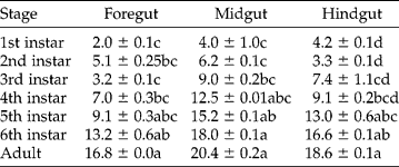
Cellulase activity in the gut regions of Z. variegatus was significantly higher (P < 0.05) in the adult stage relative to the 1st to 3rd instars, but no significant differences were found between cellulase activity in guts of the adult stage, 5th and 6th instars on one hand, and between the 1st and 3rd instars on the other (Table 1).
Cellulase activity (±SE) in the gut regions of Zonocerus variegatus during post-embryonic development (Abs/min)
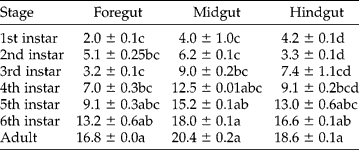
Mean values in the same column having the same letter are not significantly different, P>0.05 (SNK).
α-Glucosidase activity
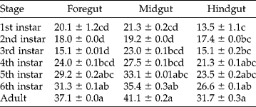
The adult stage of Z. variegatus had significantly (P < 0.05) higher α-glucosidase activity relative to other stages of development, except the 5th and 6th instars. However, no statistically significant differences were found in α-glucosidase activities of 1st to 3rd instars across the three gut regions (Table 2). The highest α-glucosidase activity (20 Abs/min) was recorded in the midgut region.
α-Glucosidase activity (±SE) in the gut regions of Zonocerus variegatus during post-embryonic development (Abs/min)
Mean values in the same column having the same letter are not significantly different, P>0.05 (SNK).
Amylase activity
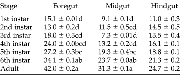
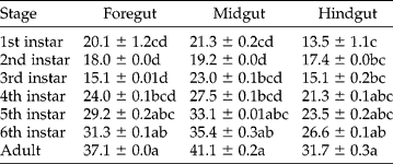
Amylase activity was detected in all the gut regions of all the stages of the insect. Amylase activity in the foregut and midgut regions was significantly higher in adults than in all the immatures, except the 6th instar (Table 3). However, no significant differences (P>0.05) were seen in amylase levels in the hindguts of the developmental stages.
Amylase activity (±SE) in the gut regions of Zonocerus variegatus during post-embryonic development (Abs/min)
Mean values in the same column having the same letters are not significantly different, P>0.05 (SNK).
Lipase activity
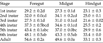
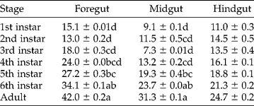
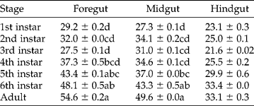
Lipase activity in the foregut (54 Abs/min) and midgut (49 Abs/min) of adult Z. variegatus was significantly higher (P < 0.05) than in the immature stages, except the 5th and 6th instars. However, no significant differences were found in the lipase activity of the hindgut across the developmental stages (Table 4).
Lipase activity (±SE) in the gut regions of Zonocerus variegatus during post-embryonic development (Abs/min)
Mean values in the same column having the same letter are not significantly different, P>0.05 (SNK).
Proteinase activity
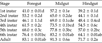
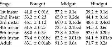
As shown in Table 5, there were significant differences (P < 0.05) in proteinase activity in the three gut regions of the insect, with the adult stage (91 Abs/min) having significantly higher activity relative to other developmental stages, except the 6th instar. However, no significant differences were found in the activities of the 1st to 3rd instars.
Proteinase activity (±SE) in the gut regions of Zonocerus variegatus during post-embryonic development (Abs/min)
Mean values in the same column having the same letter are not significantly different, P>0.05 (SNK).
Discussion
Lipase, proteinase, cellulase, α-glucosidase and amylase were present in the gut regions of Z. variegatus, as reported in previous studies (Balogun, Reference Balogun1972; Modder, Reference Modder1984; Adedire and Balogun, 1995; Woodring et al., Reference Woodring, Hoffman and Lorenz2007), which indicates that Z. variegatus is equipped with digestive enzymes able to hydrolyse a diverse diet.
The activities of the enzymes increased with age of the insect, from 1st instar to the adult stage. This increase reflects the increase in size of the gut and higher food consumption as the insect moults from one developmental stage to another, which probably stimulates enzyme production. Furthermore, Idowu and Edema (Reference Idowu and Edema2005) observed that the microbial burden in the gut regions of Z. variegatus increased with age, and that these microbial isolates produce enzymes (Idowu et al., Reference Idowu, Edema and Oyedepo2009). Similarly, Mandal et al. (Reference Mandal, Roy and Chaudhuri1981) had earlier reported that protease and lipase activities were more significant in the gut of the later instars of a large cricket Schizodactylus monstrosus. The fresh weight, total protein content and total proteolytic activities of the gut tissue of Z. variegatus were found to increase through the six nymphal instars during post-embryonic development (Obidapo, Reference Obidapo1979). It can be inferred that age significantly affects the activities of enzymes in the gut regions of Z. variegatus, as also concluded by Hill and Orchard (Reference Hill and Orchard2005) for Locusta migratoria. Higher enzyme activity in the adult stage of Z. variegatus correlates with this stage being the most voracious and destructive one of the insect (Toye, Reference Toye1982). Ademolu et al. (Reference Ademolu, Idowu and Olatunde2009) also observed significantly higher enzyme activities in the femoral muscles of adult Z. variegatus.
There were significant differences in the activities of the enzymes found in the different gut regions of Z. variegatus. This observation agrees with the findings of Mandal et al. (Reference Mandal, Roy and Chaudhuri1981), that the distribution pattern of enzyme activity varied in the different regions of the gut. The midgut of Z. variegatus had the highest enzymatic activities of the three gut regions. Wigglesworth (Reference Wigglesworth1963), Dow (Reference Dow1981) and Terra (Reference Terra1990) have demonstrated that the midgut was the major site for digestive enzyme secretion in orthopterans. The epithelium of the midgut is always simple and is composed principally of columnar and regenerative cells. In the termite Coptotermes formosanus, the largest β-glucosidase and cellulase activities were also recorded in the midgut (Jianchu et al., Reference Jianchu, Tianci, Xiaogang and Jiaan2004), followed by the foregut and the hindgut.
Although the foregut is not known to produce amylase (Lebene, Reference Lebene, Harrison and Locke1998), the present study showed that the foregut had the highest amylase activity. This might be attributed to the movement of enzymes from the midgut to the foregut. Digestive enzymes can flow forward as well as backward in the alimentary canal of the insect. For instance, the highest amylase activity in orthopterans is often found in the foregut, though the enzyme is released from the midgut endothelial cells (Hill and Orchard, Reference Hill and Orchard2005).
Enzyme activities in 1st–3rd instars across the three gut regions were not significantly (P>0.05) different. Idowu and Edema (Reference Idowu and Edema2005) earlier reported that the microbes isolated from the gut regions of 1st–3rd instars were similar, and the microbial loads (cfu) were not significantly different. The micro-organisms isolated from the gut probably contribute to enzyme production, as observed by Idowu et al. (Reference Idowu, Edema and Oyedepo2009).
Enzyme secretion is a direct response to the presence of nutrients (Terra, Reference Terra1990). In the study of Englemann (Reference Englemann1969), injection of 14C amino acids into Leucophaea maderae resulted in high protease activities in the midgut. The results of the present study corroborate this finding, as proteinase activity was significantly greater than that of the other enzymes in the gut during post-embryonic development. This might be the result of the diet provided to the insect during rearing. The insect shows preference for C. odorata and cassava (M. esculenta) during the 1st to 3rd instars and 5th instar to the adult stage, respectively. Proximate analysis (Apori et al., Reference Apori, Castro and Orskov2000) of C. odorata showed a high crude protein content (258 g/kg DM). Similarly, cassava leaves have a protein content of 49.3 g/kg DM (Ayodeji and Valentine, Reference Ayodeji and Valentine2005). In a recent related study, proteinase and lipase activities in the femoral muscles of Z. variegatus were significantly higher than those of other enzymes, reflecting the nature of its diet (Ademolu et al., Reference Ademolu, Idowu and Olatunde2009).
Cellulase activity in the gut regions of Z. variegatus was lower than that of the other enzymes. This observation corroborates Chapman (Reference Chapman1990) and Clissold et al. (Reference Clissold, Sanson and Read2004), who found very low activities of cellulase in the desert locust Schistocerca gregaria and the Australian plague locust Chortoicetes terminifera, respectively.
In conclusion, later nymphal stages and the adult stage have higher enzymatic activities, suggesting that they are more voracious and destructive than the lower instars. Thus the control of this insect pest is necessary before reaching the advanced stage of their life cycle.