





Introduction
Immune-related diseases, including infectious diseases (Refs Reference Msemburi, Karlinsky, Knutson, Aleshin-Guendel, Chatterji and Wakefield1, Reference Dugdale, Ufio, Giardina, Shebl, Coskun, Pletner, Torola, Cosar, Shapiro, Kim, Mofenson and Ciaranello2); cancer (Refs Reference Siegel, Kratzer, Giaquinto, Sung and Jemal3, Reference Bray, Laversanne, Weiderpass and Soerjomataram4); autoimmune diseases (Refs Reference Reich, Lucchinetti and Calabresi5, Reference Theofilopoulos, Kono and Baccala6); neuroinflammatory diseases (Refs Reference Scheltens, de Strooper, Kivipelto, Holstege, Chételat, Teunissen, Cummings and van der Flier7, Reference De Miguel, Khoury, Betley, Lehallier, Willoughby, Olsson, Yang, Hahn, Lu, Vest, Bonanno, Yerra, Zhang, Saw, Kaci Fairchild, Lee, Zhang, McAlpine, Contrepois, Shamloo, Elias, Rando and Wyss-Coray8); immunodeficiencies (Refs Reference Dugdale, Ufio, Giardina, Shebl, Coskun, Pletner, Torola, Cosar, Shapiro, Kim, Mofenson and Ciaranello2, Reference Yarchoan and Uldrick9, Reference Ombrello, Remmers, Sun, Freeman, Datta, Torabi-Parizi, Subramanian, Bunney, Baxendale, Martins, Romberg, Komarow, Aksentijevich, Kim, Ho, Cruse, Jung, Gilfillan, Metcalfe, Nelson, O’Brien, Wisch, Stone, Douek, Gandhi, Wanderer, Lee, Nelson, Shianna, Cirulli, Goldstein, Long, Moir, Meffre, Holland, Kastner, Katan, Hoffman and Milner10); and immunopathologies associated with organ transplantation, such as graft-versus-host disease (Refs Reference Ferrara, Levine, Reddy and Holler11, Reference Zeiser and Blazar12) and transplant rejection (Refs Reference Prendecki, Thomson, Clarke, Martin, Gleeson, de Aguiar, Edwards, Mortimer, McIntyre, Mokreri, Cox, Pickard, Lightstone, Thomas, McAdoo, Kelleher and Willicombe13, Reference O’Connell, Zhang, Menon, Yi, Schröppel, Gallon, Luan, Rosales, Ge, Losic, Xi, Woytovich, Keung, Wei, Greene, Overbey, Bagiella, Najafian, Samaniego, Djamali, Alexander, Nankivell, Chapman, Smith, Colvin and Murphy14), remain major global health challenges, contributing substantially to morbidity, mortality and societal burden. Collectively, these conditions reinforce the urgent need for sustained biomedical research, improved preventive strategies and more effective therapeutic interventions.
CD8+ T cells are a specialized class of T lymphocytes that play a critical role in adaptive immunity by directly recognizing and eliminating virus-infected and malignant tumour cells (Ref. Reference Henning, Roychoudhuri and Restifo15). Naïve CD8+ T (TN) cells, upon encountering cognate antigen in the presence of appropriate co-stimulatory signals, undergo clonal expansion and differentiation into distinct populations, including short-lived effector (TEff) cells and memory precursor (TMP) cells (Ref. Reference Henning, Roychoudhuri and Restifo15).
Immunological memory is a critical mechanism that protects the body from previously encountered pathogens and is the foundation of vaccination. There are three broad subsets of memory CD8+ T (TM) cells (Refs Reference Henning, Roychoudhuri and Restifo15, Reference Milner and Goldrath16): central memory (TCM), effector memory (TEM), and tissue-resident memory (TRM) cells.
The biology of every cell in the body, including immune cells, is governed by the precise regulation of gene expression. To date, immunological research has largely focused on the epigenetic and transcriptional mechanisms that regulate the biology of immune cells. By contrast, the post-transcriptional and translational mechanisms of gene regulation, despite their central roles in controlling mRNA fate and translational efficiency, remain underexplored in immune cells.
Furthermore, recent studies examining resting and in vitro activated CD8+ TM cells in both humans and mice have revealed a striking regulatory phenomenon: resting CD8+ TM cells express substantial levels of cytokine-encoding mRNAs but do not produce detectable levels of protein (Refs Reference Salerno, Paolini, Stark, von Lindern and Wolkers17–Reference Turner20). The mechanisms that regulate these ‘poised’ cytokine-encoding transcripts in resting TM cells remain largely uncharacterised and represent an important area for further investigations (Ref. Reference Turner20).
We are only beginning to understand the post-transcriptional mechanisms that govern immune cell function. As this understanding deepens, these insights are expected to create new opportunities to translate fundamental biological discoveries into innovative therapeutic strategies (Refs Reference Turner20, Reference Turner and Petkau21, Reference Zhu, Wheeler and Ansel22; Figure 1). In the following sections, I discuss our current understanding of how RNA-binding proteins (RBPs) regulate CD8+ T cell biology. Details of the literature search strategy and selection criteria are provided in the supplementary information. Throughout this article, findings derived from gene knockout (KO), knockdown (KD) or overexpression models are described relative to wild-type (WT) controls unless indicated otherwise. A summary of various mouse models used to study RBPs in immune cells is provided in Table 1 and Figure 1.
RNA-binding proteins regulate the function and biology of CD8⁺ T cells. RNA-binding proteins control gene expression in CD8⁺ T cells to maintain immune homeostasis. Dysregulated gene expression in these cells contributes to hyperinflammation, autoimmune disease and haematological malignancy. The development of RBP-targeted therapeutic strategies holds substantial potential for advancing innovative treatments and precision medicine for immune-related diseases.

Figure 1. Long description
At the top center is a circular diagram of a C D 8 super plus T Cell. Inside the cell, a dashed circle represents the nucleus containing a D N A double helix. An arrow points down from the D N A to an R N A strand with attached proteins, which then points down to a blue protein mass in the cytoplasm.
Two horizontal arrows point away from the cell:
* To the left, an arrow labeled Protective Function points to a bulleted list: Infectious Diseases and Cancer.
* To the right, an arrow labeled Dysregulated Gene Expression points to a bulleted list: Hyperinflammation, Autoimmune Diseases, and Hematological Malignancies.
A vertical arrow labeled R N A-Binding Proteins points down from the cell to a list of functions:
* Dynamic Expression and Function
* Regulates Effector Function and Maintains Immune Homeostasis
* Regulates Poised I f n g m R N A in Memory C D 8 super plus T Cells
* Prevents Hyperinflammation and Hyperproliferation
A final vertical arrow points down to the bottom-most bullet point: Innovative Immunotherapies and Precision Medicine.
Summary of mouse genetic models utilised to study RNA-binding proteins in immune cells

Table 1. Long description
The table consists of 14 rows of data under four headers.
* Row 1: References 18, 29, 33; Genotype C d 4-Cre plus Z f p 3 6 l 2 fl/fl; Abbreviation Z f p 3 6 l 2 c K O; Impacts C D 4 plus and C D 8 plus T cells.
* Row 2: References 28, 53, 62, 63; Genotype Z f p 3 6 minus/minus; Abbreviation Z f p 3 6 K O; Impacts all cells.
* Row 3: References 30, 32; Genotypes include C d 4-Cre plus Z f p 3 6 fl/fl, C d 4-Cre plus Z f p 3 6 l 1 fl/fl, and C d 4-Cre plus Z f p 3 6 fl/fl Z f p 3 6 l 1 fl/fl; Abbreviations Z f p 3 6 c K O, Z f p 3 6 l 1 c K O, and Z f p 3 6 / Z f p 3 6 l 1 c D K O; Impacts C D 4 plus and C D 8 plus T cells.
* Row 4: Reference 34; Genotype C d 4-Cre plus Z f p 3 6 fl/fl Z f p 3 6 l 1 fl/fl Z f p 3 6 l 2 fl/fl; Abbreviation Z f p 3 6 / Z f p 3 6 l 1 / Z f p 3 6 l 2 c T K O; Impacts C D 4 plus and C D 8 plus T cells.
* Row 5: Reference 35; Genotype C d 4-Cre plus P t b p 1 fl/fl; Abbreviation P t b p 1 c K O; Impacts C D 4 plus and C D 8 plus T cells.
* Row 6: References 36, 38; Genotype d L c k-Cre plus S r s f 1 fl/fl; Abbreviation S r s f 1 c K O; Impacts mature/post-selection C D 4 plus and C D 8 plus T cells.
* Row 7: Reference 37; Genotype C d 4-Cre plus D d x 5 fl/fl; Abbreviation D d x 5 c K O; Impacts C D 4 plus and C D 8 plus T cells.
* Row 8: Reference 39; Genotype Roquin-1 san/san (M 1 9 9 R) mutation; Abbreviation Roquin san/san; Impacts all cells.
* Row 9: Reference 43; Genotypes C p e b 4 minus/minus and C d 4-Cre plus C p e b 4 fl/fl; Abbreviations C p e b 4 K O and C p e b 4 c K O; Impacts all cells and C D 4 plus / C D 8 plus T cells.
* Row 10: Reference 44; Genotype d L c k-Cre plus Y t h d f 2 fl/fl; Abbreviation Y t h d f 2 c K O; Impacts mature/post-selection C D 4 plus and C D 8 plus T cells.
* Row 11: References 49, 72; Genotype C d 2-Cre plus Z f p 3 6 l 1 fl/fl Z f p 3 6 l 2 fl/fl; Abbreviation Z f p 3 6 l 1 / Z f p 3 6 l 2 C d 2-c D K O; Impacts thymocytes and mature T cells.
* Row 12: References 50, 51; Genotype M b 1-Cre plus Z f p 3 6 l 1 fl/fl Z f p 3 6 l 2 fl/fl; Abbreviation Z f p 3 6 l 1 / Z f p 3 6 l 2 M b 1-c D K O; Impacts B cells.
* Row 13: Reference 56; Genotype L y z 2-Cre plus Y t h d f 2 fl/fl; Abbreviation Y t h d f 2 L y z 2-c K O; Impacts myeloid cells.
* Row 14: Reference 64; Genotype L y s M-Cre plus Z f p 3 6 fl/fl Z f p 3 6 l 1 fl/fl Z f p 3 6 l 2 fl/fl; Abbreviation Myeloid-specific c T K O; Impacts myeloid cells.
Note: Genotypes, abbreviations used in this article and the major immune cell populations targeted by global and cell type-specific conditional knockout approaches are shown in this table.
RNA-binding proteins regulate CD8 + T cell biology
Global expression and functional landscapes of RNA-binding proteins in CD8+ T cells
Global transcriptomic and proteomic analysis of human CD8+ T cells revealed extensive remodelling of RBP expression during differentiation from CD8+ TN to TCM, TEM and TEff subsets (Ref. Reference Zandhuis, Nicolet and Wolkers23). The most pronounced changes occurred during the transition from TN to TEM and TEff subsets. TN cells, and to a lesser extent TCM cells, preferentially expressed translation-associated RBPs, whereas TEM and TEff cells expressed RBPs involved in RNA transport and stability. Complementary single-cell transcriptomic analysis further demonstrated that highly cytotoxic CD8+ T cells displayed a distinct RBP signature, including elevated expression of APOBEC3G, APOBEC3C and PATL2, while CD8+ T cells with low cytotoxic potential expressed higher levels of ribosomal RBPs (Ref. Reference Zandhuis, Nicolet and Wolkers23). Collectively, these findings provide valuable insights into the differentiation- and function-specific RBP expression profile in human CD8+ T cell subsets and generate testable hypotheses for functional validation.
RNA aptamer-based pull-down of in vitro-transcribed IFNG, TNF and IL2 3′ untranslated regions (UTRs), followed by mass spectrometry, identified extensive activation-dependent changes in RBP association in human CD3+ T cells (Ref. Reference Popović, Nicolet, Guislain, Engels, Jurgens, Paravinja, Freen-van Heeren, van Alphen, van den Biggelaar, Salerno and Wolkers24). In non-activated T cells, 138 RBPs were associated with these cytokine 3′ UTRs, whereas upon 2 h stimulation with PMA and ionomycin, a distinct set of 77 RBPs was identified. ZFP36L1 displayed enhanced interaction with all three cytokine 3′ UTRs upon stimulation, while several RBPs exhibited cytokine-specific increases in association, including YTHDF1 with the IFNG 3′ UTR; RRP1B with the TNF 3′ UTR; and RBM42 with the IL2 3′ UTR. In contrast, multiple RBPs showed reduced association upon stimulation, including ZC3H12D, which lost detectable association with all cytokine 3′ UTRs, as well as ZCCHC11 and PABPN1 with the IFNG 3′ UTR; DDX39A and ZNHIT6 with the TNF 3′ UTR; and DDX46 and USO1 with the IL2 3′ UTR (Ref. Reference Popović, Nicolet, Guislain, Engels, Jurgens, Paravinja, Freen-van Heeren, van Alphen, van den Biggelaar, Salerno and Wolkers24). Collectively, these findings demonstrate a global remodelling of cytokine mRNA-RBP association landscape upon human T cell activation.
A complementary study using mouse OT-I CD8+ T cells isolated from spleen and lymph nodes following in vivo OVA peptide priming ± CD134/CD137 costimulation revealed pronounced, costimulation-dependent remodelling of post-transcriptional regulatory processes (Ref. Reference Karginov, Ménoret and Vella25). Integrated transcriptomic and proteomic analysis of costimulated CD8+ T cells in this experimental model revealed strong enrichment of spliceosome-associated pathways and upregulation of 19 RBPs with established roles in alternative splicing and alternative polyadenylation. RNA sequencing analysis further identified 1,464 isoform changes across 950 genes in costimulated CD8+ T cells, comprising alternative polyadenylation (48.3%); alternative splicing (35.6%); and alternative transcriptional start site usage (16.1%), affecting transcripts encoding key regulators of CD8+ T cell function. Costimulation preferentially promoted exon skipping and 3′ UTR shortening, whereas antigen priming alone was associated with increased intron retention and enrichment of isoforms predicted to be susceptible to nonsense-mediated decay (Ref. Reference Karginov, Ménoret and Vella25). Collectively, these findings indicate that CD134/CD137 costimulation optimizes CD8+ T cell effector function by reprogramming RBP expression and transcript isoform architecture toward higher translational potential.
Together, these studies provide insight into the global dynamics of RBP-mediated post-transcriptional mechanisms in CD8+ T cells and highlight post-transcriptional regulation as a key determinant of their function.
The ZFP36 family of RNA-binding proteins regulate multiple facets of CD8+ T cell biology
Among RBPs, the ZFP36 family has been studied most extensively in the immune system, particularly for its role in post-transcriptional regulation of cytokine gene expression; however, its contribution to CD8+ T cell function and biology remains incompletely understood.
Members of the ZFP36 family belong to the CCCH-type tandem zinc finger class and are defined by their ability to bind AU-rich elements (AREs) within the 3′UTRs of target mRNAs, thereby regulating mRNA stability and translation (Refs Reference Salerno, Engels, van den Biggelaar, van Alphen, Guislain, Zhao, Hodge, Bell, Medema, von Lindern, Turner, Young and Wolkers18, Reference Lai, Carballo, Strum, Kennington, Phillips and Blackshear26). The family comprises ZFP36 (also referred to as Tristetraprolin), ZFP36L1, ZFP36L2 and ZFP36L3 (Ref. Reference Turner and Petkau21). Because expression of the Zfp36l3 gene is restricted to the placenta of specific rodent species (Ref. Reference Gingerich, Stumpo, Lai, Randall, Steppan and Blackshear27), this article uses the term ZFP36 family to refer specifically to the three conserved members: ZFP36, ZFP36L1 and ZFP36L2.
Mechanistic insights into ZFP36 family functions in CD8+ T cells have primarily been derived from studies employing gene KO strategies, including both individual and combined deletions (Table 1). This article organizes these findings according to the experimental models used and integrates them to develop a more comprehensive framework for understanding the role of the ZFP36 family in CD8+ T cell biology.
ZFP36
ZFP36 is emerging as a key regulator of CD8+ T cell biology. In mouse CD8+ TN cells, ZFP36 was induced between 2 and 8 h following activation with anti-CD3 and bone marrow-derived dendritic (BMDD) cells (Ref. Reference Moore, Blachere, Fak, Park, Sawicka, Parveen, Zucker-Scharff, Moltedo, Rudensky and Darnell28). Similarly, in mouse OT-I CD8+ TEff cells, ZFP36 expression was detected upon 6 h of OVA peptide stimulation (Ref. Reference Zandhuis, Bradarić, van der Zwaan, Hoogendijk, Popović and Wolkers29).
CD8+ T cells from Zfp36 KO mice displayed enhanced proliferation until 16 h post-activation with anti-CD3 and BMDD cells, which was likely independent of the IL-2 signalling pathway (Ref. Reference Moore, Blachere, Fak, Park, Sawicka, Parveen, Zucker-Scharff, Moltedo, Rudensky and Darnell28). Additionally, a higher percentage of virus-specific splenic CD8+ T cells isolated from Zfp36 KO mice at day 6 post-LCMV infection expressed IFN-γ and TNF upon 6 h activation with GP33-41 peptide (Ref. Reference Moore, Blachere, Fak, Park, Sawicka, Parveen, Zucker-Scharff, Moltedo, Rudensky and Darnell28). Taken together, these results from Zfp36 KO mice suggest that ZFP36 acts as a brake on both proliferative and effector cytokine output of CD8+ T cells during the early hours after in vitro activation.
In mouse OT-I CD8+ TN cells, ZFP36 induction following TCR stimulation showed limited dependence on ligand affinity (Refs Reference Petkau, Mitchell, Evans, Matheson, Salerno and Turner30, Reference Jones Evans, Mitchell, Zaucha, Petkau and Turner31). After 6 h of stimulation with N4 peptide or lower-affinity Q4, T4 and V4 variants, comparable levels of ZFP36 expression were detected despite differences in TCR signalling potency (Ref. Reference Petkau, Mitchell, Evans, Matheson, Salerno and Turner30). In a complementary study on OT-I CD8+ TM-like cells, stimulation with the high-affinity N4 peptide induced higher Zfp36 mRNA and protein expression at early timepoints (2 h) compared with lower-affinity ligands; however, this affinity dependence was transient and was not evident at later timepoints up to 6 h (Ref. Reference Jones Evans, Mitchell, Zaucha, Petkau and Turner31). Together, these findings indicate that, in this experimental system, while ZFP36 expression may transiently reflect TCR signal strength shortly after activation, it is broadly inducible downstream of TCR engagement and does not maintain affinity-dependent expression over time, suggesting that ZFP36 may help prevent inappropriate CD8+ T cell activation by low-affinity or suboptimal ligands.
Zfp36 expression in mouse CD8+ TM-like cells was induced by p38 MAPK, MEK1/2 and PKC signalling pathways, with ELK-1 identified as a key transcriptional regulator of the Zfp36 gene (Ref. Reference Jones Evans, Mitchell, Zaucha, Petkau and Turner31). These findings suggest that, in this model, pathways that support CD8+ T cell activation also induce a post-transcriptional brake that constrains the activation response. Such coupling of stimulatory and restraining processes may help calibrate the magnitude of CD8+ T cell responses and prevent excessive activation.
ZFP36L1
ZFP36L1 is also emerging as an important regulator of CD8+ T cell function. In mouse OT-I CD8+ TEff cells, similar to ZFP36, ZFP36L1 expression was detected upon 6 h of stimulation with OVA peptide (Ref. Reference Zandhuis, Bradarić, van der Zwaan, Hoogendijk, Popović and Wolkers29). Consistent with a regulatory role in effector function, Zfp36l1 sgKO OT-I CD8+ TEff cells produced higher levels of TNF, IFN-γ and IL-2 after co-culturing with B16-OVA cells for 6 h (Ref. Reference Zandhuis, Bradarić, van der Zwaan, Hoogendijk, Popović and Wolkers29).
Mechanistically, ZFP36L1 iCLIP in OT-I CD8+ T cells activated with OVA peptide for 3 h identified Il2, Nfkb1, Nfkb2, Rel, Notch1 and Irf8 mRNAs as direct targets of ZFP36L1 (Ref. Reference Petkau, Mitchell, Chakraborty, Bell, D´Angeli, Matheson, Turner, Saveliev, Gizlenci, Salerno, Katsikis and Turner32). These targets span the NF-κB, Notch and IRF pathways, suggesting that ZFP36L1 acts as a regulator of multiple signalling nodes of the TCR activation program. Consistent with this observation, Zfp36/Zfp36l1 conditional double KO CD8+ T cells exhibited increased NF-κB2 and IRF8 expression upon 24 h anti-CD3 stimulation (Ref. Reference Petkau, Mitchell, Chakraborty, Bell, D´Angeli, Matheson, Turner, Saveliev, Gizlenci, Salerno, Katsikis and Turner32).
A conserved regulatory interactome was also observed in humans, as ZFP36L1 was found to bind the 3′ UTRs of IFNG, TNF and IL2 transcripts in human CD3+ T cells, with binding increasing after 2 h of PMA and ionomycin stimulation (Ref. Reference Popović, Nicolet, Guislain, Engels, Jurgens, Paravinja, Freen-van Heeren, van Alphen, van den Biggelaar, Salerno and Wolkers24).
Mouse CD8+ TN cells that overexpressed ZFP36L1 displayed increased levels of phosphorylated STAT5 (pSTAT5) and CD25 compared with WT cells in co-culture after 48 h of stimulation with anti-CD3 and anti-CD28 (Ref. Reference Petkau, Mitchell, Evans, Matheson, Salerno and Turner30). In contrast, CD8+ TN cells isolated from the spleen and lymph nodes of Zfp36l1 conditional KO (cKO) mice displayed reduced pSTAT5 and CD25 expression compared with WT cells under the same conditions (Ref. Reference Petkau, Mitchell, Evans, Matheson, Salerno and Turner30). Together, these findings indicate that ZFP36L1 regulates IL-2 signalling in mouse CD8+ T cells in this experimental system.
ZFP36L1 expression was linked to antigen affinity in mouse CD8+ T cells (Refs Reference Petkau, Mitchell, Evans, Matheson, Salerno and Turner30, Reference Jones Evans, Mitchell, Zaucha, Petkau and Turner31). In OT-I CD8+ TN cells stimulated with OVA peptide variants for 6 h, ZFP36L1 levels scaled with ligand affinity, with the highest expression induced by the N4 peptide (Ref. Reference Petkau, Mitchell, Evans, Matheson, Salerno and Turner30). Similarly, in OT-I CD8+ TM-like cells, stimulation with N4 peptide resulted in higher Zfp36l1 mRNA and protein expression than stimulation with lower-affinity ligands across all time points examined (Ref. Reference Jones Evans, Mitchell, Zaucha, Petkau and Turner31). These results indicate that, unlike ZFP36, ZFP36L1 expression correlates with TCR ligand affinity in these experimental systems, supporting a role for ZFP36L1 as a sensor of TCR signal strength. These observations suggest a model in which more strongly stimulated cells impose greater post-transcriptional restraint on themselves relative to weakly stimulated cells by inducing both ZFP36 and ZFP36L1.
Furthermore, Zfp36l1 expression in mouse CD8+ TM-like cells was induced by p38 MAPK, MEK1/2, PKC and calcium–calcineurin–NFAT signalling pathways, with NFAT family transcription factors identified as key regulators of the Zfp36l1 gene (Ref. Reference Jones Evans, Mitchell, Zaucha, Petkau and Turner31). Notably, Zfp36l1 shares three of these upstream inputs with Zfp36 but is additionally regulated by the calcium–calcineurin–NFAT signalling pathway (Ref. Reference Jones Evans, Mitchell, Zaucha, Petkau and Turner31). This difference in upstream regulation may relate to the affinity-dependent induction of ZFP36L1 described above. These observations also indicate that both ZFP36 and ZFP36L1 are induced by TCR signalling, but through partly distinct pathways, such that post-transcriptional restraint is installed through more than one route.
ZFP36L2
ZFP36L2 regulates cytokine expression at later time points. In mouse OT-I CD8+ TEff cells, ZFP36L2 expression was induced between 24 and 48 h following stimulation with OVA peptide (Ref. Reference Zandhuis, Bradarić, van der Zwaan, Hoogendijk, Popović and Wolkers29). Consistent with its delayed expression pattern, a higher frequency of Zfp36l2 cKO OT-I CD8+ TEff cells expressed IFN-γ at 24 and 48 h of continuous stimulation with B16-OVA cells (Ref. Reference Zandhuis, Bradarić, van der Zwaan, Hoogendijk, Popović and Wolkers29). Similarly, a higher frequency of Zfp36l2 cKO CD8+ TEff cells expressed IFN-γ at 24 h following re-stimulation with anti-CD3 and anti-CD28 (Ref. Reference Zandhuis, Guislain, Popalzij, Engels, Popović, Turner and Wolkers33). Mechanistically, ZFP36L2 repressed IFN-γ production in CD8+ TEff cells through interaction with AREs in the 3′ UTR of Ifng mRNA (Ref. Reference Zandhuis, Guislain, Popalzij, Engels, Popović, Turner and Wolkers33).
Zfp36l2 cKO CD8+ T cells treated with anti-PD-1 antibody produced the highest levels of IFN-γ following 48 h of co-culture with B16-OVA cells, exceeding both untreated Zfp36l2 cKO CD8+ T cells and anti-PD-1–treated WT cells (Ref. Reference Zandhuis, Guislain, Popalzij, Engels, Popović, Turner and Wolkers33). This observation suggests that, in this melanoma co-culture system, ZFP36L2 imposes a post-transcriptional brake on cytokine production that persists even when the inhibitory PD-1–PD-L1 signalling axis is blocked. Post-transcriptional repression and PD-1–mediated inhibitory signalling may therefore act through non-redundant mechanisms, thereby providing multiple independent checkpoints. As discussed in section ‘Clinical and translational implications of RBP-mediated immune regulation: therapeutic opportunities and challenges’, a combinatorial approach targeting both brakes may therefore offer additive therapeutic benefits.
Consistent with these co-culture findings, a higher percentage of Zfp36l2 cKO CD8+ tumour-infiltrating lymphocytes (TILs), obtained by adoptive transfer of OT-I CD8+ TEff cells into a B16-OVA tumour model, expressed IFN-γ (Ref. Reference Zandhuis, Guislain, Popalzij, Engels, Popović, Turner and Wolkers33).
ZFP36L2 plays a unique role in mouse CD8+ TM cells. ZFP36L2 was shown to repress the translation of ‘poised’ Ifng transcripts in resting splenic CD8+ TM cells (Ref. Reference Salerno, Engels, van den Biggelaar, van Alphen, Guislain, Zhao, Hodge, Bell, Medema, von Lindern, Turner, Young and Wolkers18). Consistent with this, a higher percentage of resting splenic CD44+ CD8+ TM cells from Zfp36l2 cKO mice expressed IFN-γ. Notably, this 3′UTR ARE-mediated translational repression was also observed to extend beyond cytokine-encoding transcripts in mouse CD8+ TM cells (Ref. Reference Salerno, Engels, van den Biggelaar, van Alphen, Guislain, Zhao, Hodge, Bell, Medema, von Lindern, Turner, Young and Wolkers18), suggesting that, similar to ZFP36L1, ZFP36L2 regulates multiple signalling nodes associated with the effector function of CD8+ T cells.
Zfp36/Zfp36l1 double knockout
CD8+ T cells from Zfp36/Zfp36l1 conditional double KO (cDKO) mice retain ZFP36L2. These cells allow us to examine the extent to which ZFP36L2 can compensate for the combined loss of ZFP36 and ZFP36L1, while also helping define the specific roles of ZFP36 and ZFP36L1. OT-I CD8+ TN cells isolated from the spleen and lymph nodes of Zfp36/Zfp36l1 cDKO mice proliferated more extensively after 48 h of OVA peptide stimulation when cultured in isolation; however, this hyperproliferative phenotype was lost in co-culture with WT cells (Ref. Reference Petkau, Mitchell, Evans, Matheson, Salerno and Turner30).
Under co-culture conditions following 48 h of OVA peptide stimulation, a lower percentage of Zfp36/Zfp36l1 cDKO OT-I CD8+ T cells expressed CD25, and a higher percentage of these cells expressed activated caspase-3. Under the same conditions, Zfp36/Zfp36l1 cDKO OT-I CD8+ T cells expressed reduced levels of pSTAT5 and the anti-apoptotic factors MCL1 and BCL2 (Ref. Reference Petkau, Mitchell, Evans, Matheson, Salerno and Turner30). Together, these results indicate increased apoptosis and impaired IL-2 signalling in Zfp36/Zfp36l1 cDKO cells in activated co-cultures. In line with these findings, bulk RNA sequencing analysis revealed differential expression of IL-2, IL-6 and TNF signalling pathways in Zfp36/Zfp36l1 cDKO OT-I CD8+ TN cells after 6 h of OVA peptide stimulation (Ref. Reference Petkau, Mitchell, Evans, Matheson, Salerno and Turner30).
These observations are paradoxical: activated Zfp36/Zfp36l1 cDKO OT-I CD8+ T cells showed enhanced proliferation and effector programs in isolated cultures, yet reduced viability and impaired IL-2 signalling when co-cultured with WT cells. This suggests that the consequences of ZFP36/ZFP36L1 loss depend on the environment rather than being strictly cell-intrinsic. The paradoxical observations also suggest the presence of inherent safeguarding mechanisms that limit the survival of dysregulated CD8+ T cells, discussed in the following sections and illustrated in Figure 3.
Interestingly, the deletion of Suppressor of Cytokine Signalling 1 (Socs1) gene restored proliferative capacity and CD25 expression in Zfp36/Zfp36l1 cDKO cells in co-culture at 72 h post-stimulation with OVA peptide (Ref. Reference Petkau, Mitchell, Evans, Matheson, Salerno and Turner30). Moreover, adoptively transferred Zfp36/Zfp36l1/Socs1 triple KO OT-I CD8+ T cells expanded in the spleen to a similar extent as WT controls at day 7 following attenuated Listeria monocytogenes-OVA infection, and deletion of Socs1 promoted the differentiation into KLRG1+ CD8+ TEff cells (Ref. Reference Petkau, Mitchell, Evans, Matheson, Salerno and Turner30). The relevance of such combinatorial genetic deletions for engineered CD8+ T cell therapies is discussed in section ‘Clinical and translational implications of RBP-mediated immune regulation: therapeutic opportunities and challenges’.
In a complementary study, a higher percentage of Zfp36/Zfp36l1 cDKO OT-I CD8+ TN cells produced IL-2 until 16 h of OVA peptide stimulation. Mechanistically, ZFP36 and ZFP36L1 limited IL-2 production by regulating Il2 mRNA stability (Ref. Reference Petkau, Mitchell, Chakraborty, Bell, D´Angeli, Matheson, Turner, Saveliev, Gizlenci, Salerno, Katsikis and Turner32).
In addition, Zfp36/Zfp36l1 cDKO CD8+ TN cells proliferated more robustly across a range of anti-CD3 doses over 72 h and exhibited a reduced dependence on CD28 costimulation. Following anti-CD3 stimulation alone for 24 h, Zfp36/Zfp36l1 cDKO TN cells expressed higher levels of NF-κB2 and IRF8, whereas WT cells were more dependent on CD28 costimulation to achieve comparable responses (Ref. Reference Petkau, Mitchell, Chakraborty, Bell, D´Angeli, Matheson, Turner, Saveliev, Gizlenci, Salerno, Katsikis and Turner32). Together, these findings position post-transcriptional repression by ZFP36 and ZFP36L1 as part of the checkpoint mechanism that maintains the requirement for both TCR stimulation and CD28 costimulation for full CD8+ T cell activation.
Zfp36l1/Zfp36l2 double knockout
Zfp36l1/Zfp36l2 cDKO CD8+ T cells retain ZFP36. These cells allow us to examine the extent to which ZFP36 can compensate for the combined loss of ZFP36L1 and ZFP36L2, while also helping define the specific roles of ZFP36L1 and ZFP36L2. OT-I Zfp36l1/Zfp36l2 cDKO CD8+ TEff cells expressed higher levels of IFN-γ, TNF and IL-2 as early as 6 h after co-culture with B16-OVA cells (Ref. Reference Zandhuis, Bradarić, van der Zwaan, Hoogendijk, Popović and Wolkers29). Zfp36l1/Zfp36l2 cDKO OT-I CD8+ TEff cells expressed TNF for up to 48 h of continuous activation with B16-OVA cells, although TNF expression was lost by 96 h. By contrast, IFN-γ production persisted even at 96 h of stimulation. Along with sustained cytokine protein expression, Zfp36l1/Zfp36l2 cDKO OT-I CD8+ TEff cells expressed higher levels of Ifng mRNA after 72 h of continuous activation with B16-OVA cells, consistent with reduced Ifng mRNA degradation (Ref. Reference Zandhuis, Bradarić, van der Zwaan, Hoogendijk, Popović and Wolkers29).
Despite enhanced IFN-γ production, Zfp36l1/Zfp36l2 cDKO CD8+ TILs did not reduce tumour size in a B16-OVA melanoma mouse model (Ref. Reference Zandhuis, Bradarić, van der Zwaan, Hoogendijk, Popović and Wolkers29). Moreover, adoptively transferred Zfp36l1/Zfp36l2 cDKO CD8+ T cells were present at lower frequencies in the tumour-draining lymph nodes in this melanoma model and exhibited reduced viability both in vivo and in vitro (Ref. Reference Zandhuis, Bradarić, van der Zwaan, Hoogendijk, Popović and Wolkers29). These paradoxical observations suggest that enhancing effector cytokine production is not sufficient for anti-tumour immunity if viability and persistence are compromised. This has direct relevance to therapeutic strategies aimed at releasing post-transcriptional brakes in CD8+ T cells and is discussed further in section ‘Clinical and translational implications of RBP-mediated immune regulation: therapeutic opportunities and challenges’.
Mechanistically, Zfp36l1/Zfp36l2 cDKO TEff cells expressed higher levels of the stress-sensor kinase PERK in both resting and 24 h of OVA peptide stimulation conditions, suggesting the activation of the unfolded protein response (UPR) pathway (Ref. Reference Zandhuis, Bradarić, van der Zwaan, Hoogendijk, Popović and Wolkers29). In addition, resting Zfp36l1/Zfp36l2 cDKO TEff cells displayed altered cytokine receptor expression, including increased levels of IFNGR1, CD127 and IL-2RG. Following deprivation of recombinant IL-7 for 4 h and subsequent re-exposure for 20 min, Zfp36l1/Zfp36l2 cDKO CD8+ T cells expressed reduced levels of pSTAT5. Resting Zfp36l1/Zfp36l2 cDKO CD8+ T cells also expressed higher levels of DUSP4 phosphatase (Ref. Reference Zandhuis, Bradarić, van der Zwaan, Hoogendijk, Popović and Wolkers29). Together, these findings suggest impaired IL-7 signalling and activation of stress response pathways in Zfp36l1/Zfp36l2 cDKO CD8+ T cells, providing a possible explanation for their reduced persistence.
Zfp36/Zfp36l1/Zfp36l2 triple knockout
A study on conditional Zfp36/Zfp36l1/Zfp36l2 triple KO (cTKO) mice provided insights into the collective function of the ZFP36 family in CD8+ T cells (Ref. Reference Cook, Bradstreet, Webber, Kim, Santeford, Harris, Murphy, Tran, Abdalla, Schwarzkopf, Greco, Halabi, Apte, Blackshear and Edelson34). Unlike conditional single or double KO mice, cTKO mice developed severe, multi-organ inflammatory pathology and exhibited early lethality. CD8+ T cells and other immune cells infiltrated multiple organs in cTKO mice, including the eyes, kidneys, liver and brain (Ref. Reference Cook, Bradstreet, Webber, Kim, Santeford, Harris, Murphy, Tran, Abdalla, Schwarzkopf, Greco, Halabi, Apte, Blackshear and Edelson34), suggesting that ZFP36 family may regulate tissue-specific migration of immune cells.
Notably, a higher percentage of resting splenic CD8+ T cells from cTKO mice expressed IFN-γ and TNF, and these cells produced IFN-γ independent of de novo Ifng transcription. Consistent with this systemic phenotype, tissue-specific analyses revealed an increased percentage of IFN-γ+ CD8+ T cells in the kidney and liver of cTKO mice (Ref. Reference Cook, Bradstreet, Webber, Kim, Santeford, Harris, Murphy, Tran, Abdalla, Schwarzkopf, Greco, Halabi, Apte, Blackshear and Edelson34).
Bulk RNA sequencing analysis of splenic CD8+ T cells from cTKO mice identified 1,386 upregulated genes, with enrichment of IL-2-STAT5, IFN-γ response, apoptosis and proliferation-related pathways. CD8+ T cells isolated from the kidneys of cTKO mice displayed a similar pattern of pathway enrichment. In parallel, a higher percentage of splenic CD8+ T cells from cTKO mice expressed Ki67; activated caspase-3; Fas and FasL, consistent with enhanced proliferation and increased apoptosis (Ref. Reference Cook, Bradstreet, Webber, Kim, Santeford, Harris, Murphy, Tran, Abdalla, Schwarzkopf, Greco, Halabi, Apte, Blackshear and Edelson34).
Importantly, splenic CD8+ T cells from cDKO mice did not recapitulate the extent of transcriptional dysregulation observed in splenic CD8+ T cells from cTKO mice (Ref. Reference Cook, Bradstreet, Webber, Kim, Santeford, Harris, Murphy, Tran, Abdalla, Schwarzkopf, Greco, Halabi, Apte, Blackshear and Edelson34), indicating that ZFP36 family members collectively maintain gene expression in CD8+ T cells and that overlapping functions among them buffer against widespread transcriptional dysregulation.
These paradoxical findings of enhanced effector function and reduced viability of cTKO CD8+ T cells follow a pattern observed in cDKO CD8+ T cells as discussed in earlier sections. The reduced persistence of hyperactivated CD8+ T cells suggests the presence of inherent safeguarding mechanisms, discussed in the following section and illustrated in Figure 3.
A multilayered post-transcriptional checkpoint and safeguard model for ZFP36 family function in CD8+ T cells
Collectively, the studies discussed in this section demonstrate that ZFP36 family members are emerging as central regulators of CD8+ T cell biology (Figure 2). Each paralog contributes in distinct yet partially overlapping ways to optimize CD8+ T cell function and safeguard immune homeostasis. ZFP36 and ZFP36L1 act as brakes on excessive proliferation and cytokine production immediately following activation (Refs Reference Moore, Blachere, Fak, Park, Sawicka, Parveen, Zucker-Scharff, Moltedo, Rudensky and Darnell28–Reference Petkau, Mitchell, Chakraborty, Bell, D´Angeli, Matheson, Turner, Saveliev, Gizlenci, Salerno, Katsikis and Turner32).
Because the induction of ZFP36 had limited dependence on ligand affinity (Refs Reference Petkau, Mitchell, Evans, Matheson, Salerno and Turner30, Reference Jones Evans, Mitchell, Zaucha, Petkau and Turner31), it may also help prevent inappropriate CD8+ T cell activation by low-affinity or suboptimal ligands. In contrast to ZFP36, ZFP36L1 induction correlated with TCR ligand affinity in these experimental models (Refs Reference Petkau, Mitchell, Evans, Matheson, Salerno and Turner30, Reference Jones Evans, Mitchell, Zaucha, Petkau and Turner31). Together, these differing induction patterns suggest a multilayered post-transcriptional checkpoint, in which ZFP36 imposes a baseline restraint on all responding cells at early time points independent of ligand affinity, while ZFP36L1 adds a graded component that increases with signal strength. These observations also suggest a model in which more strongly stimulated cells impose greater post-transcriptional restraint on themselves relative to weakly stimulated cells by inducing both ZFP36 and ZFP36L1.
In contrast to ZFP36 and ZFP36L1, ZFP36L2 acts as a brake on excessive cytokine production at later time points following activation (Ref. Reference Zandhuis, Bradarić, van der Zwaan, Hoogendijk, Popović and Wolkers29). ZFP36L2 also plays a unique role in resting mouse CD8+ TM cells, where it acts as a translational repressor of ‘poised’ Ifng mRNA molecules (Refs Reference Salerno, Paolini, Stark, von Lindern and Wolkers17–Reference Turner20).
The ZFP36 family of RNA-binding proteins regulate multiple facets of CD8+ T cell biology.

Figure 2. Long description
At the top is a central box titled The Z F P 36 Family. Three arrows point downward to three distinct panels.
Left panel: Z F P 36. Bullet points state it is expressed between 6 and 8 hours post-T C R stimulation; regulates I f n g and T n f; regulates proliferation; and is regulated by P 38 M A P K, M E K 1 / 2, and P K C pathways, and E L K-1.
Middle panel: Z F P 36 L 1. Bullet points state it is expressed at 6 hours post-T C R stimulation; regulates I f n g, T n f, and I l 2; is a sensor of antigen affinity; regulates I L-2 signaling pathway; and is regulated by P 38 M A P K, M E K 1 / 2, P K C, and Calcium-Calcineurin-N F A T pathways, and N F A T.
Right panel: Z F P 36 L 2. Bullet points state it is expressed between 24 and 48 hours post-T C R stimulation; regulates I f n g and T n f; and regulates poised I f n g m R N A in C D 8 super plus T sub M cells.
Bottom section: Phenotypes and Characteristics Upon Gene Knockout. This section contains three sub-boxes.
1. Z f p 36 / Z f p 36 l 1 double knockout: Increased proliferation in single culture; decreased proliferation in co-culture with W T cells; increased apoptosis in co-culture with W T cells; decreased I L-2 signaling in co-culture with W T cells; and reduced dependency on C D 28 costimulation.
2. Z f p 36 l 1 / Z f p 36 l 2 double knockout: Increased I F N G, T N F, and I L-2; decreased viability; and impaired I L-7 signaling.
3. Z f p 36 / Z f p 36 l 1 / Z f p 36 l 2 triple knockout: Severe multi-organ inflammatory pathology; increased cytokine expression; increased stress response; increased proliferation; and increased apoptosis.
A. The ZFP36 family of RNA-binding proteins repress excessive pro-inflammatory cytokine production and proliferation in activated CD8+ T cells. B. Inherent safeguarding mechanisms limit the survival of dysregulated CD8+ T cells. C. Molecular mechanism of post-transcriptional regulation of gene expression by the ZFP36 family of RBPs.

Figure 3. Long description
The diagram consists of three vertical panels labeled A, B, and C.
Panel A: At the center is a C D 8 super plus T cell. To the left, an arrow points to Resting Memory C D 8 super plus T cells, noting that Z F P 36 L 2 represses translation of poised I f n g m R N A. An arrow pointing down indicates that a Low-Affinity Ligand induces Z F P 36 to prevent inappropriate activation. An arrow to the right indicates High-Affinity Ligand T C R stimulation, leading to two boxes: Z F P 36 and Z F P 36 L 1 (Induced Early, approximately 6 to 8 hours) and Z F P 36 L 2 (Induced Late, greater than or equal to 24 hours). The early induction box points up to Enforces C D 28 Costimulation. Both boxes point down to Prevents Excessive Pro-inflammatory Cytokine Production and Proliferation.
Panel B: Shows a dysregulated C D 8 super plus T cell containing Z f p 36 / Z f p 36 l 1 c D K O, Z f p 36 l 1 / Z f p 36 l 2 c D K O, or Z f p 36 / Z f p 36 l 1 / Z f p 36 l 2 c T K O. An arrow labeled Intrinsic Safeguard Mechanism points right to the outcome: Excessive Pro-inflammatory Cytokine Production and Proliferation AND Low Viability and Survival.
Panel C: A molecular diagram shows a purple m R N A strand with a 5 prime m super 7 G cap and a 3 prime poly-A tail. A teal globular protein labeled Z F P 36 family is bound to the m R N A. An arrow points to the binding site, noting that A U-rich elements can form secondary or tertiary structures recognized by R B Ps. Two dashed arrows point from the protein to m R N A decay and Translational Repression.
Studies on Zfp36/Zfp36l1 and Zfp36l1/Zfp36l2 cDKO mice revealed distinct vulnerabilities in CD8+ T cells, as defects were observed in activation, proliferation, cytokine signalling and survival (Refs Reference Zandhuis, Bradarić, van der Zwaan, Hoogendijk, Popović and Wolkers29, Reference Petkau, Mitchell, Evans, Matheson, Salerno and Turner30, Reference Petkau, Mitchell, Chakraborty, Bell, D´Angeli, Matheson, Turner, Saveliev, Gizlenci, Salerno, Katsikis and Turner32). However, the phenotype was not a severe cellular collapse, as observed in Zfp36/Zfp36l1/Zfp36l2 cTKO CD8+ T cells (Ref. Reference Cook, Bradstreet, Webber, Kim, Santeford, Harris, Murphy, Tran, Abdalla, Schwarzkopf, Greco, Halabi, Apte, Blackshear and Edelson34). This suggests that the third paralog can compensate to some extent, but not completely, for the combined loss of the other two paralogs.
Paradoxical findings in cDKO and cTKO CD8+ T cells suggest the presence of an important mechanism that safeguards immune homeostasis. Zfp36/Zfp36l1 cDKO CD8+ T cells proliferated extensively and secreted IL-2 after stimulation (Refs 30, Reference Petkau, Mitchell, Chakraborty, Bell, D´Angeli, Matheson, Turner, Saveliev, Gizlenci, Salerno, Katsikis and Turner32). These cells were also less dependent on CD28 costimulation (Ref. Reference Petkau, Mitchell, Chakraborty, Bell, D´Angeli, Matheson, Turner, Saveliev, Gizlenci, Salerno, Katsikis and Turner32). However, in activated co-cultures with WT cells, Zfp36/Zfp36l1 cDKO CD8+ T cells showed reduced fitness, decreased responsiveness to IL-2 signalling and increased apoptosis (Ref. Reference Petkau, Mitchell, Evans, Matheson, Salerno and Turner30). A higher percentage of Zfp36l1/Zfp36l2 CD8+ T cells expressed cytokines at both early and late time points after activation (Ref. Reference Zandhuis, Bradarić, van der Zwaan, Hoogendijk, Popović and Wolkers29); however, similar to Zfp36/Zfp36l1 cDKO CD8+ T cells, Zfp36l1/Zfp36l2 cDKO CD8+ T cells were not viable and exhibited reduced survival (Ref. Reference Zandhuis, Bradarić, van der Zwaan, Hoogendijk, Popović and Wolkers29). In Zfp36/Zfp36l1/Zfp36l2 cTKO CD8+ T cells, where all ZFP36 family members are lost, the phenotype is much more severe than in cDKO cells, and these cells also displayed reduced viability and increased apoptosis (Ref. Reference Cook, Bradstreet, Webber, Kim, Santeford, Harris, Murphy, Tran, Abdalla, Schwarzkopf, Greco, Halabi, Apte, Blackshear and Edelson34).
These observations suggest the presence of intrinsic safeguarding mechanisms that limit the survival of dysregulated CD8+ T cells. If post-transcriptional brakes such as the ZFP36 family members are lost, perhaps due to genetic mutations, CD8+ T cells may initially undergo excessive proliferation and produce excessive amounts of cytokines. However, these dysregulated cells also fail to survive. In this way, CD8+ T cells may have evolved protective mechanisms to eliminate themselves when they become dysfunctional and potentially harmful.
An integrative model of the role of the ZFP36 family in CD8+ T cells, based on the studies discussed in this article, is illustrated in Figure 3. Although such a model can certainly be useful for developing a broad conceptual framework, it is important to recognize that the function of RBPs in CD8+ T cells remains an emerging area of research, with a relatively limited number of studies to date. Detailed studies on the role of the ZFP36 family and other RBPs in various CD8+ T cell subsets and activation conditions in both humans and mice are therefore needed to gain a more mechanistic and comprehensive understanding of RBP function in CD8+ T cells (Figures 2 and 3).
Regulation of CD8 + T cell biology by additional RNA-binding proteins
Accumulating evidence indicates that multiple RBPs govern distinct aspects of CD8+ T cell fate, function and biology. Accordingly, the RBPs discussed below are grouped based on their predominant roles in specific biological processes in CD8+ T cells. This organizational framework provides insight into both the molecular mechanisms and therapeutic implications of RBPs in CD8+ T cell immunity.
Activation and clonal expansion
Polypyrimidine Tract Binding Protein 1 (PTBP1) was found to govern various processes in CD8+ TN cells (Ref. Reference D’Angeli, Monzón-Casanova, Matheson, Gizlenci, Petkau, Gooding, Berrens, Smith and Turner35). Following the adoptive transfer of Ptbp1 cKO and WT OT-I CD8+ TN cells and subsequent L. monocytogenes-OVA infection, Ptbp1 cKO CD8+ T cells exhibited a marked reduction in proliferation and increased apoptosis in the spleen between days 3 and 5 post-infection. Additionally, a lower percentage of Ptbp1 cKO cells expressed KLRG1 at days 4 and 5, suggesting impaired effector differentiation in this setting. These defects in proliferation and survival were recapitulated in vitro upon stimulation with anti-CD3 and anti-CD28 for 48 h (Ref. Reference D’Angeli, Monzón-Casanova, Matheson, Gizlenci, Petkau, Gooding, Berrens, Smith and Turner35).
A lower percentage of Ptbp1 cKO OT-I CD8+ TN cells expressed NR4A1, IL-2 and TNF upon 4 h OVA peptide stimulation. In addition, a lower percentage of Ptbp1 cKO cells isolated from the spleen at day 4 post-infection expressed IFN-γ and TNF upon restimulation with OVA peptide for 3 h (Ref. Reference D’Angeli, Monzón-Casanova, Matheson, Gizlenci, Petkau, Gooding, Berrens, Smith and Turner35).
Transcriptomic profiling revealed that PTBP1 regulates alternative splicing of 453 genes in CD8+ TN cells. One notable target identified was Ppp3cb, which encodes the catalytic subunit of calcineurin Aβ. Ptbp1 cKO cells expressed higher levels of exon 13–containing Ppp3cb transcripts, which could influence the calcineurin–NFAT signalling pathway (Ref. Reference D’Angeli, Monzón-Casanova, Matheson, Gizlenci, Petkau, Gooding, Berrens, Smith and Turner35). This observation offers a potential mechanistic link between PTBP1 loss and the impaired biology described above.
Collectively, these findings from this mouse model of L. monocytogenes infection suggest that PTBP1 is an important regulator of CD8+ T cell activation, expansion, survival, effector function, and differentiation.
As discussed previously, CD134/CD137 costimulation in mouse CD8+ T cells induced widespread changes in alternative polyadenylation and alternative splicing (Ref. Reference Karginov, Ménoret and Vella25). In this experimental system, TAR DNA-binding protein (TARDBP) was identified as a key RBP regulating post-transcriptional programs downstream of CD134/CD137 costimulation. In this study, Tardbp sgKO and WT OT-I CD8+ T cells were adoptively transferred into recipient mice. Following in vivo priming with OVA peptide for 4 days, in the presence or absence of costimulation, OT-I CD8+ T cells were isolated from the spleen and lymph nodes for further studies (Ref. Reference Karginov, Ménoret and Vella25).
Mouse Tardbp sgKO CD8+ T cells exhibited 233 alternative splicing events across 161 genes upon costimulation, including altered splicing of key TCR signalling regulators such as Ikzf1, Stat4 and Fyb. These splicing changes were associated with reduced expression of IKZF1 isoform II in both mouse and human CD8+ T cells (Ref. Reference Karginov, Ménoret and Vella25).
Functionally, costimulated mouse Tardbp sgKO CD8+ T cells showed reduced clonal expansion and cytokine production following 4 h restimulation with PMA and ionomycin. Similarly, human TARDBP sgKO CD8+ T cells produced lower levels of IFN-γ after 20 h PMA and ionomycin restimulation (Ref. Reference Karginov, Ménoret and Vella25).
Together, these findings identify TARDBP as a key regulator of CD134/CD137 costimulation-induced post-transcriptional programs that support optimal CD8+ T cell activation and effector responses in both humans and mice.
Serine/Arginine-Rich Splicing Factor 1 (SRSF1) was identified as a key regulator of proliferation and persistence of mouse CD8+ T cells (Ref. Reference Juarez, Su, Herbert, Teijaro and Moulton36). Srsf1 cKO mice had a reduced percentage of CD8+ T cells in peripheral lymphoid organs at both steady state and at day 8 post-LCMV infection. Splenic Srsf1 cKO CD8+ T cells from uninfected mice also exhibited reduced proliferative capacity during 48 to 96 h stimulation with anti-CD3 and anti-CD28 (Ref. Reference Juarez, Su, Herbert, Teijaro and Moulton36).
Effector and memory differentiation
DEAD-box helicase 5 (DDX5) regulated the balance between CD8+ TEff and TM subsets in a mouse model of LCMV infection (Ref. Reference Louis, Wong, Yao, Kurd, Tysl, Indralingam, Ma, Huang and Chang37). Following adoptive transfer of Ddx5 cKO and WT P14 CD8+ T cells and LCMV infection, splenic Ddx5 cKO CD8+ T cells exhibited reduced frequencies of KLRG1+CD127− TEff cells and increased frequencies of KLRG1−CD127+ TMP cells at day 7 post-infection. By day 30 post-infection, Ddx5 cKO cells showed increased frequencies of CD62L+CD127+ TCM cells with reduced frequencies of CD62L−CD127+ TEM and CD62L−CD127− terminal TEM cells. Conversely, ectopic expression of Ddx5 shifted differentiation toward effector-like subsets. At day 30 post-infection, Ddx5 cKO cells exhibited increased CD127 and CD122, suggesting an increased potential to respond to IL-7 and IL-15 cytokines (Ref. Reference Louis, Wong, Yao, Kurd, Tysl, Indralingam, Ma, Huang and Chang37).
Clustering based on transcriptional gene expression profiles showed a distinct separation between splenic Ddx5 cKO and WT CD8+ T cells at days 7 and 30 post-LCMV infection. WT CD8+ T cells were enriched for terminal effector and cytotoxicity-associated genes, whereas Ddx5 cKO CD8+ T cells upregulated TCM-associated genes. In addition, on day 30 post-infection, splenic Ddx5 cKO CD8+ T cells expressed higher levels of TCF1 and EOMES transcription factors (Ref. Reference Louis, Wong, Yao, Kurd, Tysl, Indralingam, Ma, Huang and Chang37). Collectively, these findings indicate that DDX5 limits TCM-associated transcriptional programs while promoting effector differentiation in this mouse model of LCMV infection.
SRSF1 was also found to regulate CD8+ T cell differentiation both in steady state and upon LCMV infection in mice (Refs 36, Reference Katsuyama, Li, Comte, Tsokos and Moulton38). Srsf1 cKO mice at day 8 post-LCMV infection exhibited impaired CD8+ T cell differentiation, with a reduced percentage of splenic CD8+ TEff cells and an increased percentage of splenic CD8+ TN cells (Ref. Reference Juarez, Su, Herbert, Teijaro and Moulton36); furthermore, a higher percentage of splenic CD8+ T cells isolated from LCMV-infected Srsf1 cKO mice underwent apoptosis (Ref. Reference Juarez, Su, Herbert, Teijaro and Moulton36). In a complementary study, uninfected Srsf1 cKO mice exhibited a higher percentage of splenic CD8+ TCM cells and a lower percentage of TN cells (Ref. Reference Katsuyama, Li, Comte, Tsokos and Moulton38).
Roquin (RC3H1) also governed CD8+ T cell differentiation. Mice carrying the Roquinsan/san (M199R) mutation, exhibited an increased percentage of splenic KLRG1+ CD62L− CD8+ TEff cells (Ref. Reference Chang, Lee, Hu, Davey, Duan, Cho, Karupiah, Sprent, Heath, Bertram and Vinuesa39). In a model of autoimmune diabetes, mice that received adoptive transfer of Roquinsan/san OT-I CD8+ T cells developed diabetes by day 10, and the pancreatic lymph nodes from these mice contained a higher percentage of OT-I CD8+ TEff cells (Ref. Reference Chang, Lee, Hu, Davey, Duan, Cho, Karupiah, Sprent, Heath, Bertram and Vinuesa39).
Effector function and cytokine regulation
Roquin was identified as an important regulator of effector function in mouse CD8+ T cells (Ref. Reference Chang, Lee, Hu, Davey, Duan, Cho, Karupiah, Sprent, Heath, Bertram and Vinuesa39). In Roquinsan/san mice, a higher percentage of splenic CD44+ CD8+ T cells expressed IFN-γ, and a higher percentage of TEff-like cells expressed granzyme B. Bulk RNA sequencing analysis showed upregulation of Klrg1, Eomes, Prdm1, Bcl6 and Tbx21 genes in Roquinsan/san CD8+ TN cells. Furthermore, Roquinsan/san CD44− CD8+ T cells expressed 60-fold higher levels of Ifng mRNA and a decreased rate of Ifng mRNA decay when stimulated for 16 h with anti-CD3, anti-CD28 and IL-12 (Ref. Reference Chang, Lee, Hu, Davey, Duan, Cho, Karupiah, Sprent, Heath, Bertram and Vinuesa39).
SRSF1 also regulated cytokine production in mouse CD8+ T cells. A lower percentage of splenic CD8+ T cells isolated from Srsf1 cKO mice at day 8 post-LCMV infection expressed IFN-γ following 4 h stimulation with a LCMV-specific peptide or PMA and ionomycin (Ref. Reference Juarez, Su, Herbert, Teijaro and Moulton36). Splenic CD8+ T cells isolated from uninfected Srsf1 cKO mice also demonstrated reduced cytotoxic potential ex vivo (Ref. Reference Juarez, Su, Herbert, Teijaro and Moulton36). In a complementary study, Srsf1 cKO mice were found to develop systemic autoimmunity and lupus nephritis (Ref. Reference Katsuyama, Li, Comte, Tsokos and Moulton38), and a higher percentage of splenic CD8+ T cells from uninfected Srsf1 cKO mice expressed IFN-γ upon 4 h PMA and ionomycin stimulation (Ref. Reference Katsuyama, Li, Comte, Tsokos and Moulton38). Taken together, these studies in Srsf1 cKO mice suggest that the role of SRSF1 in CD8+ T cell biology is context-dependent, with distinct outcomes observed under conditions of chronic autoimmunity and acute viral infection. Further studies are needed to delineate the mechanisms underlying these context-specific outcomes.
In human CD8+ TEff cells, DExD-box helicase 21 (DDX21) was required for 3′UTR ARE-dependent translation of IL2 mRNA upon the activation of mTOR signalling pathway (Ref. Reference Jurgens40).
Antitumor immunity and tumour microenvironment adaptation
Endoribonuclease ZC3H12A (Regnase-1) governed persistence, differentiation and function of CD8+ TILs in mice (Refs Reference Wei, Long, Zheng, Dhungana, Lim, Guy, Wang, Wang, Qian, Xu, KC, Saravia, Huang, Yu, Doench, Geiger and Chi41, Reference Zheng, Wei, Zebley, Jones, Dhungana, Wang, Mavuluri, Long, Fan, Youngblood, Chi and Geiger42). Adoptively transferred Zc3h12a sgKO CD8+ T cells displayed enhanced persistence and antitumor effects in a B16-OVA melanoma mouse model (Ref. Reference Wei, Long, Zheng, Dhungana, Lim, Guy, Wang, Wang, Qian, Xu, KC, Saravia, Huang, Yu, Doench, Geiger and Chi41). Integrative bulk RNA sequencing and ATAC sequencing analysis of Zc3h12a sgKO CD8+ TILs demonstrated enhanced expression of genes associated with TN and TM cells, including Tcf7, and Bach2. Mechanistically, Regnase-1 functioned as a post-transcriptional regulator of Batf expression (Ref. Reference Wei, Long, Zheng, Dhungana, Lim, Guy, Wang, Wang, Qian, Xu, KC, Saravia, Huang, Yu, Doench, Geiger and Chi41). BATF was essential for the antitumour function of CD8+ TILs in this model. In addition, combinatorial deletion of Regnase-1 with PTPN2 or SOCS1 further improved antitumour efficacy of CD8+ TILs in a B16-F10 melanoma mouse model (Ref. Reference Wei, Long, Zheng, Dhungana, Lim, Guy, Wang, Wang, Qian, Xu, KC, Saravia, Huang, Yu, Doench, Geiger and Chi41).
Complementary studies extend the role of Regnase-1 to CAR-T cell immunotherapy (Refs Reference Wei, Long, Zheng, Dhungana, Lim, Guy, Wang, Wang, Qian, Xu, KC, Saravia, Huang, Yu, Doench, Geiger and Chi41, Reference Zheng, Wei, Zebley, Jones, Dhungana, Wang, Mavuluri, Long, Fan, Youngblood, Chi and Geiger42). In mice bearing huCD19-Ph + B cell acute lymphoblastic leukaemia (B-ALL), adoptive transfer of Zc3h12a sgKO CD8+ CAR-T cells improved primary tumour control and reduced tumour relapse (Refs Reference Wei, Long, Zheng, Dhungana, Lim, Guy, Wang, Wang, Qian, Xu, KC, Saravia, Huang, Yu, Doench, Geiger and Chi41, Reference Zheng, Wei, Zebley, Jones, Dhungana, Wang, Mavuluri, Long, Fan, Youngblood, Chi and Geiger42).
Collectively, these studies suggest that targeting Regnase-1 may enhance the efficacy of CD8+ T cell-based cancer immunotherapies. The benefit of combined Regnase-1/SOCS1 deletion in CD8+ T cells parallels observations in Zfp36/Zfp36l1/Socs1 triple-KO CD8+ T cells described earlier, suggesting that the effects of relieving post-transcriptional restraint can be further enhanced by removing other negative regulators of cytokine signalling.
Cytoplasmic polyadenylation element binding protein 4 (CPEB4) was essential for the ability of CD8+ TILs to adapt to endoplasmic reticulum (ER) stress imposed by the tumour microenvironment (TME) in a B16-F10 melanoma mouse model (Ref. Reference Fernández-Alfara, Sibilio, Martin, Tusquets Uxó, Malumbres, Alcalde, Chanes, Cañellas-Socias, Palomo-Ponce, Batlle and Méndez43). Both global and T cell-specific Cpeb4 KO mice bearing B16-F10 melanoma exhibited reduced CD8+ T cell tumour infiltration, increased primary tumour growth, enhanced metastatic burden and reduced survival. A lower percentage of Cpeb4 cKO CD8+ TILs expressed IFN-γ, TNF and granzyme B. Cpeb4 cKO CD8+ T cells expressed higher levels of genes involved in ER stress and UPR pathways upon activation with anti-CD3, anti-CD28 and IL-2 for 48 h. RNA immunoprecipitation sequencing of CPEB4 identified transcripts of key genes involved in ER stress and UPR pathways as direct targets of CPEB4 in activated CD8+ T cells. Notably, pharmacological reduction of ER stress with the chemical chaperone tauroursodeoxycholic acid increased metabolic fitness and IFN-γ production in activated Cpeb4 cKO CD8+ T cells compared with untreated controls (Ref. Reference Fernández-Alfara, Sibilio, Martin, Tusquets Uxó, Malumbres, Alcalde, Chanes, Cañellas-Socias, Palomo-Ponce, Batlle and Méndez43). Together, these findings suggest that CPEB4 enables CD8+ TILs to sustain antitumor responses and withstand ER stress within the TME in this B16-F10 melanoma mouse model.
YTH N6-methyladenosine RNA-binding protein F2 (YTHDF2) was identified as a key regulator of CD8+ TEff differentiation, mitochondrial fitness and tumour control in mouse tumour models (Ref. Reference Zhang, Luo, Yang, Wu, Zhao, Pei, Zhang, Chen, Lei, Shi, Zhao, Chen, Wu, Zeng, Ju, Qiu, Liu, Shen, Chen, Chen, Deng, Xu and Hou44). A lower percentage of CD8+ TILs expressed IFN-γ and granzyme B in Ythdf2 cKO mice with MC38 colorectal carcinoma. CD8+ TILs from these mice also exhibited lower proliferation and increased apoptosis. Notably, Ythdf2 cKO mice with MC38 colorectal carcinoma did not respond efficiently to PD-1 blockade therapy (Ref. Reference Zhang, Luo, Yang, Wu, Zhao, Pei, Zhang, Chen, Lei, Shi, Zhao, Chen, Wu, Zeng, Ju, Qiu, Liu, Shen, Chen, Chen, Deng, Xu and Hou44).
Mechanistically, YTHDF2 regulated m6A-dependent mRNA decay of mitochondrial transcripts to limit metabolic stress in activated CD8+ T cells (Ref. Reference Zhang, Luo, Yang, Wu, Zhao, Pei, Zhang, Chen, Lei, Shi, Zhao, Chen, Wu, Zeng, Ju, Qiu, Liu, Shen, Chen, Chen, Deng, Xu and Hou44). Furthermore, YTHDF2 was found to localize to the nucleus upon CD8+ T cell activation, where its interactions with transcriptional repressors IKZF1 and IKZF3 modulated gene expression. Complementing these mouse data, single-cell RNA sequencing analysis revealed that YTHDF2 expression in CD8+ TILs correlated with cytotoxic function and enhanced clinical responsiveness to immune checkpoint blockade therapy in human cancers (Ref. Reference Zhang, Luo, Yang, Wu, Zhao, Pei, Zhang, Chen, Lei, Shi, Zhao, Chen, Wu, Zeng, Ju, Qiu, Liu, Shen, Chen, Chen, Deng, Xu and Hou44). Together, these findings suggest that YTHDF2 is a key regulator of CD8+ TIL fitness and effector function and raise the possibility that YTHDF2 expression could serve as a biomarker of responsiveness to checkpoint immunotherapy.
Using complementary in vitro chronic stimulation and in vivo mouse tumour models, La ribonucleoprotein 4 (LARP4) was identified as an integral regulator of CD8+ T cell exhaustion within the TME in mice (Ref. Reference Liu, Ni, Li, Yang, Sekielyk, Snow, Zhang, Zhang, Paul, Han, Kates, Liu, Zhang, Huang, Xu, Saibil, Mak, Han and Xu45). PD-1+ TIM-3+ CD8+ terminally exhausted T (TEX) cells within B16-OVA tumours of mice following adoptive transfer of OT-I CD8+ T cells exhibited increased translational efficiency of nuclear-encoded oxidative phosphorylation (OXPHOS) mRNAs, as examined by low-input translatome profiling, compared with PD-1+ TIM-3− CD8+ progenitor exhausted T (TPEX) cells isolated from the tumour-draining lymph nodes. Notably, LARP4 mRNA targets were enriched among these translationally upregulated transcripts in TEX cells. Furthermore, adoptively transferred Larp4 sgKO OT-1 CD8+ T cells accumulated preferentially within B16-OVA tumours but not tumour-draining lymph nodes. Larp4 sgKO CD8+ TILs comprised higher frequencies of TPEX and KLRG1+ TEff-like cells and produced increased IFN-γ, TNF and IL-2, resulting in improved tumour control and survival in this model (Ref. Reference Liu, Ni, Li, Yang, Sekielyk, Snow, Zhang, Zhang, Paul, Han, Kates, Liu, Zhang, Huang, Xu, Saibil, Mak, Han and Xu45).
Complementing these findings, a higher percentage of Larp4 sgKO CD8+ T cells were IFN-γ+ TNF+ and a lower percentage of these cells were PD-1+ TIM-3+ upon chronic in vitro stimulation with anti-CD3 for 8 days under hypoxic conditions. Additionally, following prolonged tumour antigen stimulation, Larp4 KD in CAR-T cells similarly reduced exhaustion marker expression, mitochondrial reactive oxygen species and OXPHOS gene expression, thereby enhancing anti-tumour efficacy in both lymphoma and solid tumour mouse models (Ref. Reference Liu, Ni, Li, Yang, Sekielyk, Snow, Zhang, Zhang, Paul, Han, Kates, Liu, Zhang, Huang, Xu, Saibil, Mak, Han and Xu45). Together, these findings establish LARP4-driven translational upregulation of OXPHOS pathway as a key driver of mitochondrial dysfunction and exhaustion in CD8+ TILs across multiple mouse tumour models.
Collectively, RBPs are increasingly recognized as core regulators of CD8+ T cell biology, influencing differentiation, cytokine production, proliferation, stress adaptation and persistence (Figure 4). RBPs regulate gene expression through diverse post-transcriptional mechanisms – alternative splicing, nucleus-to-cytoplasm export, RNA modifications and epitranscriptomic regulation, targeted mRNA decay and translational regulation, thereby making them versatile regulators of gene expression. Future studies defining RBP interaction networks and cooperative functions may reveal new strategies to fine-tune CD8+ T cell responses by targeting mRNA-RBP and RBP-RBP interactions (further discussed in section ‘Clinical and translational implications of RBP-mediated immune regulation: therapeutic opportunities and challenges’; Figure 4).
RNA-binding proteins collectively regulate the function and biology of CD8⁺ T cells.

Figure 4. Long description
The diagram consists of two panels.
Top Panel:
At the center is a circular C D 8 super plus T cell containing a nucleus with a D N A helix, an R N A strand, and a protein mass. Arrows point outward from the cell to two columns of boxes.
Left Column:
- C P E B 4: Regulates adaptation to endoplasmic reticulum stress in the tumor microenvironment.
- Y T H D F 2: Regulates differentiation, mitochondrial fitness, and tumor control.
- L A R P-4: Regulates mitochondrial fitness and C D 8 super plus T cell exhaustion.
- Regnase-1: Regulates persistence, differentiation, and function of C D 8 super plus T I L s.
- D D X 5: Regulates differentiation.
Right Column:
- P T B P 1: Regulates activation, expansion, survival, effector function, and differentiation.
- T A R D B P: Regulates C D 1 3 4 / C D 1 3 7 costimulation-induced post-transcriptional programs.
- S R S F 1: Regulates proliferation, differentiation, and effector function.
- Roquin-1: Regulates differentiation and cytokine gene expression.
- D D X 2 1: Regulates I L 2 expression.
Bottom Panel:
A central C D 8 super plus T cell is surrounded by four directional arrows pointing to specific protein groups and biological processes.
- Upward arrow: Activation and Clonal Expansion, involving Z F P 3 6 Family, P T B P 1, T A R D B P, and S R S F 1.
- Rightward arrow: Effector Function and Cytokine Regulation, involving Z F P 3 6 Family, D D X 2 1, and Roquin-1.
- Downward arrow: Tumor Microenvironment Adaptation and Exhaustion, involving C P E B 4, Regnase-1, Y T H D F 2, and L A R P-4.
- Leftward arrow: Effector and Memory Differentiation, involving D D X 5, Roquin-1, and S R S F 1.
Comparative review of RNA-binding protein function across the immune system reveals knowledge gaps in CD8 + T cells
RBPs, including ZFP36 family members, Regnase family members, YTHDF2, METTL3 and DDX1, have emerged as key regulators of gene expression across diverse immune cell lineages and subsets, including macrophages (Refs Reference Tiedje, Diaz-Muñoz, Trulley, Ahlfors, Laaß, Blackshear, Turner and Gaestel46–Reference Vogel, Bell, Galloway, Ahlfors and Turner49); mast cells (Ref. 50); NK cells (Ref. 51); CD4+ T cells (Refs Reference Makita, Takatori, Iwata, Tanaka, Furuta, Ikeda, Suto, Suzuki, Ramos and Nakajima52, Reference Stoecklin, Tenenbaum, Mayo, Chittur, George, Baroni, Blackshear and Anderson53); and B cells (Refs Reference Newman, Ahlfors, Saveliev, Galloway, Hodson, Williams, Besra, Cook, Cunningham, Bell and Turner54–Reference Käfer, Schmidtke, Schrick, Montermann, Bros, Kleinert and Pautz58). Since this article focuses on CD8+ T cells, a comprehensive discussion of RBP function across the innate and adaptive immune systems is beyond its scope. Nevertheless, studies in other immune cells provide a valuable comparative framework for identifying important knowledge gaps in CD8+ T cell biology and highlighting research questions for future investigations.
-
1. What post-translational modifications regulate the structure, function and activity of ZFP36 family members in CD8+ T cells?
In LPS-stimulated bone marrow-derived macrophages (BMDMs), WT ZFP36 preferentially bound UGUGUGUGU or GCUUCGGG motifs, whereas the phosphorylation-resistant mutant ZFP36AA strongly associated with UUUAUUUAUU sequences, indicating phosphorylation-dependent RNA-binding specificity (Ref. Reference Tiedje, Diaz-Muñoz, Trulley, Ahlfors, Laaß, Blackshear, Turner and Gaestel46). ZFP36AA showed increased binding to the 3′UTRs of cytokine- and chemokine-encoding transcripts, including Tnf and Cxcl10, suggesting that MK2-mediated phosphorylation weakens ZFP36–mRNA interactions (Ref. Reference Tiedje, Diaz-Muñoz, Trulley, Ahlfors, Laaß, Blackshear, Turner and Gaestel46).
However, the mechanistic details of post-translational modification-dependent regulation of ZFP36, ZFP36L1 and ZFP36L2 target selection; subcellular localization; mRNA decay activity; and translational repression activity in CD8+ T cells remain unknown.
-
2. What are the mechanisms by which ZFP36 family members regulate CD8+ T cell differentiation?
In mouse CD4+ Treg cells, ZFP36L2 destabilized Ikzf2 mRNA, which encodes the Helios transcription factor, thereby regulating Treg identity and suppressive capacity (Ref. 47). Furthermore, ZFP36L1 played a unique role in mouse marginal zone B cell identity by regulating the expression of the transcription factors IRF8 and KLF2 (Ref. 48).
The mechanistic details of whether and how ZFP36 family members regulate the transcription factor networks governing CD8+ T cell development, identity and differentiation remain to be determined.
-
3. Do ZFP36 family members regulate cell-cycle progression and genomic stability during clonal expansion and re-stimulation of memory CD8+ T cells?
ZFP36L1 and ZFP36L2 also play an important role in T cell development. Double Negative-stage 3 (DN3) cells from Zfp36l1/Zfp36l2 Cd2-cDKO mice displayed increased proliferation along with upregulation of Cyclin D3, Cyclin E2 and Myc. Zfp36l1/Zfp36l2 Cd2-cDKO DN3 cells also exhibited higher apoptosis and higher levels of DNA damage response-associated factors, including pH2AFX, pCHEK1 and MDM2 (Ref. 49).
In the context of B cells, a lower percentage of Zfp36l1/Zfp36l2 Mb1-cDKO late pre-B cells expressed P27 (CDKN1B); furthermore, these cells displayed defective V(D)J recombination and increased apoptosis (Ref. 50). Mouse Zfp36l1/Zfp36l2 Mb1-cDKO germinal centre (GC) B cells, particularly light zone B cells, accumulated in G2-M phase, experienced higher replication stress and underwent apoptosis (Ref. 51).
Together, these results suggest that ZFP36L1 and ZFP36L2 safeguard T and B cell development in these mouse models by post-transcriptionally restraining cell-cycle progression and preserving genome stability by maintaining appropriate DNA damage response signalling.
It remains unknown whether ZFP36 family members similarly regulate cell-cycle progression and protect CD8+ TN cells from replication stress during clonal expansion, or CD8+ TM cells from replication stress during memory recall. It also remains unknown whether the loss of viability and increased apoptosis observed in activated Zfp36/Zfp36l1 cDKO (Ref. Reference Petkau, Mitchell, Evans, Matheson, Salerno and Turner30); Zfp36l1/Zfp36l2 cDKO (Ref. Reference Zandhuis, Bradarić, van der Zwaan, Hoogendijk, Popović and Wolkers29) and Zfp36/Zfp36l1/Zfp36l2 cTKO (Ref. Reference Cook, Bradstreet, Webber, Kim, Santeford, Harris, Murphy, Tran, Abdalla, Schwarzkopf, Greco, Halabi, Apte, Blackshear and Edelson34) CD8+ T cells result from replication stress and genomic instability.
-
4. Does ZFP36 target pre-mRNA transcripts from cytokine-encoding genes in the nucleus, thereby ‘marking’ them for subsequent degradation in the cytoplasm in CD8+ T cells?
In LPS-activated mouse RAW 264.7 macrophages, ZFP36 was discovered to bind pre-mRNA transcripts of pro-inflammatory cytokine genes, including Tnf, in the nucleus, and the corresponding mature mRNAs are subsequently degraded in the cytoplasm (Ref. 47).
It remains unknown whether this mechanism holds true in CD8+ T cells as well. Additionally, the mechanistic details of how ZFP36 could potentially interact with factors associated with the entire life cycle of an mRNA molecule, including splicing, maturation, export from the nucleus to the cytoplasm and degradation in activated CD8+ T cells, remain unknown.
-
5. Does the ZFP36 family regulate anti-inflammatory cytokine expression in regulatory CD8+ T cells?
In BMDMs, the anti-inflammatory cytokine IL-10 is regulated by ZFP36, with higher Il10 mRNA stability observed in LPS-activated BMDMs from ZFP36 KO mice (Ref. 48).
Studies of the ZFP36 family in CD8+ T cells have primarily focused on pro-inflammatory cytokines, including IFN-γ, TNF and IL-2. However, it remains unknown whether ZFP36 family members regulate IL-10 or other anti-inflammatory cytokines in regulatory CD8+ T cells (Ref. Reference Vuttaradhi, Vermelle, Choi, Niu, Tang, Azzi and Cantor(60)) and the mechanisms underlying such regulation remain unclear.
-
6. Do epitranscriptomic regulation and mRNA decay machinery work together in CD8+ T cells?
In human CD8+ T cells from PBMCs, upon activation, mRNA stability was governed by the AREs positioned near RRACH m6A sites in the 3’UTR (Ref. Reference Gameiro, Foskolou, Butt, Martens, Kuret Hodnik, Ruiz de los Mozos, Zandhuis, Hozjan, Kot, Faraway, Vermeulen, Wolkers, Johnson and Ule(61) ).
Tumour-infiltrating myeloid cells in multiple human cancers expressed YTHDF2 mRNA (Ref. 49). Ythdf2 Lyz2-cKO mice displayed reduced tumour and metastatic burden across multiple tumour models, partly through a CD8+ T cell-dependent mechanism (Ref. 49). Mechanistically, YTHDF2 promoted decay of Stat1 mRNA via m6A recognition, regulated IFNγ–STAT1 signalling and suppressed antitumoral function (Ref. 49). Additionally, YTHDF2 was found to regulate m6A-dependent mRNA decay of mitochondrial transcripts to limit metabolic stress in activated mouse CD8+ T cells (Ref. Reference Zhang, Luo, Yang, Wu, Zhao, Pei, Zhang, Chen, Lei, Shi, Zhao, Chen, Wu, Zeng, Ju, Qiu, Liu, Shen, Chen, Chen, Deng, Xu and Hou44).
These studies suggest that epitranscriptomic regulation and mRNA decay machinery work together. However, mechanistic details of cross-talk between epitranscriptomic regulation, mRNA decay and translational regulation machinery need to be further investigated in resting and activated CD8+ T cells in the context of both viral infections and anti-tumour immunity.
-
7. Does METTL3 function as a translational repressor of cytokine production in CD8+ T cells?
A higher percentage of activated Mettl3 KD mouse mast cells expressed TNF, IL-6 and IL-13 and displayed enhanced degranulation, suggesting that METTL3 restrains cytokine responses in activated mast cells (Ref. 50).
The mechanism by which METTL3 regulates mRNA decay and translation of cytokine-encoding mRNA molecules in activated CD8+ T cells remains to be studied.
-
8. Does KSRP regulate cytokine secretion in CD8+ T cells?
The KSRP RBP regulated the expression of various cytokines, including IL-5, IL-9, IL-10 and IL-13, in activated mouse CD4+ T cells (Ref. 53). The role of KSRP in post-transcriptional regulation of cytokine gene expression in resting and activated CD8+ T cells remains unknown.
-
9. What is the role of DDX1 in CD8+ T cell biology?
Within the GCs, DEAD-box helicase 1 (DDX1) regulated MYC+ B cell proliferation by modulating tRNA splicing and translational capacity (Ref. 57) and enabled class-switch recombination by remodelling switch-region G-quadruplex RNAs (Ref. 58). The role of DDX1 in CD8+ T cell function and biology remains unknown.
-
10. Does splicing of retained introns in cytokine-encoding transcripts contribute to cytokine production in CD8+ T cells?
In human NK92 cells, IL-2-dependent splicing of IFNG transcripts was identified as a novel post-transcriptional mechanism essential for IFN-γ production and efficient effector function (Ref. 51).
Whether TCR stimulation or IL-2 signalling triggers splicing events of retained introns in cytokine-encoding transcripts to enhance protein production in mouse and human CD8+ T cells, along with the details of RBPs involved in this mechanism, remains to be investigated.
Collectively, these studies establish RBPs as central post-transcriptional regulators across the immune system and highlight the need for further systematic studies in CD8+ T cells (Table 1).
Clinical and translational implications of RBP-mediated immune regulation: therapeutic opportunities and challenges
Accumulating evidence that RBPs regulate immune cell function has important implications for diseases ranging from autoimmunity and chronic inflammation to haematological malignancies.
RBPs in immunopathology and haematological malignancies
The ZFP36 family members are emerging as key factors that restrain inflammatory pathology. Zfp36 KO mice developed a TNF-driven systemic autoimmune-like disease characterized by cachexia, dermatitis, arthritis and autoantibody production, which was significantly prevented by anti-TNF therapy in young mice (Refs Reference Taylor, Carballo, Lee, Lai, Thompson, Patel, Schenkman, Gilkeson, Broxmeyer, Haynes and Blackshear62, Reference Phillips, Kedersha, Shen, Blackshear and Anderson63). Similar systemic hyperinflammatory phenotypes were observed in T cell-specific (Ref. Reference Cook, Bradstreet, Webber, Kim, Santeford, Harris, Murphy, Tran, Abdalla, Schwarzkopf, Greco, Halabi, Apte, Blackshear and Edelson34) and myeloid-specific (Ref. Reference Snyder, Huang, Burkholder, Donahue, Mahler, Bortner, Lai and Blackshear64) cTKO mice, which exhibited early lethality.
By contrast, Zfp36/Zfp36l1 cDKO mice survived influenza infection without signs of immunopathology (Ref. Reference Petkau, Mitchell, Chakraborty, Bell, D´Angeli, Matheson, Turner, Saveliev, Gizlenci, Salerno, Katsikis and Turner32), and Zfp36l1/Zfp36l2 cDKO mice were resistant to experimental autoimmune encephalitis induction due to reduced infiltration and impaired fitness of CD4+ T cells in the central nervous system (Ref. Reference Cook, Bradstreet, Webber, Kim, Santeford, Harris, Murphy, Tran, Abdalla, Schwarzkopf, Greco, Halabi, Apte, Blackshear and Edelson34). Interestingly, stabilizing Zfp36 mRNA (Ref. Reference Patial, Curtis, Lai, Stumpo, Hill, Flake, Mannie and Blackshear65) or preventing ZFP36 phosphorylation (Ref. Reference Ross, Naylor, O’Neil, Crowley, Ridley, Crowe, Smallie, Tang, Turner, Norling, Dominguez, Perlman, Verrills, Kollias, Vitek, Filer, Buckley, Dean and Clark66) protected mice against autoimmune diseases. Furthermore, adenoviral Zfp36 delivery reduced periodontal bone loss following Aggregatibacter actinomycetemcomitans challenge in rats (Ref. Reference Patil, Liu, Zhao, Coatney, Li, VanTubergen, D’Silva and Kirkwood67).
In humans, genetic variation in the ZFP36 gene may contribute to susceptibility to rheumatoid arthritis (Ref. Reference Carrick, Chulada, Donn, Fabris, McNicholl, Whitworth and Blackshear68); dysregulated ZFP36L1 and ZFP36L2 expression in airway epithelial cells contributed to severe asthma (Ref. Reference Rynne, Ortiz-Zapater, Bagley, Zanin, Doherty, Kanabar, Ward, Jackson, Parsons, Rosenblatt, Adcock and Martinez-Nunez69); and a homozygous nonsense mutation in the RC3H1 gene in a patient was associated with hemophagocytic lymphohistiocytosis-like disease with hyperinflammation (Ref. Reference Tavernier, Athanasopoulos, Verloo, Behrens, Staal, Bogaert, Naesens, de Bruyne, van Gassen, Parthoens, Ellyard, Cappello, Morris, van Gorp, van Isterdael, Saeys, Lamkanfi, Schelstraete, Dehoorne, Bordon, van Coster, Lambrecht, Menten, Beyaert, Vinuesa, Heissmeyer, Dullaers and Haerynck70).
RBPs also play critical roles in haematological malignancies (Ref. Reference Hodson, Screen and Turner71). Zfp36l1/Zfp36l2 Cd2-cDKO mice developed T-ALL, and notably, Zfp36l1/Zfp36l2 Cd2-cDKO mice that received a NOTCH1-blocking antibody did not develop thymic tumours (Ref. Reference Hodson, Janas, Galloway, Bell, Andrews, Li, Pannell, Siebel, MacDonald, de Keersmaecker, Ferrando, Grutz and Turner72). Furthermore, enforced expression of ZFP36L1 in primary thymic tumour cells cultured with OP9-DL1 cells substantially reduced tumour cell survival (Ref. Reference Hodson, Janas, Galloway, Bell, Andrews, Li, Pannell, Siebel, MacDonald, de Keersmaecker, Ferrando, Grutz and Turner72).
In human CD19+ B cell chronic lymphocytic leukaemia cells, high cytoplasmic nucleolin stabilized BCL2 mRNA, leading to elevated BCL2 protein expression and apoptosis resistance (Ref. Reference Otake, Soundararajan, Sengupta, Kio, Smith, Pineda-Roman, Stuart, Spicer and Fernandes73). On the other hand, ZFP36L1 destabilized BCL2 transcripts and regulated apoptosis in various human cancer cell lines (Ref. Reference Zekavati, Nasir, Alcaraz, Aldrovandi, Marsh, Norton and Murphy74). In Myc-driven lymphoma, MYC was found to suppress Zfp36 transcription, leading to increased stability of ARE-containing transcripts, including Ccnd1. Furthermore, restoring ZFP36 expression suppressed Myc-driven lymphomagenesis in mice, resulting in extended survival (Ref. Reference Rounbehler, Fallahi, Yang, Steeves, Li, Doherty, Schaub, Sanduja, Dixon, Blackshear and Cleveland75). Additionally, the cytotoxic granule-associated RBP TIA-1 is now recognized as a clinically relevant biomarker of cytotoxic CD8+ TILs in both solid tumours and haematological cancers (Refs Reference Zlobec, Karamitopoulou, Terracciano, Piscuoglio, Iezzi, Muraro, Spagnoli, Baker, Tzankov and Lugli76, Reference Morice, Kurtin, Tefferi and Hanson77, Reference Matutes, Coelho, Aguado, Morilla, Crawford, Owusu-Ankomah and Catovsky78).
Despite these advances, the contribution of RBPs to immune dysregulation in metabolic disorders (Ref. Reference DiMeglio, Evans-Molina and Oram79), cardiovascular diseases (Ref. Reference Sagar, Liu and Cooper80), neuroinflammatory (Ref. Reference Reich, Lucchinetti and Calabresi5) and neurodegenerative disorders (Ref. Reference Scheltens, de Strooper, Kivipelto, Holstege, Chételat, Teunissen, Cummings and van der Flier7), and barrier-tissue inflammatory disorders (Ref. Reference Theofilopoulos, Kono and Baccala6) remains largely unexplored.
Modulating the activity of ZFP36 family members in CD8+ T cells
Despite compelling evidence that ZFP36 family members regulate CD8+ T cell effector function and anti-tumour immunity, their pharmacological targeting presents substantial challenges that must be carefully considered. To date, no small molecules that selectively inhibit or activate the expression or activity of ZFP36, ZFP36L1 or ZFP36L2 have been reported. Although inhibitors of MK2 kinase (Ref. Reference Gaur, Mensah, Stricker, Adams, Parton, Cedzik, Connarn, Thomas, Horan, Schafer, Mair, Palmisano and Ramírez-Valle81), as well as activators of PP2A phosphatase (Ref. Reference Ross, Naylor, O’Neil, Crowley, Ridley, Crowe, Smallie, Tang, Turner, Norling, Dominguez, Perlman, Verrills, Kollias, Vitek, Filer, Buckley, Dean and Clark66), can indirectly modulate ZFP36 activity through a reduction in ZFP36 phosphorylation, these agents lack selectivity for the ZFP36 family. Additionally, ZFP36 family members are expressed in various immune and non-immune cells. Therefore, systemic delivery, whether through oral or intravenous routes, could carry a high risk of off-target effects that may outweigh potential therapeutic benefits.
One potentially promising strategy is the ex vivo engineering or modulation of CD8+ T cells, similar to approaches used in CAR-T cell therapy. In this setting, CD8+ T cells could be modified in vitro to alter the activity or expression of ZFP36 family members before being transferred into a patient. In a recent study, singular disruption of ZFP36 in human CAR-T cells failed to enhance antitumor function, likely owing to functional redundancy among family members (Ref. Reference Mai, Boyce, Mehta, Reff, Scholler, Sheppard and June82). Furthermore, studies from genetic knockout mouse models demonstrated that although disruption of ZFP36 family members in CD8+ T cells can enhance cytokine production, these cells are not viable and do not survive (Refs Reference Zandhuis, Bradarić, van der Zwaan, Hoogendijk, Popović and Wolkers29, Reference Petkau, Mitchell, Evans, Matheson, Salerno and Turner30, Reference Cook, Bradstreet, Webber, Kim, Santeford, Harris, Murphy, Tran, Abdalla, Schwarzkopf, Greco, Halabi, Apte, Blackshear and Edelson34). Therefore, therapeutic strategies may need to involve combinatorial targeting. For example, deletion of ZFP36 family members along with other RBPs such as Regnase-1 and Roquin-1 may increase cytokine production (Refs Reference Wei, Long, Zheng, Dhungana, Lim, Guy, Wang, Wang, Qian, Xu, KC, Saravia, Huang, Yu, Doench, Geiger and Chi41, Reference Zheng, Wei, Zebley, Jones, Dhungana, Wang, Mavuluri, Long, Fan, Youngblood, Chi and Geiger42, Reference Mai, Johnson, Reff, Fan, Scholler, Sheppard and June83, Reference Behrens, Edelmann, Raj, Kronbeck, Monecke, Davydova, Wong, Kifinger, Giesert, Kirmaier, Hohn, de Jonge, Pisfil, Fu, Theurich, Feske, Kawakami, Wurst, Niessing and Heissmeyer84) whereas targeting regulators such as SOCS1 may improve cell survival (Ref. Reference Petkau, Mitchell, Evans, Matheson, Salerno and Turner30). Additionally, therapeutic strategies coupling existing cancer therapies, such as PD-1 checkpoint blockade therapy, with RBP-targeted approaches remain to be explored.
The development of small-molecule modulators of RBPs based on the structural and biochemical properties of mRNA–RBP and RBP–RBP interactions remains an evolving area of drug discovery (Refs Reference Wu85, Reference Miao, Porter, Lopez-Pajares and Khavari86). The structural and biochemical features governing ZFP36 family interactions with target mRNAs in CD8+ T cells remain incompletely characterized, limiting the current ability to evaluate the feasibility for direct and selective small-molecule modulation.
Potential strategies to enhance ZFP36 family activity in CD8+ T cells include stabilizing mRNA–ZFP36 interactions; preventing inhibitory phosphorylation of ZFP36 family proteins; increasing ZFP36 family expression; or limiting protein degradation. In inflammatory diseases, transient and tissue-specific enhancement (Ref. Reference Zhao, Ukidve, Kim and Mitragotri87) of ZFP36 family activity may offer greater therapeutic precision and a more favourable safety profile than systemic modulation.
It is important to note that although these approaches appear conceptually promising, they require rigorous and careful validation. Development of RBP-targeted therapeutic strategies would need to examine specificity, stability, efficacy, durability, potential side effects and safety in detail.
Conclusion
The biology and protective functions of CD8+ T cells are governed by tightly regulated gene expression programs and disruption of these programs can lead to diverse immunopathologies and haematological malignancies. RBPs play a key role in the post-transcriptional and translational regulation of gene expression. Recent studies, as discussed in this article, have provided mechanistic insights into the dynamic expression, regulation and functional diversity of RBPs in immune cells. Despite these significant advances, our understanding of RBPs as key post-transcriptional and translational checkpoint regulators in the innate and adaptive immune systems remains incomplete. In addition to the many knowledge gaps discussed in this article, the structural, biochemical and biophysical properties governing RBP–mRNA and RBP–RBP interactions in immune cells remain to be fully characterized.
From a translational and clinical perspective, RBP-targeted therapeutic strategies aimed at restoring balanced gene-expression programs in CD8+ T cells represent a promising new class of interventions for immune-related diseases. Disease- and patient-specific validation of these strategies may further support their potential contribution to precision medicine. However, substantial work remains before such approaches can be translated into safe and effective clinical therapies.
Abbreviations
- KD
-
refers to cells in which genes were knocked down in vitro using siRNA-mediated RNA interference.
- sgKO
-
refers to cells in which genes were knocked out in vitro using CRISPR-Cas9-mediated genome editing with single-guide RNAs (sgRNAs) and the Cas9 endonuclease. CD8⁺ TN cells refers to naïve CD8⁺ T cells. CD8⁺ TEff cells refers to short-lived effector CD8⁺ T cells. CD8⁺ TMP cells refers to memory precursor CD8⁺ T cells. CD8⁺ TM cells refers to memory CD8+ T cells. CD8⁺ TCM cells refers to central memory CD8⁺ T cells. CD8⁺ TEM cells refers to effector memory CD8⁺ T cells. CD8⁺ TRM cells refers to tissue-resident memory CD8⁺ T cells. RBPs: abbreviation for RNA-Binding Proteins. AREs: abbreviation for AU-rich elements. “KO mice” refers to mice with a global knockout of the indicated gene. “cKO mice” refers to mice in which the indicated gene was conditionally knocked out in specific cell types. BMDD cells: abbreviation for bone marrow-derived dendritic cells. BMDMs: abbreviation for bone marrow-derived macrophages. TILs: abbreviation for tumor-infiltrating lymphocytes. GC B cells: abbreviation for germinal center B cells. “sgKO” refers to cells in which genes were knocked out in vitro using CRISPR-Cas9-mediated genome editing with single-guide RNAs (sgRNAs) and the Cas9 endonuclease.“KD” refers to cells in which genes were knocked down in vitro using siRNA-mediated RNA interference.
Literature Search Strategy and Selection Criteria
Relevant and up-to-date literature was critically analyzed to obtain mechanistic insights into the roles of RNA-binding proteins (RBPs) in CD8+ T cells. A structured literature search was conducted using PubMed, PubMed Central (PMC), Europe PMC, and Google Scholar, collectively providing broad coverage of molecular biology and cellular immunology research. Search strategies were adapted to the syntax and functionality of each database.
Supplementary material
The supplementary material for this article can be found at http://doi.org/10.1017/erm.2026.10075.
Acknowledgements
The author thanks Dr. Sudha Rao (Executive Director) and Dr. Raja M. (Chief Executive Officer), Genotypic Technology Pvt. Ltd., Bengaluru, Karnataka, India, for their generous support, guidance and encouragement.
The author thanks Professor Dr. Samir K. Brahmachari (Scientific Advisor, Genotypic Technology Pvt. Ltd.) for their support, guidance, encouragement and valuable feedback on the manuscript.
The author thanks Shobha Vijayaraghavan (Accenture, Bengaluru, Karnataka, India); Dr. Meenal Vyas, Dr. Pooja Vaid and Dr. Amol Date (Genotypic Technology Pvt. Ltd.) for their valuable feedback on the manuscript.
The author thanks the editor, editorial team and the anonymous reviewers for their thoughtful comments and valuable feedback on the manuscript.
Author contributions
Conceptualization: A.B.R.K.; Investigation: A.B.R.K.; Methodology: A.B.R.K.; Visualization: A.B.R.K.; Writing – original draft: A.B.R.K.; Writing – review and editing: A.B.R.K.
Competing interests
The author declares no conflicts of interest. This work was conducted independently, and no external funding was applied for or received.
Independence of work
This scholarly work was conducted independently by the author alongside full-time professional responsibilities, driven by a deep and enduring interest in RNA biology and immunology.
Author responsibility and institutional disclaimer
All scientific ideas, interpretations and conclusions presented in this manuscript are exclusively those of the author and do not necessarily represent the views of Genotypic Technology Pvt. Ltd., University of Rochester Medical Center or any individuals acknowledged.
Scope and literature coverage
This narrative review presents a selective synthesis of the literature based on the author’s scholarly interpretation, informed by structured literature searches and scientific expertise. While every effort has been made to include key studies, the author acknowledges that some important contributions may not be cited due to limitations of scope and space. The author recognizes and appreciates the collective efforts of researchers in the field of RNA immunology.
Gene and protein nomenclature
Gene and protein nomenclature in this manuscript follows the conventions of the Human Genome Organisation (HUGO) Gene Nomenclature Committee (HGNC) and the Mouse Genome Informatics (MGI) resource. Human gene symbols are written in uppercase italics (example: ZFP36), with proteins in uppercase roman (ZFP36). Mouse gene symbols are written with only the first letter uppercase and in italics (example: Zfp36), with proteins in uppercase roman (ZFP36).
Declaration of generative AI and AI tools
During the preparation of this manuscript, the author used GPT-5.5 (ChatGPT, OpenAI) and Grammarly for language editing to enhance clarity and readability. All scientific ideas, methodological design, literature review, analysis, interpretation and scholarly conclusions are entirely the author’s own original work. The author carefully reviewed and edited the text after using these AI tools and takes full responsibility for the content of the article.


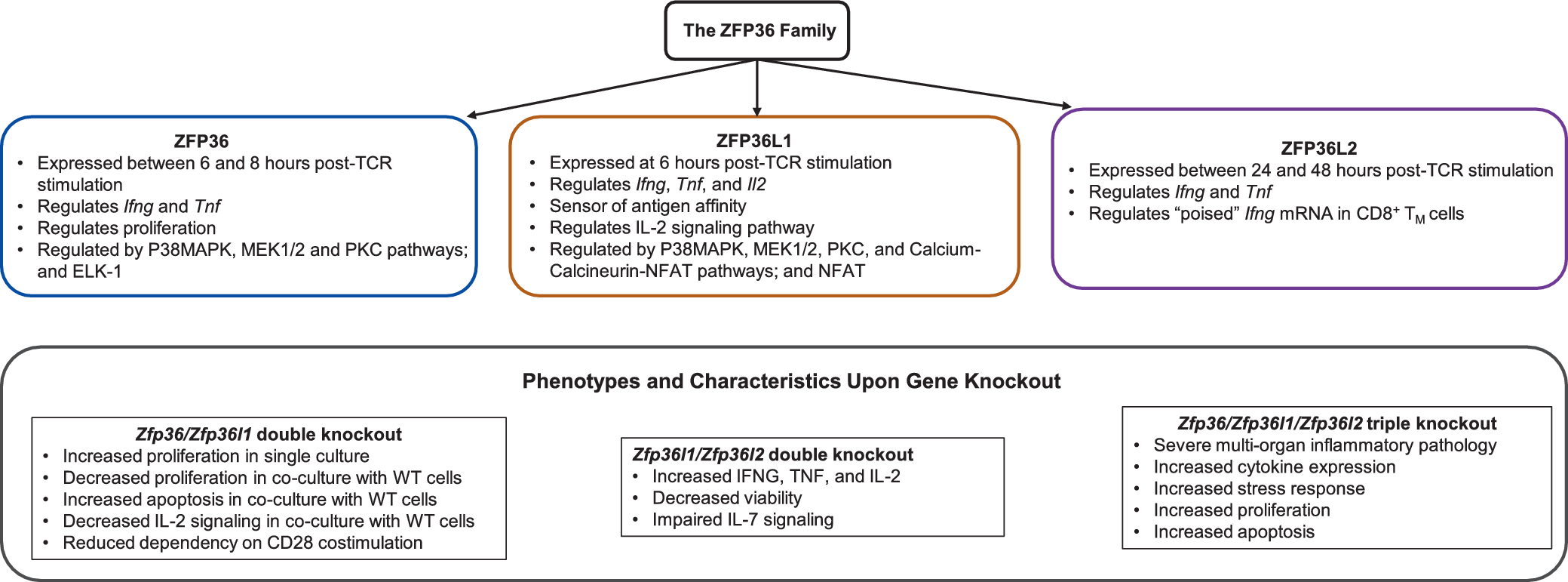


 Open access

