Case report
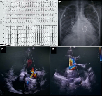
A 11-year-old girl presented with low grade fever, dyspnoea on exertion NYHA class II and multiple episodes of haemoptysis for 2 days. She had rheumatic heart disease with severe mitral regurgitation and mild mitral stenosis, for which she underwent mitral valve replacement 2 months back with a 27 mm SJM mechanical mitral valve. There was no history of cough with expectoration or thromboembolic manifestation. On examination she was tachypnoeic, tachycardic and had bilateral crepitations on auscultation. Based on the initial clinical evaluation by the paediatric medicine team, a working diagnosis of lower respiratory tract infection and infective endocarditis was made and started investigating in those lines. Electrocardiogram revealed left ventricular hypertrophy and sinus tachycardia (Fig 1a). Chest X ray revealed features suggestive of pulmonary venous hypertension. However, there was no features of pneumonic patch, pleural effusion or any cavitary lesion in the X ray (Fig 1b). Screening echocardiography showed a gradient of 8 mm Hg across mitral valve at a heart rate of 130 bpm. She was referred to us for a detailed echocardiographic evaluation to rule out infective endocarditis.
(a) Electrocardiogram on presentation showing sinus tachycardia and left ventricular hypertrophy (b) Chest X ray posteroanterior view showing features suggestive of pulmonary venous hypertension (c) Colour doppler across the mitral valve in apical 4 chamber view (d) Colour doppler across the mitral valve in parasternal long axis view showing eccentric turbulent jet towards the ventricular septum.

Patient was not willing to undergo transoesophageal echocardiography; hence transthoracic echocardiography was done. The mean diastolic gradient across mitral valve was 9 mmHg at heart rate of 126 bpm, which is not too much given the presence of tachycardia. On first glance there was no turbulence in colour doppler across the mitral valve in apical four chamber view. However, in the parasternal long axis view an eccentric turbulent jet could be seen directed towards the interventricular septum (Figure 1c and d). Mobility of the leaflets could not be clearly ascertained. On analysis of the mitral inflow continuous wave doppler, pressure half time was more than 200 m/second, which suggested obstruction across prosthetic valve. Left ventricle was dilated with left ventricular ejection fraction of 35%. We did cine fluoroscopy, which revealed mobility of one leaflet was completely restricted, while the other one was partially restricted (supplementary video 1). Her complete blood count and antistreptolysin O titres were normal, blood culture was sterile and serum procalcitonin was negative. She had sub therapeutic international normalised ratio levels (1.3), which was probably the cause for prosthetic heart valve thrombosis.
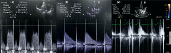
Since patient had ongoing haemoptysis, we were initially sceptical about going ahead with thrombolytic therapy. Cardiac surgeons were also in favour of managing the patient medically. Warfarin was discontinued and was started on heparin since the day of admission. Since there was no evidence to suggest pneumonia or infective endocarditis, we hypothesised that the haemoptysis was due to rupture of bronchial veins as a consequence of acute elevation of left atrial pressure secondary to obstructive prosthetic heart valve thrombosis. She was already on heparin for 2 days without any improvement in symptoms and continuation of heparin might aggravate haemoptysis. Hence, we decided to administer low dose slow infusion of thrombolytic, in an effort to improve the success of thrombolysis and reduce bleeding. She was started on tenecteplase 7.5 mg (0.25 mg/kg) over 6 hours. However, after administration of half the dosage, she had a bout of haemoptysis. The infusion was discontinued, and echocardiography was repeated. The mean diastolic gradient reduced from 9 to 4 mmHg (Fig 2b). But the mobility of one of the leaflets was still completely restricted. Over the next 24 hours the frequency and volume of haemoptysis had reduced and so we decided to give a repeat dose of tenecteplase 7.5 mg over 6 hours.
(a) Continuous wave doppler across the mitral valve on presentation (b) Continuous wave doppler across the mitral valve after initial Tenecteplase infusion (c) Final Continuous wave doppler across the mitral valve showing E & A waves and normal gradient.

Repeat fluoroscopy showed normal mobility of both the leaflets (Supplementary video 2). The mean diastolic gradient reduced even further to 1.5 mmHg (Fig 2c). Tachycardia and haemoptysis resolved in the next 24 hours. She was started on warfarin, aspirin, beta blockers, angiotensin converting enzyme inhibitor and spironolactone. On 2 weeks follow up she is doing well and international normalised ratio in therapeutic range.
Discussion
Prosthetic heart valve thrombosis is a major complication of mechanical heart valves. The annual incidence of heart valve thrombosis is around 0.3–1.3 per patient year, which is even higher in developing nations to the tune of 6% in the first 6 months of surgery. Reference Roudaut, Serri and Lafitte1,Reference Talwar, Kapoor and Velayoudam2 The common clinical manifestations of prosthetic heart valve thrombosis are heart failure, breathlessness or systemic embolisation. A significant number of cases also get detected on routine echocardiography or fluoroscopy. Patients can rarely present with fever, haemoptysis or infective endocarditis. Reference Roudaut, Serri and Lafitte1 Obstructive mitral valve thrombosis leads to sudden rise in left atrial pressure, leading to acute dilatation and rupture of bronchial veins manifesting as haemoptysis, analogous to pulmonary apoplexy in severe mitral stenosis. The first line of investigation is transthoracic echocardiography. In most instances, the visibility of leaflets is not good due to poor echocardiographic windows and acoustic shadowing. Colour doppler might show turbulent flow across the stenotic valve. The mean diastolic gradient will be significantly elevated. However, a word of caution in those with left ventricular dysfunction or bradycardia. Elevated left ventricular end-diastolic pressure in presence of left ventricular dysfunction leads to reduced difference in left atrium to left ventricle pressures, resulting in reduced mean diastolic gradient in presence of significant stenosis as seen in our patient. Pressure half time > 200 m/second and dimensional velocity index calculated as VTIprMV/VTILVOT more than 2.5 suggests significant prosthetic valve obstruction. Reference Zoghbi, Chambers and Dumesnil3 Transoesophageal echocardiography and multidetector computed tomography helps in differentiating between thrombus and pannus, which is vital in determining further course of treatment.
Management of patients presenting in NYHA class I/II includes initial heparinisation and if not responding proceed to thrombolysis. Reference Baumgartner, Falk and Bax4 Thrombolytics that have been widely studied are streptokinase and alteplase, which are the ones recommended in the ESC heart failure guidelines too. Because of widespread usage of tenecteplase these days in the management of myocardial infarction, relative ease of administration and less expensive than alteplase, tenecteplase is a potential option for thrombolysis in prosthetic heart valve thrombosis. There are a few observational studies which have shown tenecteplase to be effective in dosage of 1 mg/kg IV bolus. Reference Kumar Jha, Bansal and Issar5,Reference Sharma, Singh and Mishra6 There is only a single study which studied infusion of tenecteplase 0.5 mg/kg over 24 hours. Reference Kathirvel, Justin Paul and Prathap Kumar7 Studies by Ozkan et al. have shown that using lowering the dose and prolonging the infusion with alteplase lead to higher success rates with reduced bleeding. Reference Özkan, Gündüz and Gürsoy8,Reference Özkan, Gündüz and Biteker9 Our patient had ongoing haemoptysis as a consequence of prosthetic heart valve thrombosis, which needed to be treated keeping in mind the bleeding risk associated with thrombolysis. Hence, we decided to give 0.25 mg/kg over 6 hours Tenecteplase infusion.
To the best of our knowledge there are no previous reports of similar clinical presentation managed successfully with tenecteplase infusion. For patients presenting with prosthetic heart valve thrombosis, with high bleeding risk low dose tenecteplase infusion might be a safe and effective treatment strategy especially in resource limited settings. Also monitoring therapeutic success with echocardiographic gradient alone might be misleading, as seen in our case. Contour of the continuous wave doppler trace provides valuable clue to the function of the prosthetic heart valve.
Acknowledgements
None.
Financial support
This research received no specific grant from any funding agency, commercial or not-for-profit sectors.
Conflict of interest
None.
Ethical standards
The authors assert that all procedures contributing to this work comply with the ethical standards as mentioned in the Helsinki Declaration of 1975, as revised in 2008.
Supplementary material
To view supplementary material for this article, please visit https://doi.org/10.1017/S1047951121000603