Introduction
Following the advent of human in vitro fertilization (Steptoe & Edwards, Reference Steptoe and Edwards1978), much attention has been given to identifying first embryo morphology and, later, oocyte morphology as prognostic tools (Elder & Dale, Reference Elder and Dale2011); less attention has been given to sperm morphology. The spermatozoon delivers the haploid male genome to the oocyte, introduces the centrosome and triggers the oocyte egg into activity.
The sperm head may be considered in three parts: (1) the nucleus with a haploid set of chromosomes, in which deoxyribonucleic acid (DNA) is packaged into a volume that is typically less than 10% of the volume of a somatic cell nucleus (Dadoune, Reference Dadoune2003; Elder & Dale, Reference Elder and Dale2011); (2) the acrosome, a large Golgi-derived secretory vesicle on the proximal hemisphere of the head that contains an array of hydrolytic enzymes used for digesting the zona pellucida during penetration (Gerton, Reference Gerton and Hardy2002; Yoshinaga & Toshimori, Reference Yoshinaga and Toshimori2003); and (3) the perinuclear theca, a rigid capsule composed of disulphide bond stabilized structural proteins amalgamated with various other protein molecules (Oko, Reference Oko1995).
Human spermatozoa exhibit a wide range of shapes. Several studies have indicated that sperm morphology best predicts the outcome of natural fertilization (Kruger et al., Reference Kruger, Acosta, Simmons, Swanson, Matta and Oehninger1988; Bartoov et al., Reference Bartoov, Eltes, Pansky, Lederman, Caspi and Soffer1999), intra-uterine insemination (Berkovitz et al., Reference Berkovitz, Eltes, Soffer, Zabludovsky, Beyth, Farhi, Levran and Bartoov1999), conventional in vitro fertilization (IVF) (Kruger et al., Reference Kruger, Acosta, Simmons, Swanson, Matta and Oehninger1988; Mashiach et al., Reference Mashiach, Fisch, Eltes, Tadir, Ovadia and Bartoov1992) and intracytoplasmic sperm injection (ICSI) (Palermo et al., Reference Palermo, Joris, Devroey and Van Steirteghem1992; Bartoov et al., Reference Bartoov, Berkovitz, Eltes, Kogosowski, Menezo and Barak2002), and several techniques have been described that provide valuable information on the morphology and pathological features of spermatozoa. In a classical clinical evaluation, human sperm are fixed, stained and analysed by optical microscopy. Recently, several novel techniques have been developed for the identification of more detailed features of the cells. Differential interference contrast microscopy, scanning near-field optical microscopy, electrostatic force microscopy, atomic force microscopy and scanning thermal microscopy (Akaki et al., Reference Akaki, Nagayasu, Nakano and Aikawa2002; Bartoov et al., Reference Bartoov, Berkovitz, Eltes, Kogosowski, Menezo and Barak2002; Rothery et al., Reference Rothery, Gorelik, Bruckbauer, Yu, Korchev and Klenerman2003). Most of these techniques involve biochemical processing that requires specific equipment and may also alter the vitality of the sperm analysed.
The sperm cell is almost transparent in conventional bright field microscopy, as its optical proprieties differ slightly from the surrounding liquid, generating little contrast. However, a light beam that passes through a spermatozoon undergoes a phase change, in comparison with the surrounding medium, the amplitude of which depends on the light source, the thickness and the integral refractive index of the object itself. A qualitative visualization of this phase contrast may be obtained by contrast interference microscopy (phase contrast or Nomarski/Zernicke interferential contrast microscopy). Recently, efforts have been renewed to improve differential interferometric contrast methods in order to provide quantitative information in microscopy (Bon et al., Reference Bon, Maucort, Wattellier and Monneret2009; Kou et al., Reference Kou, Waller, Barbastathis and Sheppard2010). On the other hand, over the last few years, digital holography in microscopy has been established as a valid non-invasive, quantitative, label-free, high-resolution, phase-contrast imaging technique. Furthermore, this technique has been applied successfully to image a variety of cell types (Carl et al., Reference Carl, Kemper, Wernicke and von Bally2004; Marquet et al., Reference Marquet, Rappaz, Magistretti, Cuche, Emery, Colomb and Depeursinge2005; Charrière et al., Reference Charrière, Marian, Montfort, Kuehn, Colomb, Cuche, Marquet and Depeursinge2006; Kemper et al., Reference Kemper, Carl Schnekenburger, Bredebusch, Schäfer, Domschke and von Bally2006; Di Caprio et al., Reference Di Caprio, Gioffrè, Saffioti, Grilli, Ferraro, Puglisi, Balduzzi, Galli and Coppola2010) to obtain additional information about their structure. In a recent preliminary study in human spermatozoa using digital holographic microscopy (DHM), a statistically significant difference in phase shift was observed when comparing normal sperm with oligoasthenozoospermic sperm (Crha et al., Reference Crha, Zakova, Huser, Ventruba, Lousova and Pohanka2011), however the authors did not measure standard morphological parameters using this technique, nor investigate the characteristics of sperm with nuclear vacuoles. One of the main advantages of DHM is that high-resolution three dimensional (3-D) quantitative sample imaging can be automatically produced by numerical refocusing of a two dimensional (2-D) image at different object planes without any mechanical scanning (Dubois et al., Reference Dubois, Joannes and Legros1999; Ferraro et al., Reference Ferraro, Grilli, Alfieri, De Nicola, Finizio, Pierattini, Javidi, Coppola and Striano2005). Moreover, using a single acquired image it is possible both to reduce the size of the mass storage devices required for image saving and to achieve a fast image transfer.
In this study, we have compared semi-automated digitally enhanced Nomarski microscopy (DESA) with DHM to study morphometrical, morphological and volumetrical measurements in normal and vacuolated human spermatozoa.
Materials and methods
Specimen collection and analytical procedures
Ejaculates were collected by masturbation from 15 males scheduled to undergo IVF at the Centro Fecondazione Assistita (CFA), Naples. After liquefaction, 0.5–1.0 ml of each specimen was processed using a double-density-gradient centrifugation method (Percoll, Sydney IVF). The final pellets were resuspended in Ham's F-10 (Gibco) and used to prepare slides for DESA. Briefly, a semen aliquot that contained 2 × 10⁶ spermatozoa was washed twice by centrifugation with phosphate-buffered saline (PBS). Then the pellet was suspended in 100 μl of 2% formaldehyde in PBS and fixed for 10 min at room temperature. A 10-μl aliquot was then spotted onto a clean microscope slide, allowed to air dry and mounted with PBS:glycerol (1:1) (v/v).
In order to compare the same cells by both DESA and DHM, a grid of 20 × 20 circles (with a radius of 100 μm) was placed over the microscope. This grid was made by a photolithographic process that allows the transfer of the shape of the grid from a mask to a photo-sensitive polymer. At the end of this process, the whole surface of the slide was covered with a 1 μm-thick photo-sensitive material except the circles. A 10-μl aliquot was than spotted on the grid and finally a cover slide was placed to seal all. Only cells that fell in the circles of the grid were analysed (Fig. 1).
(A) A circle of a grid of 20 × 20 circles (with a radius of 100 μm) placed over microscope slide. (B) Differential interference contrast micrograph of a sperm head. (C) Pseudocolour plot of the same spermatozoon.

Slides were first examined under immersion oil using an inverted microscope (TI-DH; Nikon Instruments Italia) equipped with Nomarski optics enhanced by digital imaging to achieve a magnification of up to ×1500. The images were captured by a colour video camera for high-quality image production and analysed using image processing software (NIS-Element Documentation, Nikon).
Principles of DHM
In holography, an object is illuminated by a collimated, monochromatic, coherent light with wavelength λ. The object scatters the incoming light forming a complex wavefield (the object beam):
 \begin{equation}
O{\rm x,y = }\left| {O{\rm x,y}} \right|e^{j\varphi({\rm x,y})}\end{equation}
 \begin{equation}
\varphi ({\rm x,y}) = \frac{{2{\rm \pi }}}{\lambda}\ .\ {\rm OPD}\end{equation}
Image reconstruction
The image reconstruction procedure allows the retrieval of a discrete version of the complex optical wavefront present on the surface of the object under test. This optical wavefront is obtained by a numerical back propagation of spatially filtered product between the acquired hologram and a numerical replica of the reference beam (Cuche et al., Reference Cuche, Marquet and Depeursinge1999; Coppola et al., Reference Coppola, Ferraro, Iodice, De Nicola, Finizio and Grilli2004; Ferraro et al., Reference Ferraro, Coppola, Alfieri, De Nicola, Finizio and Pierattini2004). Thus, the reconstruction procedure allows the simultaneous determination of both the intensity and, especially, the phase distribution ϕ(m,n) of the optical wavefront of the specimen. Where φ(m,n) is the discretized version of the phase distribution ϕ(x,y) and m,n are positive integer numbers that identify the m-th row and n-th column of the pixels matrix of the CCD (CMOS) camera. By inverting Equation (2) and considering a homogeneous material with refractive index nc, from the reconstructed phase distribution, the thickness distribution s(m,n) of the object under investigation can be obtained as follows:
 \begin{equation}
{\rm OPD}_{{\rm m,n}} = \frac{\lambda }{{2{\rm \pi }}}\varphi ({\rm m,n}) = \frac{\lambda }{{2{\rm \pi }}}\arctan \frac{{{\mathop{\rm Im}\nolimits} [\rm Qm,n]}}{{{\mathop{\rm Re}\nolimits} [\rm Qm,n]}}\end{equation}
Finally, the possibility offered by digital holography to manage the phase of the reconstructed image allows the removal and/or compensation of any unwanted wavefront variations, such as optical aberrations (spherical, coma, tilt) and slide deformations (Ferraro et al., Reference Ferraro, De Nicola, Finizio, Coppola, Grilli, Magro and Pierattini2003).
Statistical analysis
Statistical analyses were carried out by Student's t-test. Probability values lower than or equal to 0.05 were considered to be significant.
Results
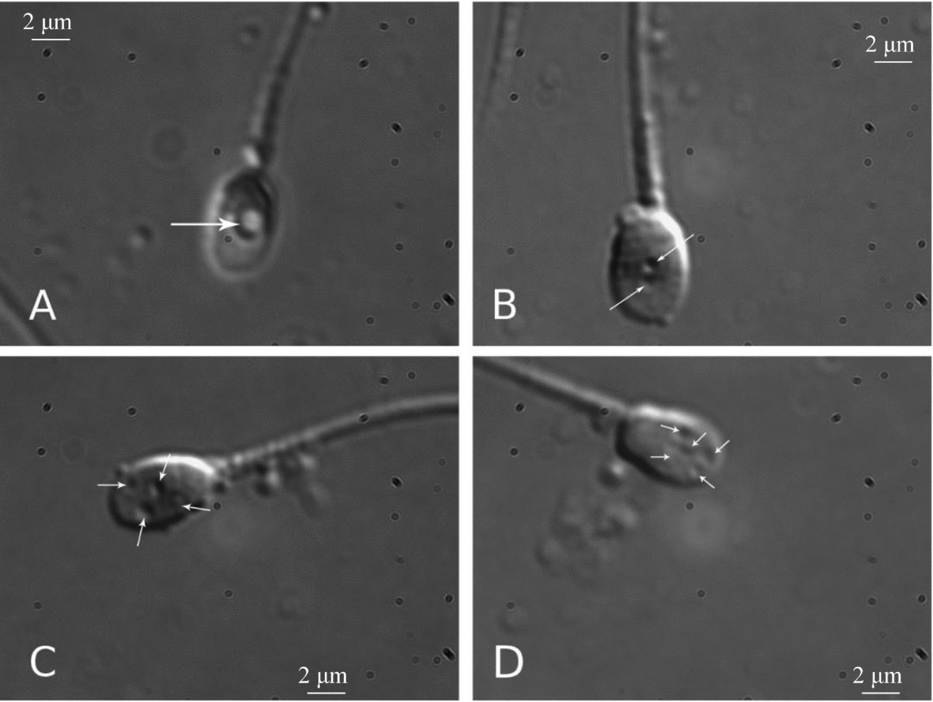
In total, 2000 digitalized sperm heads were analysed for six primary parameters (length, width, perimeter, area, number and size of vacuoles). DESA analysis revealed that the mean values for length, width, perimeter and area of the sperm head were 5.18 ± 0.64 μm (mean ± standard deviation (SD)) (range: 3.63–7.87), 3.53 ± 0.45 μm (range: 2.37–5.62), 13.75 ± 1.35 μm (range: 9.64–19.68), 14.12 ± 3.03 μm2 (range: 7.77–25.82). In the 15 ejaculates, 62.8% of spermatozoa with one or more vacuoles were found. The number of vacuoles per sperm ranged from 0–8 (mean: 2.01 ± 1.78) measuring 0.03–5.90 μm2 in area (Fig. 2).
High-power light microscope micrograph of sperm heads (×1500). (A) Crater-like appearance of nuclear vacuoles (arrow). (B–D) Spermatozoa with one or more vacuoles (arrows).
We used DHM to evaluate the morphology of 200 of the aforementioned spermatozoa (Fig. 3). Figure 3A is an example of an acquired hologram, with the fringe pattern highlighted in the inset. In Fig. 3B a pseudocolour plot of the phase-contrast map reconstruction of a human spermatozoon is shown. The colour bar shows the value in rad of the phase difference, which depends on the optical density and thickness of the biological sample. Figure 3C illustrates the quantitative reconstructed morphology obtained by applying Equation 3 to the phase-map contrast.
(A) Acquired hologram, a region is enhanced in order to show the interference pattern (inset). (B) Pseudocolour plot of a phase-contrast map for a human spermatozoon. (C) Pseudo three-dimensional (3-D) representation of a human spermatozoon image reconstructed by digital holographic microscopy (DHM).
It is important to note that this 3-D image is obtained from the reconstruction of a single acquired hologram, without the use of any mechanical scanning, allowing us to carry out numerical analyses of the six primary sperm parameters mentioned above. No significant differences were observed in the gross morphometric values of the sperm cells analysed (using DESA or DHM; Table 1).
Mean morphometric values of normal sperm heads obtained by DESA and DHM techniques
Mean ± standard deviation (SD) is presented. Significance is calculated with Student's t-test. P-values <0.05 are considered significant. DESA, digitally enhanced Nomarski microscopy; DHM, digital holographic microscopy.
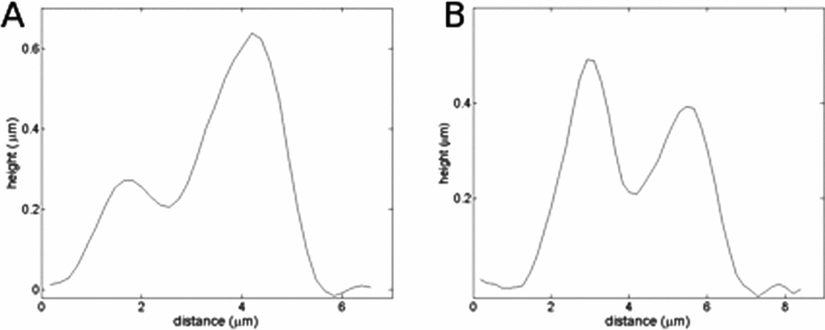
In Fig. 4A and Fig. 4B we show the quantitative profiles of a spermatozoon along the lines AA′ and BB′ illustrated in Fig. 3A, respectively. These profiles show, point by point, a quantitative value of the phase shift due to spermatozoan structure. Quantitative phase shift information from DHM allows us to calculate the volume/mass of the sperm head. In Fig. 5, for example, an isoline plot relative to different heights of the sample is displayed. For each region defined by the isolines, the occupied area and the relative volume has been estimated numerically. The analysis revealed that the mean value for volume in normal sperm is 8.03 ± 0.72 μm3.
Profile plot along the lines (A) AA′ and (B) BB′, reported in Fig. 3B.
Isolines plot of the reconstructed spermatozoon image.
In Fig. 6 we show a quantitative comparison between a control spermatozoon and a spermatozoon with vacuoles. Figure 6A,B illustrates the height profile along the major axis of the sperm head for the defect-free spermatozoon and the spermatozoon with vacuoles, respectively. In Fig. 6E the two profiles are shown together to stress the differences. It is worth note that both the shapes and the point-by-point value of the height are different. In particular, the spermatozoon with vacuoles has a distinct depression in the profile (see the arrow in the figure). The profile of the normal spermatozoon is higher than that of the spermatozoon with vacuoles, whereas their 2-D dimensions (such as area, and axes length) are similar. The difference in height difference implies a volume difference between the normal spermatozoon and the spermatozoon with vacuoles.
Comparison between a defect-free spermatozoon (left column) and a spermatozoon with vacuoles (right column). Results for a defect-free spermatozoon are reported as a quantitative three-dimensional representation (A), phase-contrast map (C), or as an isolines plot (F). For a spermatozoon with vacuoles, defects are reported as a quantitative three-dimensional representation (B), phase-contrast map (D), or as an isolines plot (G). (E) Comparison between the profile of the spermatozoon with vacuoles (curve indicated by arrow) and one without defects.
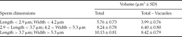
Table 2 shows three distinct groups of spermatozoa defined using two morphometric variables, head length and head width. The mean values of the volume for the three subpopulations were 5.76 ± 0.73 μm3 (for length < 2.9 μm and width < 4.2 μm), 8.24 ± 0.78 μm3 (for 2.9 < length < 3.7 μm and 4.2 < width < 5.3 μm), 10.13 ± 0.81 μm3 (for length > 3.7 μm and width > 5.3 μm). Mean values of the total volume of the spermatozoa minus the vacuoles volume are also reported.
Mean volumetric values of vacuolated sperm clustered in three different subpopulations
SD, standard deviation.
Discussion
Here we have used digital holographic microscopy as a novel approach for a more advanced morphological analysis of human spermatozoa, in particular to measure the head volume in the presence and absence of vacuoles.
In human sperm, the presence of vacuoles has been related to poor outcome in ART (Berkovitz et al., Reference Berkovitz, Eltes, Lederman, Peer, Ellenbogen, Feldberg and Bartoov2006), an increase in DNA fragmentation (Franco et al., Reference Franco, Baruffi, Mauri, Petersen, Oliveira and Vagnini2008; Wilding et al., Reference Wilding, Coppola, Di Matteo, Palagiano, Fusco and Dale2011) and abnormal chromatin packaging (Franco et al., Reference Franco, Mauri, Petersen, Massaro, Silva, Felipe, Cavagna, Pontes, Baruffi, Oliveira and Vagnini2012).
Nuclear vacuoles have been described as a crater defect in the spermatozoa of stallion (Johnson & Hurtgen, Reference Johnson and Hurtgen1985), as a pouch formation (Bane & Nicander, Reference Bane and Nicander1965), a diadem defect (Blom, Reference Blom and Blom1977) or a nuclear sperm defect (Miller et al., Reference Miller, Hrudka, Cates and Mapletoft1982) in bull spermatozoa, and as a crater defect (Johnson & Truitt-Gibert, Reference Johnson and Truitt-Gibert1982) or a pouch formation (Bane & Nicander, Reference Bane and Nicander1965) in boar spermatozoa. The defect is believed to originate during spermiogenesis, as vacuoles have been found in both early and late spermatids (Johnson & Truitt-Gibert, Reference Johnson and Truitt-Gibert1982). Nuclear vacuoles were shown by electron microscopy to be narrow-mouthed invaginations of the nuclear membrane into the nucleoplasm often filled with an amorphous, cytoplasmic material (Barth & Oko, Reference Barth and Oko1989). The predominant locations of the vacuoles are the apical region and the acrosome-postacrosomal sheath junction, but they have also been found throughout the sperm head (Barth & Oko, Reference Barth and Oko1989).
Our results show that spermatozoa with vacuoles had a reduced volume probably due to variation of the inner structure of the sperm head with loss of material. We suggest that vacuolated spermatozoa with normal length and width (Bartoov et al., Reference Bartoov, Berkovitz, Eltes, Kogosowski, Menezo and Barak2002) should be avoided for selection during the ICSI or IMSI procedure, until we acquire more data on the integrity and volume normal viable spermatozoon.
Recently, we have employed a microfluidic system with DHM on unstained bovine spermatozoa in their natural physiological surroundings (Di Caprio et al., Reference Di Caprio, Gioffrè, Saffioti, Grilli, Ferraro, Puglisi, Balduzzi, Galli and Coppola2010). This situation raises the possibility to use the same technique for a more complete analysis of human spermatozoa, with the additional possibility of sorting cells according to specific morphological criteria.