Introduction
Cryptosporidiosis is the disease caused by the protozoan parasite Cryptosporidium. It is an important zoonotic pathogen which affects many species including humans (Chalmers and Davies, Reference Chalmers and Davies2010; Fayer, Reference Fayer2010; Chalmers and Katzer, Reference Chalmers and Katzer2013). In the UK cryptosporidiosis is a common disease in livestock (APHA and SRUC, 2014), although clinical disease is usually limited to neonatal livestock and is caused by Cryptosporidium parvum (Tzipori et al., Reference Tzipori, Smith, Halpin, Angus, Sherwood and Campbell1983); older animals are believed to mostly be infected with other species of the parasite (C. andersoni in adults and C. ryanae and C. bovis in calves of several months old) and tend not to show clinical signs (Anderson, Reference Anderson1987; Esteban and Anderson, Reference Esteban and Anderson1995; Fayer et al., Reference Fayer, Santin and Trout2008; Santin et al., Reference Santin, Trout and Fayer2008). In livestock and humans, the disease usually causes self-limiting watery diarrhoea, loss of appetite and abdominal pain (Tzipori and Ward, Reference Tzipori and Ward2002; Klein et al., Reference Klein, Kleinova, Volek and Simunek2008). Farm animals that have recovered from cryptosporidiosis have in some cases been shown to have slower growth rates than uninfected animals (de Graaf et al., Reference De Graaf, Vanopdenbosch, Ortega-Mora, Abbassi and Peeters1999; Sweeny et al., Reference Sweeny, Ryan, Robertson, Yang, Bell and Jacobson2011a, Reference Sweeny, Ryan, Robertson and Jacobson2011b). The economic losses associated with Cryptosporidium have not been estimated but costs attributable to diarrhoeal disease in calves have been estimated to be at least £34 per affected calf (Gunn and Stott, Reference Gunn and Stott1997).
Cryptosporidium oocysts are transmitted between hosts via the fecal–oral route. The oocysts can survive for several months in the environment (Robertson et al., Reference Robertson, Campbell and Smith1992; Jenkins et al., Reference Jenkins, Trout, Higgins, Dorsch, Veal and Fayer2002) and are highly resistant to commonly used disinfectants, making them difficult to eradicate (Carpenter et al., Reference Carpenter, Fayer, Trout and Beach1999; Weir et al., Reference Weir, Pokorny, Carreno, Trevors and Lee2002). Infected hosts can shed huge numbers of oocysts per day (over 10 × 1010), which are immediately infective to other humans or animals (Nydam et al., Reference Nydam, Wade, Schaaf and Mohammed2001). There are currently over 30 recognized species of Cryptosporidium and many more genotypes (Fayer, Reference Fayer2010; Chalmers and Katzer, Reference Chalmers and Katzer2013) but the most common species in humans and calves is C. parvum (Brook et al., Reference Brook, Christley, French and Hart2008; Chalmers and Giles, Reference Chalmers and Giles2010; Chalmers et al., Reference Chalmers, Smith, Hadfield, Elwin and Giles2011a, Reference Chalmers, Smith, Elwin, Clifton-Hadley and Giles2011b; Gormley et al., Reference Gormley, Little, Chalmers, Rawal and Adak2011; Xiao and Feng, Reference Xiao and Feng2009). It is known that young calves become infected with C. parvum very soon after birth but it is not known if they pick up the infection from their dams, the environment or via another route.
At present, the only preventative or therapeutic products licensed in the UK for Cryptosporidium in cattle are Halocur® [halofuginone lactate (MSD Animal Health)] and Parofor® crypto [paromomycin (Huvupharma)]. Halocur® must be administered carefully as it is toxic at only twice the recommended dose and contra-indicated in dehydrated animals (MSD Animal Health Data Sheet, 2012). Parofor® crypto must only be given to animals with a confirmed diagnosis of Cryptosporidium and should not be given after the onset of diarrhoea (Huvepharma Data Sheet). Both should be given for 7 days consecutively. Due to the lack of suitable therapeutics or prophylaxis to control cryptosporidiosis in calves, it is important to understand transmission routes in order to develop better management strategies.
The studies described here aimed to determine the species and subtypes of Cryptosporidium present in calves and the variation in occurrence throughout the first year of life. Although some studies have previously been carried out to determine the prevalence of Cryptosporidium in calves in the UK these have mostly used less sensitive techniques and were unable to speciate or subtype the parasites. The present longitudinal study is the first to examine a population of calves from birth to sexual maturity on a farm in the UK and to investigate dams as a possible source of infection for calves.
Materials and methods
Sample collection
Calves
Fecal samples were collected from 25 calves (Study-1) and 30 calves born to cows and heifers (Study-2) from the day of birth until 6 weeks of age, three times per week (Monday, Wednesday and Friday). The calves were kept under the normal working conditions of this farm regarding housing, feeding and veterinary treatment. The calves were kept with their mothers for 1 or 2 days in a straw-bedded pen with other dams and new-born calves then moved to a small group of 5–8 calves in a straw-bedded pen adjacent to the calving pens for 1 week. All calves in both studies were seen to suckle their dams within the first few hours of life. Following removal from their dams calves were fed on calf milk replacer by an automatic feeder and housed in larger indoor straw-bedded pens with approximately 20 calves in each pen for the remaining 5 weeks, calves 1–14 were kept in one pen and calves 15–25 were kept in another pen (Study-1). Both pens contained other calves that were not included in this study. All calves wore a transponder to enable the recording of daily feed intake, so that it was easy for stock-workers to determine if a calf was inappetant and to intervene accordingly. Calves also had ad lib access to hay and pelleted calf feed. Calves older than 6 weeks were moved and housed in a separate straw-bedded pen in larger groups where they had ad lib access to water, silage and pelleted feed. Samples were collected from the same calves again at 3, 6, 9 and 12 months of age (Study-1) and at 6 months from calves in Study-2. The samples were aliquoted into 7 mL tubes and stored at −20 °C for further processing.
Adult cattle (Study-2)
Fecal samples were collected from 29 in-calf adult dairy cattle (heifers and cows) three times per week for up to 10 weeks pre-calving, in total 209 samples were collected. The cows were kept under the normal working conditions of this farm and as such were housed in cubicle sheds before calving, in straw courts at calving and in slatted cubicle sheds after calving when they returned to the milking herd. Cows were observed in the cattle shed until they defecated and then the entire motion was collected in a plastic bag. Due to the method of collection, it was not always possible to collect a sample from each cow at each sampling point. After collection, the samples were transferred to the laboratory where the entire motion was mixed and a sub-sample aliquoted into a 125 mL sample pot and stored at 4 °C.
Fecal consistency
The consistency of the collected samples was assessed (Study-1 only) and assigned one of three possible scores: normal – forms a pat (1), soft – forms a puddle (2), diarrhoeic – entirely liquid, flows freely (3). For statistical analysis as a binary outcome, scores 1 and 2 were combined and contrasted with score 3.
Sample processing and DNA extraction
Parasite DNA was extracted from the feces using a Macherery-Nagal NucleoSpin® Tissue Kit according to the manufacturer's recommendations. Except that 10 × freeze–thaw cycles in liquid nitrogen were added prior to the overnight incubation. The DNA was eluted in 100 µL dH2O and stored at −20 °C.
Samples from older calves (>6 months) and adults were concentrated prior to DNA extraction.
Sample concentration (older calves)
Briefly, 3 g feces were resuspended in 25 mL dH2O and left to settle for approximately 5 min. Once large particles had settled the supernatant was removed and added to another tube. This step was repeated and the tube containing the supernatant was centrifuged at 1000 × g for 5 min, the supernatant was poured off and the resultant pellet used for DNA extraction (Brook et al., Reference Brook, Christley, French and Hart2008).
Sample concentration (adults)
Samples were processed to concentrate Cryptosporidium oocysts from a 50 g starting sample by acid flocculation and salt flotation as described by Wells et al. (Reference Wells, Thomson, Ensor, Innes and Katzer2016). This resulted in a fecal pellet which was used for DNA isolation.
PCR amplification of the 18S rRNA gene
A previously described nested PCR protocol (Xiao et al., Reference Xiao, Escalante, Yang, Sulaiman, Escalante, Montali, Fayer and Lal1999) was used to amplify a ~840 bp fragment of the 18S rRNA gene. Each 25 µL reaction contained 10 × PCR buffer (45 mm Tris-HCl pH 8.8, 11 mm (NH4)2SO4, 4.5 mm MgCl2, 4.4 µ m EDTA, 113 µg mL-1 BSA, 1 mm each of the four deoxyribonucleotide triphosphates) (Burrells et al., Reference Burrells, Bartley, Zimmer, Roy, Kitchener, Meredith, Wright, Innes and Katzer2013), 0.5 units BioTaq (Bioline, UK), 10 µ m of forward and reverse primers [AL1687 (EF) and AL1691 (ER); and AL1598 (IF) and AL3032 (IR)] and 3 µL DNA in the primary round and 1 µL primary PCR product in the secondary round. The total volume was made up to 25 µL with dH2O. In each PCR run, one set of positive control DNA and negative controls consisting of dH2O were included. All reactions were carried out in triplicate.
Cycling conditions were 3 min at 94 °C, followed by 35 cycles of 45 s at 94 °C, 45 s at 55 °C and 1 min at 72 °C. The final extension was 7 min at 72 °C. Secondary round amplification products (~3 µL) were visualized following electrophoresis on a 1.5% agarose gel stained with GelRed™ (Biotium, UK) on an AlphaImager 2000.
Species identification by nssm-PCR (nested species-specific multiplex-PCR)
For the differentiation of Cryptosporidium species, the primary PCR products from the 18S rRNA nested PCR were amplified using a nested species-specific multiplex PCR (nssm-PCR) (Thomson et al., Reference Thomson, Innes, Jonsson and Katzer2016) which can distinguish the four most commonly detected bovine species of Cryptosporidium.
The primary PCR products were diluted with 50 µL dH2O and 1 µL of the dilution used as a template in the secondary round. Secondary PCR reactions contained 2.5 µL 10 × PCR buffer, 0.5 units BioTaq (Bioline), 10 µ m of each primer (AL1598, AL3032, CaF, CrF, CphF and CbF) and 1 µL template.
Cycling conditions were 3 min at 94 °C, followed by 35 cycles of 45 s at 94 °C, 45 s at 56 °C and 1 min at 72 °C. The final extension was 7 min at 72 °C. Secondary PCR products (~2 µL) were visualized following electrophoresis on a 1.5% agarose gel stained with GelRed™ (Biotium) on an AlphaImager 2000.
PCR amplification of the GP60 gene for C. parvum subtyping
A nested PCR designed to amplify a 450 bp fragment of the GP60 gene (Brook et al., Reference Brook, Anthony, French and Christley2009) was used on a selection of C. parvum-positive samples to identify their subtypes. Each 25 µL reaction contained 10 × PCR buffer, 0.5 units BioTaq (Bioline), 10 µ m of forward and reverse primers (GP60 1F and GP60 1R) and (GP60 2F and GP60 2R) with 3 µL DNA in the primary round and 1 µL primary PCR product in the secondary round. Cycling conditions were 3 min at 94 °C, followed by 35 cycles of 45 s at 94 °C, 45 s at 55 °C and 1 min at 72 °C. The final extension was 7 min at 72 °C. Secondary PCR products (~2 µL) were visualized following electrophoresis on a 1.5% agarose gel stained with GelRed™ (Biotium) on an AlphaImager 2000.
Sequencing of GP60-positive samples
GP60-positive samples were sequenced in the forward and reverse orientation. DNA sequencing was carried out by GATC Biotech (Köln, Germany). The sequences were analysed using ChromasLite (version 2.01) and the subtypes named as according to (Sulaiman et al., Reference Sulaiman, Hira, Zhou, Al-Ali, Al-Shelahi, Shweiki, Iqbal, Khalid and Xiao2005).
Statistical analysis
Statistical analyses were carried out using R (R Core Team 2016). Response variables (diarrhoea/not diarrhoea, C. parvum positive/negative) were coded as binary data. Generalized linear models (GLM) with a binomial error structure and a logit link function were used. Initially, a maximal model was constructed with all variables and interaction terms were first fitted for the effects examined in each study. These models were reduced by successively dropping out non-significant interaction terms. None of the interaction terms were significant, so additive models were used. To more accurately model the effect of time on the response variables, generalized additive models (GAM) were constructed using a smoothed term for days. The GAM and GLM were compared according to the Akaike information criterion (AIC) and the percentage of deviance explained. In all cases, the GAM provided a lower (and hence better) AIC. For Study-1, when the response variable was C. parvum positivity, the factors included in the model were date of birth (DOB), pen and days after birth. In Study-1, when the response variable was diarrhoea, effects were days postpartum and C. parvum positivity of the sample. In Study-2 (calves), the factors were DOB and days postpartum. In Study-2 (adults), the only factor was the number of days prepartum. For the calf data in Study-1 and Study-2, to determine the effects of the factors of interest, GAMs were run on full data-sets including all time-points, however, for clarity of presentation, GAM figures are presented for only the first 6 weeks (Study-1) or 7 weeks (Study-2).
Results
Cryptosporidium prevalence in calves
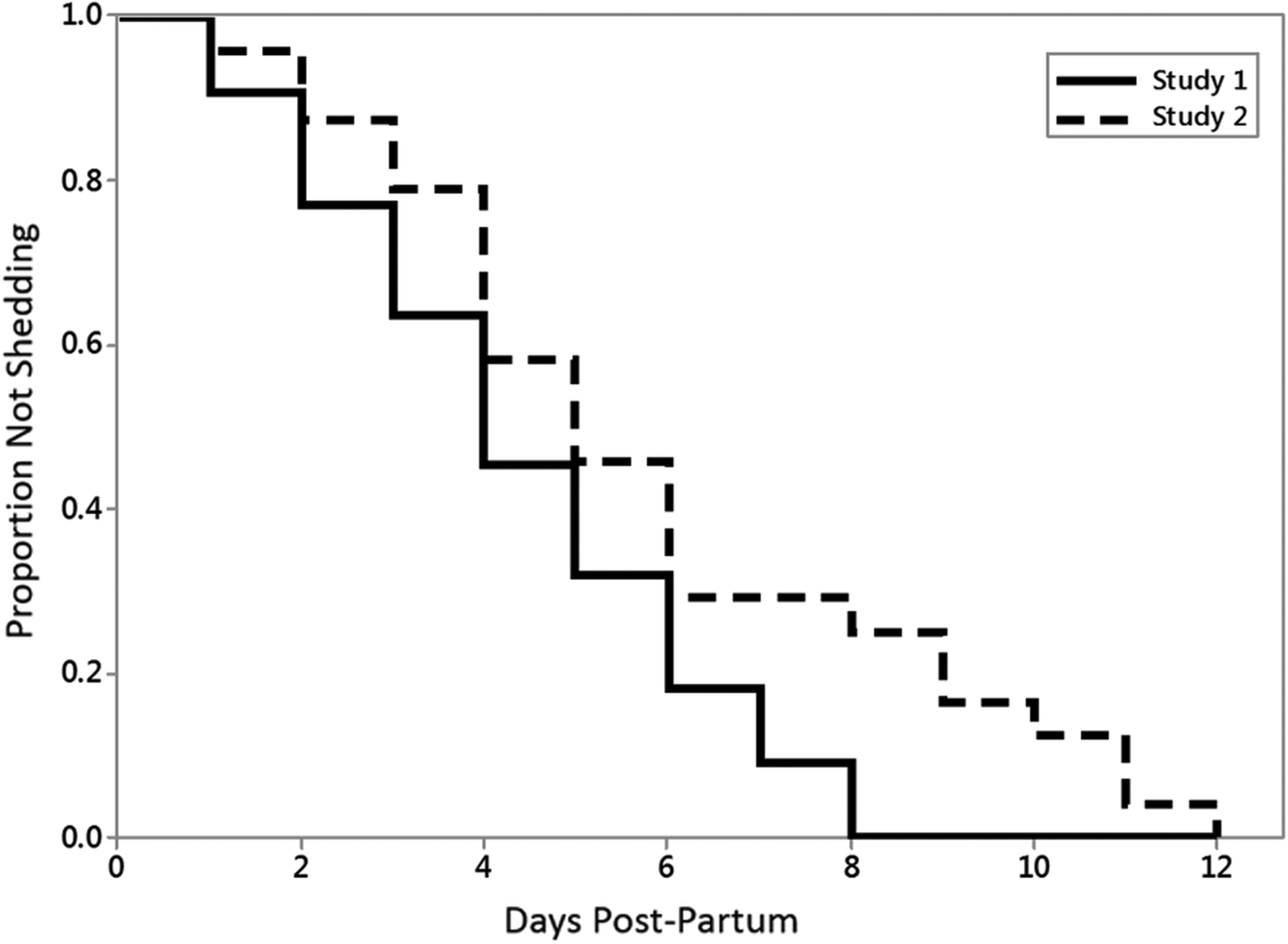
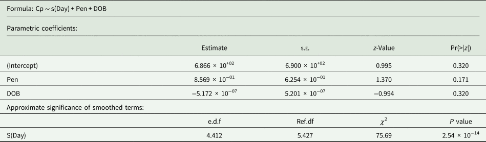
In total 384 fecal samples were collected from 25 calves in Study-1; 351 samples from the first 6 weeks, 17 from calves at 3 months of age, 16 from calves at 6 months of age. Three hundred and thirty-one samples were collected in total from 30 calves in Study-2; 315 from the initial collection period and 16 from the same calves at 6 months of age. All 715 samples were screened for the presence of Cryptosporidium parasite DNA using the 18S PCR and overall the percentage of positive samples was 64.6% (n = 248) in Study-1 and 57.4% (n = 190) in Study-2. The age of earliest detection of Cryptosporidium parasite DNA was 1 day old, and the longest interval from birth to onset of shedding was 12 days. Figure 1 shows that by Day-6, the majority of the calves in both studies had begun shedding. The GAM from Study-1 for the effects of date of birth, pen, days postpartum explained 23% of the deviance in the probability of C. parvum positivity and is shown in Table 1.
Kaplan–Meier curve showing the proportion of calves not having shed Cryptosporidium by days postpartum. It can be seen that the first record of shedding occurred on Day 1 and that by Day 8, all calves in Study-1 had shed Cryptosporidium on at least one occasion. Calves in Study-2 had all shed at least once by Day 12.

Study-1: Generalized additive model (GAM) for the effects of date of birth, pen and days after birth on the probability of any sample being positive for C. parvum

R 2(adj) = 0.268 Deviance explained = 23% UBRE = 0.067016 Scale est. = 1 n = 352.
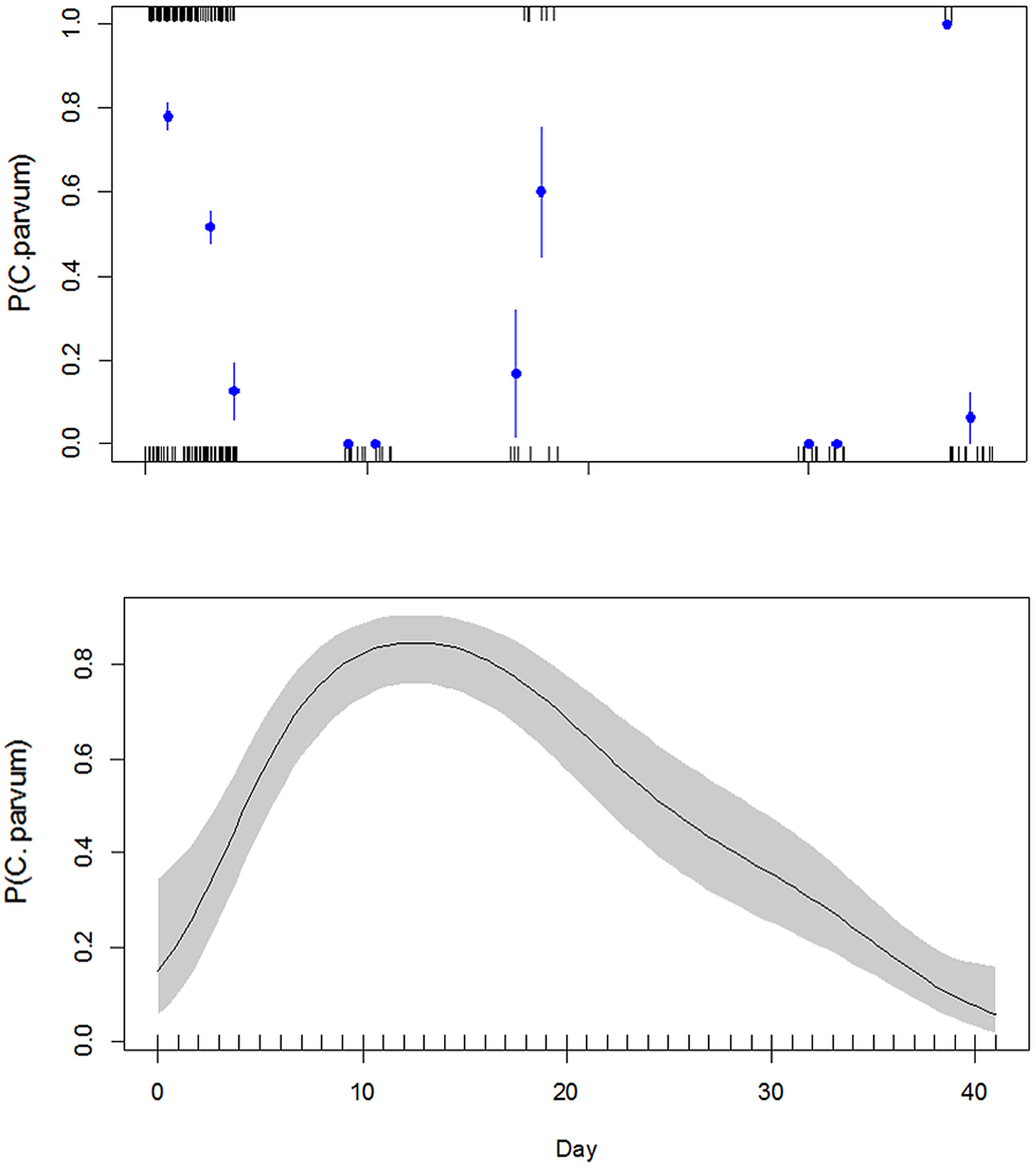
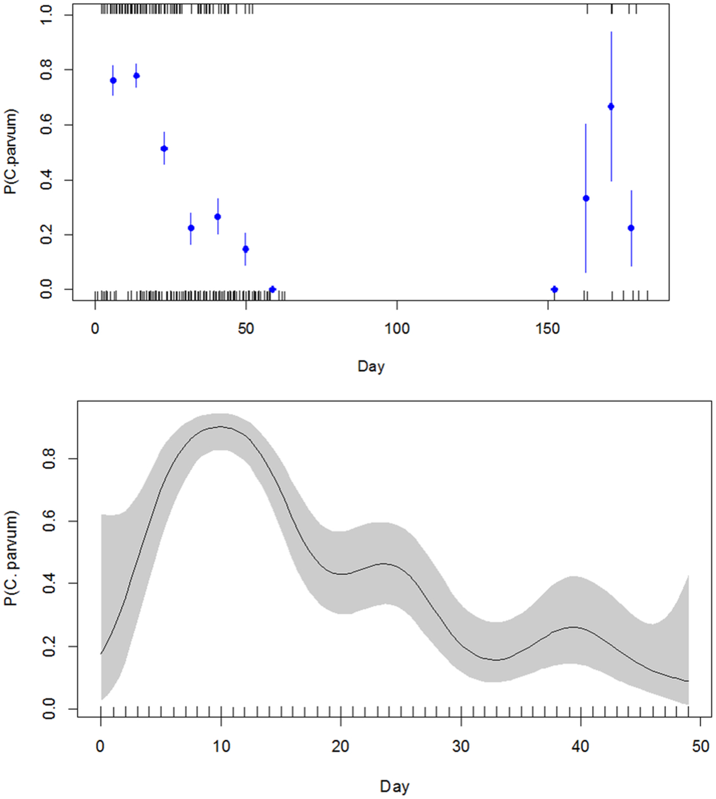
Neither pen nor date of birth significantly affected the period from birth to the onset of shedding of Cryptosporidium (P = 0.17 and 0.32, respectively). However, days postpartum had a highly significant effect (P < 0.0001). Figure 2 shows the predicted probability of any sample being positive for C. parvum for the entire study period and below shows the GAM for the first 40 days postpartum. The GAM from Study-2 (Fig. 3) is very similar to that from Study-1, explains 24% of the deviance and is shown in Table 2. Date of birth did not significantly affect the probability of shedding on any day (P = 0.43) but the day after birth was highly significant (P < 0.0001).
Study-1: Above – Estimated probabilities and standard errors for C. parvum positivity for samples obtained from throughout the entire Study-1 period. The tick marks on the lower axis represent the sampling occasions with negative results (i.e. not positive for C. parvum) tick marks on the upper axis indicate sampling occasions with positive results (i.e. positive for C. parvum). Below – Predicted (solid line) and 95% confidence interval of the probability of any sample being positive for Cryptosporidium by each day of the first 6 weeks of life from calves in Study-1.

Study-2: Above – Estimated probabilities and standard errors for C. parvum positivity for samples obtained from throughout the entire Study-2 period. The tick marks on the lower axis represent the sampling occasions with negative results (i.e. not positive for C. parvum) tick marks on the upper axis indicate sampling occasions with positive results (i.e. positive for C. parvum). Below – Predicted (solid line) and 95% confidence interval of the probability of any sample being positive for Cryptosporidium by each day of the first 7 weeks in Study-2 (below).

Study-2: Generalized additive model (GAM) for the effects of date of birth and days after birth on the probability of any sample being positive for C. parvum

R 2(adj) = 0.277 Deviance explained = 23.8% UBRE = 0.10676 Scale est. = 1 n = 339.
Prevalence of Cryptosporidium in adult cattle
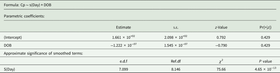
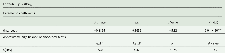
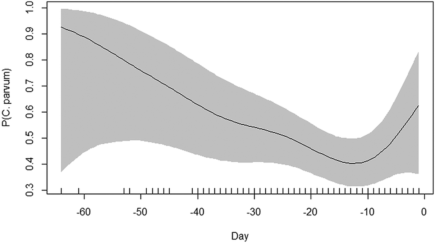
Most samples were collected from adult cows during the 4-week period prior to calving. All 209 samples were tested for the presence of Cryptosporidium parasite DNA by 18S PCR following oocyst concentration. Overall 27.3% (n = 57) of samples tested positive for Cryptosporidium. Only samples from four cows did not test positive at any point throughout the study, the remaining 24 (82.2%) cows tested positive on at least one occasion. The GAM for the probability of shedding C. parvum is shown in Table 3. The model explained only 5% of the deviance and there was no significant effect of time on the probability of adult cattle shedding C. parvum in the 100 days prior to calving (P = 0.104) (Fig. 4).
Study-2: Generalized additive model (GAM) for the effect of day prepartum on the probability of any sample being positive for C. parvum

R 2(adj) = 0.0385 Deviance explained = 4.56% UBRE = 0.21096 Scale est. = 1 n = 182.
GAM showing the effect of ‘Day’ on the probability of C. parvum positivity in adult cows in the 100 days leading to parturition. Tick marks on the lower margin reflect the time of sampling. Black line is the predicted probability and grey shading is the 95% confidence interval.

Speciation of Cryptosporidium by nssm-PCR
All Cryptosporidium-positive samples were speciated using an nssm-PCR (Thomson et al., Reference Thomson, Innes, Jonsson and Katzer2016) and four distinct species were identified (C. parvum, C. bovis, C. ryanae and C. andersoni).
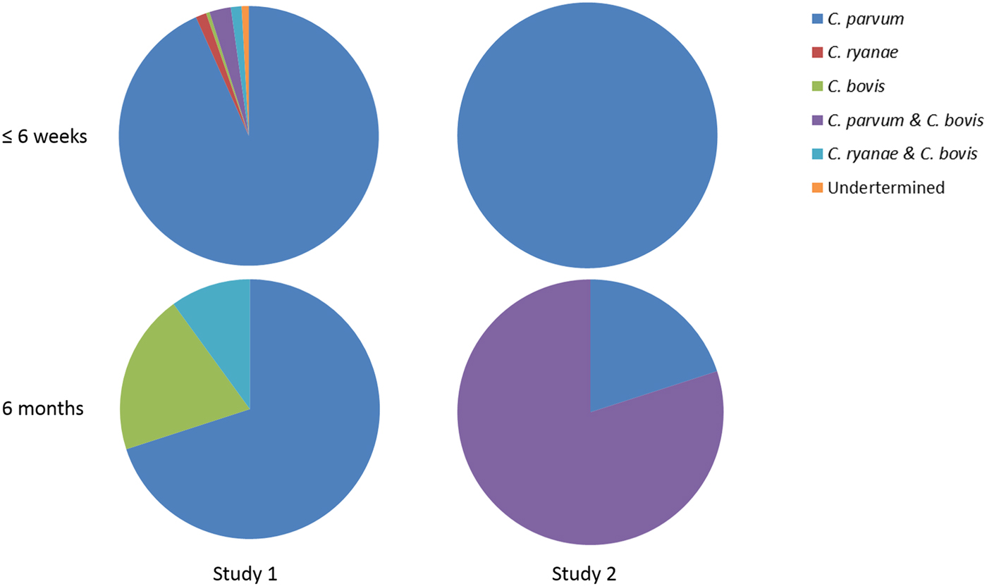
In Study-1 C. parvum was identified in 217 samples, C. bovis in ten samples and C. ryanae in 6 samples from calves ⩽6 weeks. Nine of these were as mixed infections; C. parvum and C. bovis (n = 6) and C. bovis and C. ryanae (n = 3). At 3 and 9 months only C. ryanae and C. bovis were detected in two samples each at 3 months and as either single [C. ryanae (n = 2), C. bovis (n = 3)] or mixed infections (n = 2) at 9 months. However, at 6 months of age, the predominant species detected was C. parvum, with 7/10 of the positive samples being identified as this species.
Two calves shed C. parvum at 12 months of age, one as a mixed infection with C. bovis. Cryptosporidium andersoni was not detected at all, and for three of the samples, it was not possible to determine the species by either nssm-PCR or sequence analysis of the 18S rRNA gene.
In Study-2, only C. parvum (n = 178) was detected in calves ⩽6 weeks old while mixed infections of C. bovis and C. parvum (n = 4) and C. bovis alone (n = 1) were detected in calves at 6 months of age.
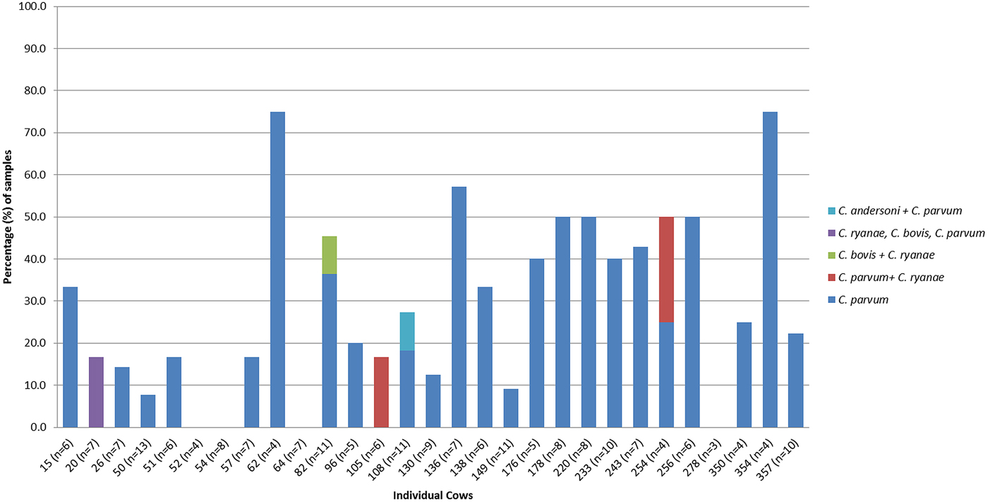
All four species were identified in samples from adult cattle from Study-2, C. parvum alone was detected in 52 (91.2%) samples, C. ryanae (mixed with C. parvum) in two samples, C. bovis (mixed with C. ryanae) in one sample, C. andersoni (mixed with C. parvum) in one sample and one sample containing C. parvum, C. ryanae and C. bovis (Fig. 5).
Species of Cryptosporidium detected in fecal samples from each of the adult cattle. The species that was most frequently detected in adult cattle was C. parvum. Four cows did not test positive for any Cryptosporidium species.

In both studies, C. parvum was the predominant species detected in fecal samples from calves ⩽6 weeks old, with seven calves from Study-1 showing evidence of mixed infections. Mixed infections of C. parvum and C. bovis were detected in calves between 3 and 5 weeks of age and C. bovis and C. ryanae were detected together only in 6-week-old animals. No mixed infections were detected in calves from Study-2 (Fig. 6). Cryptosporidium parvum was also the most commonly detected species at 6 months of age in both studies with seven and five calves shedding this species in Study-1 and Study-2, respectively (Fig. 6).
Species shed by calves at ⩽6 weeks of age and at 6 months. Cryptosporidium parvum was the most prevalent species in calves ⩽6 weeks of age and at 6 months of age.

Subtyping of C. parvum by GP60 PCR
To determine the gp60 subtypes, 363 samples that were positive for C. parvum from both studies were subjected to a nested PCR to amplify a fragment of the GP60 gene. Three hundred and six of these were positive at the GP60 locus, a subset (n = 111) from calves in both studies and 17 positive samples from adult cattle were sent for sequence analysis.
In calves from both studies, two distinct gp60 subtypes were identified. In all of the samples from the calves ⩽6 weeks, the subtype was IIaA19G2R1 (n = 101) and in the samples from the same calves at 6 months the subtype was IIaA15G2R1 (n = 10). Four different gp60 subtypes were identified in adult cattle; these were IIaA15R1 (n = 2), IIaA15G2R1 (n = 11), IIaA18G2R1 (n = 3) and IIaA19G2R1 (n = 1).
Fecal consistency
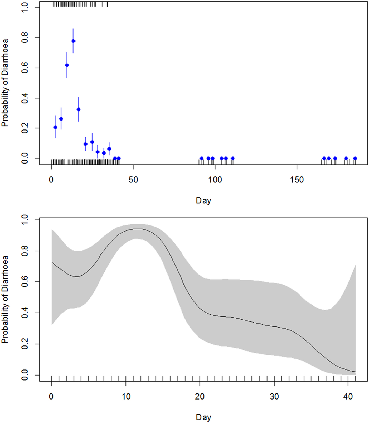
All samples collected from calves in Study-1 were scored for fecal consistency (1 = normal, 2 = soft, 3 = diarrhoeic). In the first 6 weeks of life, 21.4% (n = 75) of the samples collected were scored as diarrhoeic. The majority of these (n = 69) occurred in the first 3 weeks of life, with a peak in diarrhoeic samples (n = 17, 89.5%) at 12–14 days of age. By week 6 none of the samples were scored as diarrhoeic or soft. All of the samples that were collected at 3, 6, 9 or 12 months of age were scored as normal; no soft or diarrhoeic samples were seen. Cryptosporidium bovis and C. ryanae were not isolated from any soft or diarrhoeic samples, although some samples that were negative for Cryptosporidium were scored as either diarrhoeic or soft and C. parvum was detected in normal feces. The GLM for diarrhoea indicated a significant effect of day and of C. parvum positivity on the probability of diarrhoea (P = 0.00013), but only explained 18% of the deviance in probability. However, the GAM (Fig. 7), which allows for smoothed, non-linear effects of time, provided a significantly better model, explaining 30% of the deviance and showed a highly significant effect of day on the probability of diarrhoea (P < 0.0001) but only a weak, non-significant effect of C. parvum positivity (P = 0.11). The use of the GAM thus enabled the confounding effects of day and C. parvum positivity to be separated. Fecal scores were not recorded for samples collected from calves or adults from Study-2.
Above – Probability plot of the likelihood of diarrhoea by day postpartum for the entire period of the study. Dots represent probability estimates with error bars representing the standard errors of the estimates. The tick marks on the lower axis represent the sampling occasions with negative results (i.e. not diarrhoea) tick marks on the upper axis indicate sampling occasions with positive results (i.e. diarrhoea). Below – GAM for effect of ‘Day’ on the probability of diarrhoea for the first 40 days postpartum. Black line represents the predicted probability of diarrhoea and the grey shading covers the 95% confidence interval.

Discussion
Though there are many species of Cryptosporidium described only four are generally found in cattle, in this study all four of the common cattle adapted species were identified. Cryptosporidium andersoni is one of the largest of the Cryptosporidium species and usually infects the abomasum of adult cattle, this species does not typically cause diarrhoea but has been associated with reduced milk yield (Anderson, Reference Anderson1987). Cryptosporidium ryanae and C. bovis are similar in size to C. parvum and are smaller than the oocysts of C. andersoni, these species, as with C. parvum tend to infect the small intestine of susceptible hosts. Only C. ryanae has not been reported in humans (Zahedi et al., Reference Zahedi, Paparini, Jian, Robertson and Ryan2016).
In the UK, and worldwide, cryptosporidiosis is frequently reported as a major cause of neonatal calf diarrhoea (APHA and SRUC, 2014), which may have long-term impacts on the health of infected calves (Gunn and Stott, Reference Gunn and Stott1997; Thomson et al., Reference Thomson, Hamilton, Hope, Katzer, Mabbott, Morrison and Innes2017). It is well documented that the zoonotic species C. parvum is the most commonly detected species in young calves (Santin et al., Reference Santin, Trout, Xiao, Zhou, Greiner and Fayer2004, Reference Santin, Trout and Fayer2008; Rieux et al., Reference Rieux, Paraud, Pors and Chartier2013b) except in a few reports from Sweden (Silverlas and Blanco-Penedo, Reference Silverlas and Blanco-Penedo2013) and China (Cai et al., Reference Cai, Guo, Pan, Li, Wang, Tang, Feng and Xiao2017) in which C. bovis was identified as the most common species in pre-weaned (⩽8 weeks old) calves. In this study C. parvum was detected in 59.3% (n = 395) samples from calves 6 weeks old or younger, C. bovis and C. ryanae were also detected in calves of this age but at much lower levels (1.9%, n = 13) and only in calves from Study-1. In general, only C. parvum is associated with diarrhoea, this finding was confirmed again in this study.
Previous reports suggest an age-related distribution of Cryptosporidium species in cattle that C. parvum is most frequent in pre-weaned calves, that the most common species in calves older than 2 months are C. bovis and C. ryanae (Santin et al., Reference Santin, Trout, Xiao, Zhou, Greiner and Fayer2004, Reference Santin, Trout and Fayer2008; Langkjaer et al., Reference Langkjaer, Vigre, Enemark and Maddox-Hyttel2007), and that adult cattle are usually infected with C. andersoni. Consistent with this, pre-weaned calves in the present study were infected with C. parvum, those of 3 and 9 months of age were infected with C. bovis and C. ryanae (Study-1); however, at 6 months of age (Study-1 and Study-2) the calves were mostly infected with C. parvum. This inconsistent pattern of infection is very unusual and C. parvum in older calves has only been reported in three previous studies (Huetink et al., Reference Huetink, van der Giessen, Noordhuizen and Ploeger2001; Santin et al., Reference Santin, Trout, Xiao, Zhou, Greiner and Fayer2004; Follet et al., Reference Follet, Guyot, Leruste, Follet-Dumoulin, Hammouma-Ghelboun, Certad, Dei-Cas and Halama2011) although in the study by Follet et al. (Reference Follet, Guyot, Leruste, Follet-Dumoulin, Hammouma-Ghelboun, Certad, Dei-Cas and Halama2011) the calves examined were between 31 days and 6 months of age and it is not stated in exactly which age group C. parvum was detected. Most other studies examining calves in this age group have reported C. bovis and C. ryanae (Langkjaer et al., Reference Langkjaer, Vigre, Enemark and Maddox-Hyttel2007; Silverlas et al., Reference Silverlas, Naslund, Bjorkman and Mattsson2010; Follet et al., Reference Follet, Guyot, Leruste, Follet-Dumoulin, Hammouma-Ghelboun, Certad, Dei-Cas and Halama2011), occasionally C. andersoni (Santin et al., Reference Santin, Trout, Xiao, Zhou, Greiner and Fayer2004), C. ubiquitum (Follet et al., Reference Follet, Guyot, Leruste, Follet-Dumoulin, Hammouma-Ghelboun, Certad, Dei-Cas and Halama2011) and C. suis-like (Langkjaer et al., Reference Langkjaer, Vigre, Enemark and Maddox-Hyttel2007).
It is well known that young calves can become infected with, and begin shedding, C. parvum very soon after birth (Santin et al., Reference Santin, Trout and Fayer2008; Silverlas et al., Reference Silverlas, Naslund, Bjorkman and Mattsson2010; Silverlas and Blanco-Penedo, Reference Silverlas and Blanco-Penedo2013; Rieux et al., Reference Rieux, Paraud, Pors and Chartier2013b). In these studies, the earliest detection of C. parvum DNA in feces was at 1 day of age. At this point it is unlikely that this animal was actively infected with the parasite as the prepatent period of C. parvum is 2–7 days (Tzipori et al., Reference Tzipori, Smith, Halpin, Angus, Sherwood and Campbell1983); however, it does demonstrate that calves can ingest the parasite very early in life.
The reported prevalence of Cryptosporidium in adult cattle varies markedly between studies from 0 to 80% (Lorenzo Lorenzo et al., Reference Lorenzo Lorenzo, Ares-Mazas and Villacorta Martinez dM1993; Atwill et al., Reference Atwill, Harp, Jones, Jardon, Checel and Zylstra1998; Atwill and Pereira, Reference Atwill and Pereira2003; Silverlas and Blanco-Penedo, Reference Silverlas and Blanco-Penedo2013; Smith et al., Reference Smith, Clifton-Hadley, Cheney and Giles2014; Wells et al., Reference Wells, Shaw, Hotchkiss, Gilray, Ayton, Green, Katzer, Wells and Innes2015) but many of these studies did not use the most sensitive techniques for oocyst detection and so prevalence, in some studies, may have been under-reported. Many of the previous studies report that the most common species of Cryptosporidium in adult cattle is C. andersoni (Feng et al., Reference Feng, Alderisio, Yang, Blancero, Kuhne, Nadareski, Reid and Xiao2007; Santin et al., Reference Santin, Trout and Fayer2008; Fayer et al., Reference Fayer, Santin and Dargatz2010; Ralston et al., Reference Ralston, Thompson, Pethick, McAllister and Olson2010). This species is only very rarely detected in young calves (Kvac et al., Reference Kvac, Hromadova, Kvetonova, Rost and Sak2011; Gong et al., Reference Gong, Cao, Deng, Li, Huang, Lan, Xiao, Zhang, Feng, Zhang, Wang, Guo, Wu and Peng2017) suggesting that adult cattle are likely not a source of infection for neonatal calves. However, in addition to the present study, other recent work has shown that adult cattle can shed C. parvum (Wells et al., Reference Wells, Shaw, Hotchkiss, Gilray, Ayton, Green, Katzer, Wells and Innes2015) and that a large percentage of adults may be shedding this species. In the present study, 86.2% (n = 25) of the cows tested positive for C. parvum and the work by Wells et al. (Reference Wells, Shaw, Hotchkiss, Gilray, Ayton, Green, Katzer, Wells and Innes2015) showed that 80.0% (n = 24) of adult cattle from four different farms were shedding Cryptosporidium with 96.0% of Cryptosporidium-positive samples being identified as C. parvum.
One possible source of infection for calves which has been proposed is transmission at birth from their mother. This theory has been examined previously but most of the studies were carried out prior to the development of molecular typing tools which allow the identification of subtypes of C. parvum. Therefore, it is important to revisit adult cattle as a source of infection for neonatal calves. Subtype analysis of C. parvum-positive samples from calves and adults in this study revealed that, on this farm, there seems to be an age-related separation in subtypes in cattle of different ages. In young calves (⩽6 weeks of age) the only gp60 subtype to be identified was IIaA19G2R1 while in samples from the same calves at 6 months of age the only subtype detected was IIaA15G2R1 in both Study-1 and Study-2. Adult cattle shed four subtypes, including both of the subtypes identified in pre-weaned and 6-month-old calves. The most prevalent subtype in the adult cattle was IIaA15G2R1, which is the most frequently reported gp60 subtype in animals and humans worldwide (Imre and Darabus, Reference Imre and Darabus2011), but this subtype was not identified in any calves ⩽6 weeks of age. The subtype found in calves aged 6 months was only identified in one sample from an adult cow, and is much less frequently reported in cattle than IIaA15G2R1 (Couto et al., Reference Couto, Lima and Bomfim2013; Rieux et al., Reference Rieux, Chartier, Pors, Delafosse and Paraud2013a; Hijjawi et al., Reference Hijjawi, Mukbel, Yang and Ryan2016). Subtype IIaA19G2R1 has also been detected in pre-weaned calves on the farm where these studies were carried out for several years and was the only one we identified in young calves, despite the fact that only one adult cow showed evidence of this subtype. It is quite possible that young calves were infected with multiple gp60 subtypes but that the methods used in the present study were not sensitive enough to detect them. Grinberg et al. (Reference Grinberg, Biggs, Dukkipati and George2013) demonstrated that when using next generation sequencing methods compared with Sanger sequencing, up to ten distinct gp60 subtypes were identified in samples in which Sanger sequencing had only identified one. In the present study, two samples were sequenced from five cows each and three of these cows were shedding different gp60 subtypes at each sample point. This may indicate that many of the other animals in this study could have been shedding multiple subtypes which were simply not detected. It also highlights the importance of carrying out longitudinal studies rather than point-prevalence studies in order to gain a true and clear picture of the Cryptosporidium situation on a farm.
If adult cattle are not the source from which new-born calves become infected (because they mostly shed different genotypes) then other sources of infection, including other calves, require investigation. A study by Smith et al. (Reference Smith, Clifton-Hadley, Cheney and Giles2014) showed that wildlife (small rodents and birds) living in and around farm buildings are often infected with C. parvum. In their study, the subtypes found in some of the wildlife samples were the same as those found in the cattle. It is possible that calves are first infected by environmental contamination or other host species rather than from their dams. Wells et al. (Reference Wells, Shaw, Hotchkiss, Gilray, Ayton, Green, Katzer, Wells and Innes2015) also examined some wildlife (deer) and found that they shed the same gp60 subtypes found in cattle; however, more work is probably required to confirm this.
Conclusion
The studies described here confirm that all calves shed Cryptosporidium at some point in their life and that younger animals are more susceptible to the parasite, peak shedding occurs at around 3 weeks of age and the most predominant species at this time is C. parvum. The species which cattle are infected with varies with age; and it is possible for calves to become re-infected with, and shed, different C. parvum subtypes later in life. In these studies, we have also demonstrated that adult cattle are an unlikely source of infection as only very few dams in these studies shed the C. parvum genotype identified in young calves. Although, adult cattle may shed large numbers of oocysts into the environment due to the larger volume of feces and so cannot be completely ruled out.
Author ORCIDs
Sarah Thomson, 0000-0003-0549-1104
Acknowledgements
The authors would like to thank the farm staff for allowing access to the farm and cattle in order to carry out this study.
Financial support
The authors would like to thank AHDB, EBLEX, HCC, QMS and DairyCo, for funding awarded to Sarah Thomson and The Scottish Government for funding awarded to Elisabeth Innes and Frank Katzer.
Conflict of interest
None.
Ethical standards
Not applicable.