INTRODUCTION
Species of the genus Eimeria have a complex life cycle and their pathogenicity is closely associated with their intracellular life cycle in chickens. Eimeria tenella is recognized as the most significant pathogen in its genus due to its virulence (Chapman and Shirley, Reference Chapman and Shirley2003). Namely, after the ingestion of sporulated oocysts by chickens, 4 sporocysts from an oocyst are released in the intestine. Two sporozoites released from each sporocyst enter the epithelium of the caeca. Trophozoites which were characterized as forming parasitophorous vacuoles or the first generation of schizonts parasitizes crypt epithelial cells, while the development of second-generation schizonts takes place within the lamina propria. Second-generation schizonts are the largest (more than 40 μm in diameter) among the intracellular stages and contain more than 150 merozoites (McDonald and Shirley, Reference McDonald and Shirley1987). Extensive tissue destruction, including haemorrhage, occurs during the release of the merozoites; therefore, this is the stage of most concern regarding the induction of E. tenella pathogenicity (Fernando, Reference Fernando and Long1982; McDonald and Shirley, Reference McDonald and Shirley1987). The third generation is thought to occur and be located in the crypts. The merozoites subsequently develop into the sexual forms, as micro- and macro-gametocytes, which fuse to form zygotes, and oocysts are eventually released in the feces (McDonald and Shirley, Reference McDonald and Shirley1987).
Recently, transcriptome analysis has been effectively employed in the discovery of novel and developmentally regulated genes in many parasitic protozoa (Li et al. Reference Li, Brunk, Kissinger, Pape, Tang, Cole, Martin, Wylie, Dante, Fogarty, Howe, Liberator, Diaz, Anderson, White, Jerome, Johnson, Radke, Stoeckert, Waterston, Clifton, Roos and Sibley2003; Radke et al. Reference Radke, Behnke, Mackey, Radke, Roos and White2005; Otto et al. Reference Otto, Wilinski, Assefa, Keane, Sarry, Böhme, Lemieux, Barrell, Pain, Berriman, Newbold and Llinás2010). In the case of E. tenella, the genome of which has not been completely sequenced, analyses with expressed sequence tags (ESTs) using complementary DNA (cDNAs) have been carried out in both the extracellular stages, including oocysts and sporozoites, and intracellular stages, including purified first and second merozoite probably containing schizonts (Wan et al. Reference Wan, Chong, Ng, Shirley, Tomley and Jangi1999; Ng et al. Reference Ng, Sanusi Jangi, Shirley, Tomley and Wan2002; Li et al. Reference Li, Brunk, Kissinger, Pape, Tang, Cole, Martin, Wylie, Dante, Fogarty, Howe, Liberator, Diaz, Anderson, White, Jerome, Johnson, Radke, Stoeckert, Waterston, Clifton, Roos and Sibley2003; Miska et al. Reference Miska, Fetterer and Rosenberg2008; Amiruddin et al. Reference Amiruddin, Lee, Blake, Suzuki, Tay, Lim, Tomley, Watanabe, Sugimoto and Wan2012; Novaes et al. Reference Novaes, Rangel, Ferro, Abe, Manha, de Mello, Varuzza, Durham, Madeira and Gruber2012). The results suggested that gene expression profiles differed significantly among stages. However, analyses of the most critical stages of E. tenella development, especially the schizont stages, have been rarely performed. One reason is that the E. tenella life cycle, including probable asynchronism at each stage, has not been completely clarified. Although a large number of observations of zoite development in infected caeca has been reported (Kheysin, Reference Kheysin and Todd1972; Levine, Reference Levine1973; McDonald and Rose, Reference McDonald and Rose1987; McDonald and Shirley, Reference McDonald and Shirley1987), these findings were limited to determinate periods after infection or characteristic occurrence, such as the appearance of mature schizonts. Therefore, the long-term findings of successive development and distribution of zoites in tissues at each stage remain unclear. Furthermore, difficulties have been encountered in purifying individual intracellular stages and removing the host cells without the use of treatments (i.e. enzymatic). In the analysis of gene expression, it is imperative to use a strategy that allows for isolation of pure and undegraded parasite messenger RNA.
Laser microdissection (LMD) was developed to separate specific subpopulations of cells, such as tumor or stromal cells, within a biopsy specimen, under direct microscopic visualization using a pulsed infrared laser (Emmert-Buck et al. Reference Emmert-Buck, Bonner, Smith, Chuaqui, Zhuang, Goldstein, Weiss and Liotta1996; Bonner et al. Reference Bonner, Emmert-Buck, Cole, Pohida, Chuaqui, Goldstein and Liotta1997). The excised cell populations can be applied to identify differences in RNA or protein expression. Recently, this tool has been used for parasitological investigations in parasites such as Plasmodium spp. and Schistosoma spp. (Semblat et al. Reference Semblat, Silvie, Franetich, Hannoun, Eling and Mazier2002; Sacci et al. Reference Sacci, Ribeiro, Huang, Alam, Russell, Blair, Witney, Carucci, Azad and Aguiar2005; Jones et al. Reference Jones, Higgins, Stenzel and Gobert2007), but never in intestinal protozoa. In the present study, we histologically characterized in detail the developmental stages and infection sites of E. tenella, and confirmed its synchronous development in the ceca. Furthermore, the utility of LMD for the purification of mRNAs in second-generation schizonts was assessed.
MATERIALS AND METHODS
Chickens
Two-week-old chicks (Nisseiken) were used. The chicks were maintained in wire-floored cages in coccidian-free rooms. They were allowed free access to feed and water, in which no anticoccidial or antibiotics were present, and treated in accordance with protocols approved by the Animal Care and Use Committee, NIAH (Approval nos. 10-009, 11-026).
Parasites
The E. tenella NIAH strain, which is virulent and maintained at the Laboratory of Parasitic Diseases, National Institute of Animal Health (Tsukuba, Ibaraki, Japan), was used. Eimeria tenella cells were purified by the sugar flotation method, sporulated at 28 °C in 2·5% potassium dichromate, and stored at 4 °C for up to 1 month before use.
Experimental design
Three to five 2-week-old chicks per time-point were infected with 2 × 10⁴ sporulated oocysts in 0·1 ml of distilled water by oral inoculation, anaesthetized and killed by cervical dislocation at 24 h, and at 12 h intervals after 48 h until 180 h. The infected caeca were removed, fixed in 10% buffered formalin at room temperature for 4–7 days, and cut into 3 equal parts: proximal, medial, and distal regions. Each part of 3 regions of the caeca were furthermore cut into 3–5 parts at a length of 3–5 mm, embedded in paraffin, cut as cross-sections at a thickness of 4 μm, and stained with haematoxylin and eosin. Three to five sections from each part were examined using light microscopy. The lesion types in the infected ceca were determined by counting the clusters of schizonts in each section. Some of the serial sections from 24 h to 84 h were stained using rabbit anti-E. tenella antiserum (1:1000) plus Alexa Fluor 488-conjugated goat anti-rabbit immunoglobulin G (H+ L) (1:1000; Molecular Probes, OR, USA) to detect low numbers of first-generation schizonts. The polyclonal antibody was generated by utilizing a previously described method (Vaitukaitis, Reference Vaitukaitis1981; Ortega-Mora et al. Reference Ortega-Mora, Troncoso, Rojo-Vázquez and Gómez-Bautista1992) and showed cross-reactivity with several stages, including merozoites, schizonts, gametocytes and oocysts.
LMD and reverse transcription polymerase chain reaction (RT-PCR)
For isolation of the second-generation schizonts by LMD, the infected caeca were removed at 84 h and 96 h after inoculation, based on histological observation, and washed with phosphate-buffered saline (PBS) on ice. The distal and medial regions of these samples were immediately embedded in an OCT compound (Sakura Finetek), frozen, sectioned from 10 to 20 μm with a cryostat, and mounted on membrane slides for LMD (LMD6000, Leica Microsystems). The sections were fixed by ethanol-acetic acid (19:1; v/v) for 3 min on ice, washed with RNAase-free water for 1 min, and stained with 0·1% toluidine blue for 30 sec. After 2 washes with RNAase-free water for 1 min, the sections were immediately air-dried at room temperature. All steps were carried out under RNAase-free conditions on ice. Second-generation schizonts >30 μm in diameter, as mature schizonts, developing in the lamina propria were excised under a microscope by the LMD system and collected in tubes with a total area of 8 mm2. These experiments were repeated 3 times.
For RT-PCR, total RNA was extracted from LMD-collected schizonts using a RNeasy Mini Kit (QIAGEN Sciences) following the manufacturer's instructions. RNA integrity was assessed by an Agilent Bioanalyzer (Agilent Technologies) (Roberts et al. Reference Roberts, Bowers, Sensinger, Lisowski, Getts and Anderson2009). RNA populations were assessed from the band densities in the Bioanalyzer gel-like image, using ImageJ 1.42 software (National Institute of Health, USA). Total RNA was converted into cDNA with a Takara RNA PCR Kit (AMV) (Takara Bio Inc.). First-strand reactions were used in PCR amplifications with oligonucleotides specific for the E. tenella actin gene (Ryan et al. Reference Ryan, Shirley and Tomley2000) and E. tenella microneme protein gene (Ding et al. Reference Ding, Lillehoj, Dalloul, Min, Sato, Yasuda and Lillehoj2005), of which the predicted sizes were 350 bp and 1100 bp, respectively. RNAs purified from E. tenella sporulated oocysts were used as a control.
RESULTS
Development of the first-generation stage
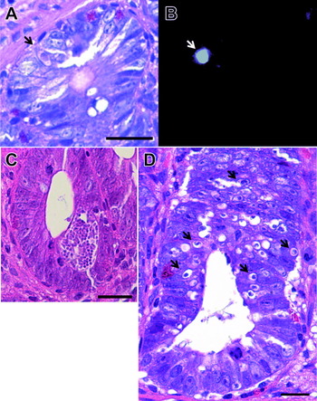
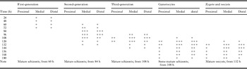
The findings of each stage are summarized in Table 1. The first-generation young schizonts were very small (approximately 5 μm in diam.) and were rarely found compared with those of other stages; thus, the anti-E. tenella antibody was useful in their detection (Fig. 1A, B). The schizonts were present within the superficial areas of the crypts from 24 h as trophozoites or immature ones; however, the number of schizonts was quite small. Mature schizonts appeared at 60 h (Fig. 1C), and at the same time, clusters of newly invaded first merozoites, or trophozoites of the second generation, were seen in the epithelium surrounding the crypts or lamina propria (Figs 1D and 2A). First- and second-generation schizonts were detected in the medial and distal regions of the caeca, but not in the proximal region. Significant pathological changes in the caeca, such as inflammatory responses, were not seen until 84 h.
Photomicrographs of first-generation schizonts of Eimeria tenella. Haematoxylin and eosin staining (A) and immunofluorescence staining with anti-E. tenella polyclonal antibody (B) at 24 h after inoculation. Arrows in A and B show trophozoites of the first generation in a serial section. A mature first-generation schizont (C) and newly invaded first-generation merozoites in epithelial cells around crypts (arrows) (D) at 60 h. All samples were obtained from the medial region of the infected caeca. Scale bars are 10 μm.

Development of asexual and sexual stages of Eimeria tenella in proximal, medial and distal regions of caeca at 24–180 h after inoculation

+, 1 to 10 clusters in the infected cecal section; + + , 10 to 30; + + +, > 30.
Development of the second- and third-generation stages
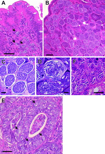
The young second-generation schizonts grew larger in the lamina propria, and the number increased from 84 h (Fig. 2A-D). Then, the lamina propria was filled with immature and mature schizonts, including many merozoites. At 96 h, after the rupture of the second merozoites, young third-generation schizonts appeared within the superficial areas of the villi around crypts (Fig. 2E, F), while many second-generation schizonts were also observed. During this period, some areas of the superficial regions, crypts, lamina propria, and submucosa were disrupted, haemorrhages had occurred, and many heterophils and some macrophages infiltrated the lamina propria and submucosa (data not shown). The mature third-generation schizonts (approximately 10 μm in diam.) were found to be smaller than those of the first and second stages. After the appearance of mature third-generation schizonts, young gametocytes were found in superficial areas of the crypts from 108 h.
Photomicrographs of second- and third-generation schizonts of Eimeria tenella. Early-stage immature schizonts at 72 h (arrows) (A), and large immature and mature schizonts at 84 h (B) of the second generation in the lamina propria. Figure 2C and D show the high magnification of immature and mature schizonts of the second generation, respectively, at 84 h. Third-generation schizonts were present in the epithelium of crypts at 96 h (arrows) (E). Figure 2F shows the high magnification of mature third-generation schizonts. All samples were obtained from the medial region of the infected caeca. Scale bars are 30 μm (A, B and E), and 10 μm (C, D, and F).

Asexual and oocyst stages
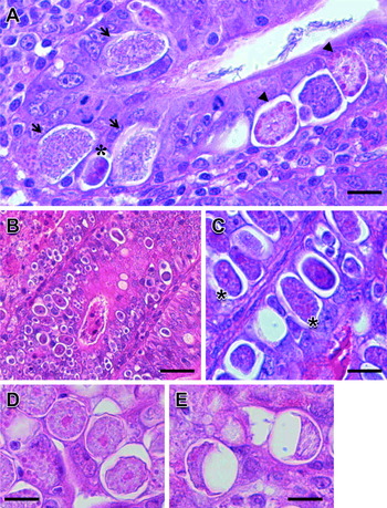
Macrogametocytes had a single nucleus at the centre of the cytoplasm with a prominent wall-forming body, and were more frequently seen than multinucleated microgametocytes at the early sexual stage (Fig. 3A). Large numbers of microgametocytes appeared at 120 h and young zygotes were observed (Fig. 3B, C). The young zygotes were characterized by a central nucleus and the formation of an oocyst wall (Fig. 3C). During this period, few second-generation schizonts were observed and the tissue destroyed by the release of second-generation merozoites was fibrosed. Lymphocytes but not heterophils were observed in the lamina propria and submucosa. Both mature and immature oocysts were observed from 132 h. The formation of oocyst walls disrupted formalin penetration during fixation, and these zoites were characterized by a distorted oocyst shape (Fig. 3D, E). During E. tenella development, the medial and distal regions of the caeca were mostly parasitized, and the sexual stage after the third generation were seen in all regions, including the proximal region.
Photomicrographs of sexual and oocyst stages of Eimeria tenella in the epithelium of crypts. Arrowheads, arrows, and asterisks in (A) show macrogametocytes with a prominent wall-forming body, microgametocytes, and zygotes or early oocysts, respectively, at 120 h after inoculation. At 132 h, (B) shows zygotes or early oocysts in the epithelium, and (C) shows the high magnification of zygotes or early oocysts (arrowheads). (D and E) High magnification of immature oocysts and mature oocysts, respectively, which are characterized by distorted oocyst walls at 168 h. All samples were obtained from the medial region of the infected caeca. Scale bars are 30 μm (A) and 10 μm (B, C, D, and E).

Application of LMD for isolation of schizonts
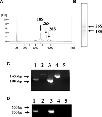
At 84 h and 96 h after inoculation, mature second schizonts were microscopically identified and successfully excised from the lamina propria in 16 μm sections by LMD. Subsequently, total RNA was extracted from the schizonts. Agilent Bioanalyzer software was used to evaluate RNA quality. This software assigns an RNA integrity (RIN) score from 10 (highest integrity) to 2 (lowest integrity) based on the entire electrophoretic trace, including ribosomal bands and degradation products of the RNA sample. Our samples contained 0·83 ± 0·15 μg of RNA (estimated total number of 10 000 to 15 000 schizonts) and had an RIN score of 6·3 ± 0·1. Two distinct peaks and 2 bands, which were thought to be 18S small subunit ribosomal RNA (SSU rRNA) and 26S large subunit ribosomal RNA (LSU rRNA), were present (Fig. 4A, B), consistent with previous reports indicating that the size of E. tenella LSU rRNA is 26S, which is distinct from that of mammals (Schaap et al. Reference Schaap, Arts, van Poppel and Vermeulen2005). The average 26S LSU rRNA:18S SSU rRNA ratio was 0·55 ± 0·1. A peak of 28S LSU rRNA, which represents RNA derived from chicken cells, was detected at low levels. According to band density, the population of chicken RNA was estimated to be 15·3% of total RNA. Using RT-PCR, mRNAs specific for the E. tenella actin and microneme 2 genes were successfully amplified (Fig. 4C, D), and the >2·0 kb products of the microneme protein 2 gene, which were amplified from genomic DNA, were detected in control samples in the absence of reverse transcriptase (Fig. 4C). Therefore, a small amount of genomic DNA was present in samples purified using an RNA purification kit.
Evaluation of RNA quality from isolated schizonts by Agilent Bioanalyzer software and RT-PCR using the cDNA. Figure 4A and B show an electropherogram and Agilent Bioanalyzer 2100 gel-like image of total RNA of isolated second-generation schizonts, respectively. The x-axis on the electropherogram represents amplicon size (bp), while the y-axis represents the measurement response in fluorescence units (FU). 18S shows the 18S small subunit ribosomal RNA (SSU rRNA), and 26S and 28S indicate the 26S large subunit ribosomal RNAs (LSU rRNA), which are thought to be derived from Eimeria tenella, and 28S LSU rRNA from chicken hosts, respectively. Figure 4C and D show the results of RT-PCR using primers for the E. tenella microneme protein 2 and actin genes, respectively. Lanes 1 and 2, cDNA converted from the second-generation schizonts isolated by LMD with and without a reverse transcriptase, respectively. Lanes 3 and 4, cDNA from sporulated E. tenella oocysts with and without a reverse transcriptase, respectively. Lane 5, reaction buffer as a negative control.

DISCUSSION
The development of Eimeria spp. schizonts in caeca is closely related to their pathogenicity. Transcriptome analyses of these stages could lead to the identification of new target molecules for disease control. We first observed the successive development of E. tenella in caeca by conventional microscopy, confirmed the infection sites in caeca and determined in detail the parasite populations at each stage, according to timelines based on previous characterizations (McDonald and Shirley, Reference McDonald and Shirley1987; McDonald and Rose, Reference McDonald and Rose1987; Daszak et al. Reference Daszak, Ball, Pittilo and Norton1993; Ball et al. Reference Ball, Daszak, Pittilo and Norton1995). Our findings showed that the developmental stages were synchronous, which is likely associated with the arrival of excysted sporozoites in the caeca, establishment of infection, and a subsequent active period of oocyst shedding. Therefore, difficulties were encountered in purifying only 1 generation of schizonts using previously reported methods (Geysen et al. Reference Geysen, Ausma and Vanden Bossche1991), as those of other stages could be purified concurrently.
There are a number of reports about schizont purification using enzymes or discontinuous gradient centrifugation (Geysen et al. Reference Geysen, Ausma and Vanden Bossche1991; Ouarzane et al. Reference Ouarzane, Labbé and Péry1998). However, RNA is not generally stable, and treatments such as the above might affect the gene expression of zoites. Here, we assessed the purification of RNAs at specific developmental stages of E. tenella by LMD. The RNAs were not degraded and were >80% in quality; however, contamination with host cell RNA occurred due to the close proximity of chicken cells and schizonts, resulting in their concurrent isolation. Using the previously developed centrifugal method, it was almost impossible to remove the host cells and to isolate only 1 developmental stage. Moreover, we attempted to purify the second-generation schizonts (probably containing mature and immature schizonts) at 84 h by centrifugation with a Percoll gradient (Geysen et al. Reference Geysen, Ausma and Vanden Bossche1991), but >90% of total RNA was found to be derived from chicken cells (data not shown). Thus, we could remarkably reduce RNA contamination from host cells and successfully isolate only mature second-generation schizonts. Eimeria tenella-specific genes were amplified using RT-PCR. The presence of genomic DNA indicated the necessity of DNase treatment to remove contaminating DNA, thereby obtaining increased RNA purity. Taken together, LMD appears to be effective for the isolation of schizonts, at different stages or maturity, from infected lamina propria and purification of schizont RNAs.
In conclusion, we isolated stable RNA from E. tenella second-generation schizonts using LMD. The second-generation schizonts were very large and developed in the lamina propria, which differs from the infection sites of other stages; thus, they are easily isolated from the host tissue. Although further improvement is needed for parasite isolation at other stages, our results have revealed LMD as a useful tool for gene expression analyses of the intracellular stages of E. tenella infection.
ACKNOWLEDGMENTS
A part of this study was supported by Drs Takashi Minowa and Shoko Kajiwara (‘Nanotechnology Network Project’ of the Ministry of Education, Culture, Sports, Science and Technology (MEXT), Japan). We thank Mr Masami Hashizume (Histo Science Laboratory Co., Ltd, Japan) for advice on the pathological findings. This work was partly supported by Grants-in-aid for Scientific Research from the Ministry of Education, Culture, Sports, Science and Technology (grant number 22700773 to M. M.).