Introduction
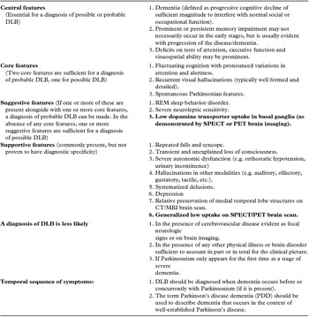
DLB is considered to be the second most common form of neurodegenerative disorders after AD (Geser et al., Reference Geser, Wenning, Poewe and McKeith2005; Zupancic et al., Reference Zupancic, Mahajan and Handa2011), affecting as many as 100,000 people in the UK and up to 1.3 million in the USA. Prevalence estimates range from 0% to 5% in the general population, and up to 30.5% of all dementia cases (Zaccai et al., Reference Zaccai, McCracken and Brayne2005), and these are largely influenced by the clinical criteria used for DLB diagnosis. Thus, the latest 2005 revised International Consensus Criteria (McKeith et al., Reference McKeith2005; Table 1) for DLB identify substantially more DLB patients than the 1996 original criteria (McKeith et al., Reference McKeith1996; Vann Jones and O’Brien, Reference Vann Jones and O’Brien2013).
Consensus criteria for DLB (McKeith et al., Reference McKeith2005)

However, nearly half of DLB cases remain undiagnosed (Palmqvist et al., Reference Palmqvist, Hansson, Minthon and Londos2009), and many patients with DLB are deprived from having an early and adequate treatment of their distressing symptoms. These include a combination of motor and cognitive problems, and behavioral disturbances (e.g. confusion, hallucinations in various modalities, problems interpreting visual information, and sleeping difficulties). Their presence leads consequently to greater dependence earlier in the course of the disease and the need for increased level of care (Ferman et al., Reference Ferman2004; Galvin et al., Reference Galvin2010). In addition, patients with DLB have higher prevalent rates for physical problems including delirium, fall-related injuries, pneumonia and decubital lesions and higher mortality than patients with other forms of dementia, including AD (Hanyu et al., Reference Hanyu, Sato, Hirao, Kanetaka, Sakurai and Iwamoto2009; Santangelo et al., Reference Santangelo2010).
DLB: accurate and early diagnosis
Accurate and early diagnosis of DLB is important for both patients and their caregivers, since the neuropsychiatric symptoms require specific management. Thus, the DLB clinical symptoms have a differential response to pharmacological treatment, with visual hallucinations (VH) best responding to cholinesterase inhibitors (Ballard et al., Reference Ballard, Aarsland, Francis and Corbett2013). DLB patients are especially vulnerable to antidopaminergic and anticholinergic side effects from most conventional neuroleptics that are used for treatment of behavioral and psychiatric symptoms of dementia. In particular, 50% of DLB patients are sensitive to develop extrapyramidal symptoms and potentially fatal complications of neuroleptic sensitivity (reviewed in Baskys, Reference Baskys2004; Bosboom and Wolters, Reference Bosboom and Wolters2004). Even selective serotonin re-uptake inhibitors (e.g. sertraline), used for treatment of depression and anxiety, have been shown to result in adverse extrapyramidal side in DLB (Kim et al., Reference Kim2012). The latter findings are of a particular relevance, since depression and anxiety are highly prevalent in DLB (Boot et al., Reference Boot2013).
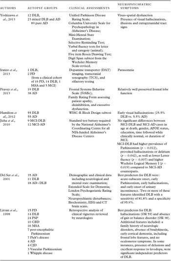
At present, the early diagnosis of DLB can be facilitated by several neuropsychological discriminants (Table 2). These include: impaired visuo-spatial function, presence of VH, illusions, and extrapyramidal tract signs (Yoshizawa et al., Reference Yoshizawa, Vonsattel and Honig2013), parasomnia, as verified via presence of idiopathic rapid-eye-movement (REM) sleep behavior disorder (Iranzo et al., Reference Iranzo2013) and relatively well preserved frontal lobe function (Peavy et al., Reference Peavy2013). However, the diagnosis of DLB in routine clinical practice still poses a difficulty for clinicians, irrespective of the progression at clinical presentation. In this respect, having a method with high sensitivity and specificity to diagnose this dementia syndrome would be a marked improvement on the working clinical diagnosis and subsequent management of patients with suspected DLB (Kemp et al., Reference Kemp, Clyde and Holmes2011).
Early neuropsychiatric discriminants for DLB diagnosis based on correlative clinico-neuropathological studies

Abbreviations: DLB, dementia with Lewy bodies; AD, Alzheimer's disease; PD, Parkinson's disease; PDD, Parkinson's disease dementia; MCI, mild cognitive impairment; MSA, multiple system atrophy; PSP, progressive supranuclear palsy, CBD, cortico-basal degeneration.
Biomarkers to facilitate diagnosis of DLB in clinical setting
The greatest advances in peripheral biomarkers research have been made in AD (Blennow et al., Reference Blennow2010; Hampel et al., Reference Hampel2011; Shoji, Reference Shoji2011). Some of them have been recommended for the diagnosis of probable AD, based on the underlying pathophysiological process of the disease, e.g. biomarkers of amyloid (Aβ) processing [e.g. low cerebrospinal fluid (CSF) Aβ42 and positive position emission tomography (PET) amyloid imaging] and biomarkers of downstream neuronal degeneration or injury [e.g. elevated CSF tau, both total and phosphorylated (p-tau)], decreased 18fluorodeoxyglucose (FDG) uptake on PET in temporo–parietal cortex; and disproportionate atrophy on structural magnetic resonance imaging (MRI) in medial, basal, and lateral temporal lobe, and medial parietal cortex (McKhann et al., Reference McKhann2011).
A recent review (Sharma et al., Reference Sharma2013) offered a detailed classification of potential biomarkers for Parkinson's disease (PD) that can also be easily applied to various forms of dementia, including DLB. These biomarkers are largely based on clinical staging and neurobiological characteristics, and include biochemical, physiological, genetic, morphological, immunological, and pathological categories. In contrast to PD, in DLB the course of the disease, including the temporal appearance of clinical symptoms and neurobiological changes is poorly understood. The preclinical stage is largely under-investigated, and the diagnosis is frequently made rather late in the course of the disease. Furthermore, the clinical symptoms are not necessarily related to the hallmarks of the disease, α-synuclein inclusions (Lewy bodies; LBs) and neurites (Mukaetova-Ladinska et al., Reference Mukaetova-Ladinska2013). Thus, both LBs, loss of choline acetyltransferase (ChAT) activity (Samuel et al., Reference Samuel, Alford, Hofstetter and Hansen1997) and neurofibrillary pathology (Oinas et al., Reference Oinas2009) have been inconsistently linked to cognitive impairment in DLB (Gómez-Tortosa et al., Reference Gómez-Tortosa, Irizarry, Gómez-Isla and Hyman2000; Stern et al., Reference Stern2001). Similarly, the clinical symptoms of DLB appear to better correlate with co-morbid (e.g. neurofibrillary tangles) than diffuse Lewy body pathology (Oinas et al., Reference Oinas2009). Furthermore, presence of VH, a core feature of DLB is linked to a various extent of pathology, including: amygdalar atrophy (Brooks and Halliday, Reference Brooks and Halliday2009), increase in α-synuclein LBs in fronto–temporal lobes and transenthorinal cortex (Harding et al., Reference Harding, Broe and Halliday2002a, Gallagher et al., Reference Gallagher2011), thalamic nuclei (Brooks and Halliday, Reference Brooks and Halliday2009), limbic system (Harding et al., Reference Harding, Stimson, Henderson and Halliday2002b) and anterior cingulate cortex (Gallagher et al., Reference Gallagher2011; Ferman et al., Reference Ferman2013), as well as widespread neocortical neurofibrillary pathology (Oinas et al., Reference Oinas2009). Interestingly, neurofibrillary pathology alone increases the likelihood of developing VH by 3–4 fold, depending whether it is restricted to the medial temporal lobe or widespread in the neocortical association and primary areas (Oinas et al., Reference Oinas2009).
Patients with DLB, irrespectively of the presence of VH, may display similar extent of LBs, neuronal cell loss and spongioform change in various parts of the geniculo-striate and extrastriate visual pathways and amygdala (Yamamoto et al., Reference Yamamoto2006), with only substantia nigra having somewhat higher LB score in DLB patients with VH (Yamamoto et al., Reference Yamamoto2006). These correlative clinico-neuropathological findings clearly support the molecular heterogeneity of the disease, and warrant further in-depth analysis of larger samples of patients to identify highly sensitive and specific molecular markers for DLB, that will better match the clinical symptoms and correspond to the progression of the disease process.
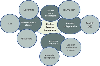
To date, a number of neuroimaging techniques have been studied to aid the clinical diagnosis of DLB, all with varying results. Among them, regional cerebral blood flow, based on specific cerebral blood flow patterns, shows some promise in identifying DLB patients. In one of the latest work in this field, Colloby et al. (Reference Colloby2013) provided further evidence for the robust diagnostic accuracy of Tc 9 9m-exametazime brain blood flow single-photon emission computed tomography (SPECT) to distinguish DLB from other forms of dementia, e.g. AD, with 87% diagnostic accuracy (81% sensitivity and 88% specificity) (Figures 1a and 1b). Similarly, MRI has demonstrated distinct cortical thinning (predominantly affecting inferior parietal, posterior cingulate, and fusiform gyrus; Watson et al., Reference Watson, Colloby, Blamire and O’Brien2014) and cerebellar grey matter loss (Colloby et al., Reference Colloby, O’Brien and Taylor2014) in DLB patients, whereas functional MRI revealed potential role of attention-executive networks (Peraza et al., Reference Peraza2014) and thalamic involvement in fluctuating cognition in DLB (Delli Pizzi et al., Reference Delli Pizzi2014). Other promising imaging biomarkers are those associated with the core DLB neuropathological and biochemical features, e.g. changes in neurotransmitters underlying cognitive and non-cognitive clinical symptoms in DLB, autonomic dysfunction and/or amyloid polypeptides (Figure 2). In the current study, we review the latest advances in molecular imaging biomarkers that are applicable to DLB, based on their neuropathological and neurobiochemical properties, and discuss their potential use in clinical setting. Since there were a number of recent extensive reviews on regional cerebral blood flow using Tc99m-hexamethylpropyleneamineoxime (HMPAO), N-isopropyl-p-(123I)iodoamphetamine (IMP) tomography and Tc99m-ethyl cysteine dimer (Mak et al., Reference Mak, Su, Williams and O’Brien2014) and metabolic abnormalities, as detected by 18F-Fludeoxyglucose (18F-FDG; Davison and O’Brien, Reference Davison and O’Brien2014), including a systematic review (Yeo et al., Reference Yeo, Lim, Khan and Pal2013), we did not include these imaging modalities in the current study.
Tc99m Exametazime SPECT and 123I-FPCIT SPECT images in healthy aging and DLB. Tc99m Exametazime SPECT images (a measure of cerebral perfusion) in a healthy older individual (a) and a patient with DLB (b). Note the presence of occipital hypoperfusion in DLB (arrows). Also shown are 123I-FPCIT SPECT (DaTSCAN) images depicting normal uptake (c) which is a typical pattern in healthy participants and patients with Alzheimer's disease, as well as abnormal uptake (d) that is associated with either DLB or Parkinson's disease. Figure 1 courtesy of Prof. John O’Brien, University of Cambridge and Dr. Sean Colloby, IoN, Newcastle University.

Molecular imaging biomarkers for DLB. Detailed explanation is found in the text.

Methods
This narrative review is based on a comprehensive search that included Medline, PsychInfo, Embase, and PubMed databases for studies published until 15.09.2014. The key words used in the current search were: DLB, PD, Parkinson's disease dementia (PDD), Lewy Body disease (LBD), imaging, biomarker, ligand, probe, neuroradiology, neurotransmitter, tau protein, amyloid, beta-amyloid, α-synuclein, glia, neuroinflammation, synapse, vesicular monoamine transporter (VMAT). All abstracts were critically reviewed and the full text of relevant papers was obtained.
Molecular imaging biomarkers for DLB diagnosis
NEUROTRANSMITTER BASED IMAGING BIOMARKERS: EVALUATION OF NIGROSTRIATAL DOPAMINERGIC INTEGRITY
Presynaptic dopaminergic imaging
Although some neuroradiological studies demonstrated the usefulness of straight neuroradiological imaging (e.g. 3D T1-weighted MRI and FDG-PET) to diagnose DLB (Warr and Walker, Reference Warr and Walker2012; Nakatsuka et al., Reference Nakatsuka, Imabayashi, Matsuda, Sakakibara, Inaoka and Terada2013), the use of presynaptic dopaminergic imaging has been now widely used to confirm the clinical diagnosis of DLB. This mode of imaging is based on the visualization of low presynaptic dopamine transporter activity as an indicator of nigrostriatal neurodegeneration (Figure 2).
The revised 2005 Consensus Diagnostic Criteria for DLB (McKeith et al., Reference McKeith2005) also include the presence of low dopamine transporter up-take in the basal ganglia (as demonstrated by PET and SPECT imaging) as one of the features of possible and probable DLB (Table 1). The imaging of presynaptic dopaminergic terminal functions with SPECT and PET has currently the greatest evidence to support the probable diagnosis of DLB and differentiate DLB from other forms of dementia (McKeith et al., Reference McKeith2007). Furthermore, the in vivo findings of presynaptic dopaminergic imaging correlate well with neuropathological findings at autopsy, having a high sensitivity of 88% and a specificity of 100% for this imaging procedure to distinguish between DLB and AD (reviewed in Tatsch, Reference Tatsch2008).
The diagnostic accuracy of presynaptic dopaminergic imaging with 123I-FP-CIT (DaTSCAN) in DLB has been investigated in-depth over the last several years (O’Brien et al., Reference O’Brien2009; Sinha et al., Reference Sinha, Firbank and O’Brien2012; Walker and Cummings, Reference Walker and Cummings2012; Mortimer et al., Reference Mortimer, Likeman and Lewis2013; Figures 1c and 1d). In a post-mortem series, 123I-FP-CIT had higher sensitivity (88%) and specificity (100%) to differentiate DLB from non-Lewy body dementias than clinical diagnosis alone (75% and 42%, respectively; Cummings et al., Reference Cummings2011). More importantly, the reduced uptake is a result of nigral dopaminergic neuronal loss, but not additional pathology, e.g. neurofibrillary tangles, amyloid plaques and/or LBs (Colloby et al., Reference Colloby, McParland, O’Brien and Attems2012). The data from a recent meta-analysis of four studies, with a total of 419 patients with diagnostic uncertainty regarding dementia subtypes, provide further support for the DaTSCAN use in clinical practice. Thus, the estimated pooled sensitivity of DaTSCAN in differentiating DLB from non-DLB dementias was 86.5% and specificity 93.6% (Papathanasiou et al., Reference Papathanasiou, Boutsiadis, Dickson and Bomanji2012).
The mesocortical dopaminergic deficits, as visualized with 123I-FP-CIT correlate with some of the neuropsychiatric symptoms in DLB, including VH, delusions, depression, and apathy (Roselli et al., Reference Roselli2009). The latter finding is similar to that described for apathy in people with AD, where the relationship between apathy and dopamine transporter levels is independent from motor activity (David et al., Reference David2008). These findings suggest that some degree of dopaminergic neuronal loss occurs in other forms of dementia, and may be linked to some of the behavioral psychiatric symptoms characteristic for the dementia syndrome. Not surprisingly, one third of patients with fronto–temporal lobe dementia and 10% of those with AD have abnormal DaTSCANs (Bittner et al., Reference Bittner2011; Morgan et al., Reference Morgan2012).
Similarly, some patients fulfilling the clinical criteria for probable DLB have normal DaTSCANs (Siepel et al., Reference Siepel2013). Thus, about 10%–15% of DLB patients will have normal DaTSCANs (Morgan et al., Reference Morgan2012; Siepel et al., Reference Siepel2013). Furthermore, the latest reports of 123I-N-(3-iodoprop-2E-enyl)-2-β-carbomethoxy-3β-(4-methylphenyl) nortropane (123I-PE2I) and SPECT argue that cognitive, motor and behavioral clinical sympatomatology in DLB may not necessarily be associated with a reduction in striatal dopamine transporter despite its firm involvement in DLB pathology (Ziebell et al., Reference Ziebell2013). Overall, these findings suggest cautious interpretation of DaTSCANs alongside clinical phenomenology to aid the DLB diagnosis, especially in newly diagnosed DLB.
Vesicular monoamine transporter (VMAT)
Our recent neuropathological studies confirmed extensive synaptic changes, affecting predominantly the synaptic proteins involved in the exocytosis in patients with DLB (Mukaetova-Ladinska et al., Reference Mukaetova-Ladinska2009; Reference Mukaetova-Ladinska2013). The lower levels of t-SNARE proteins in DLB suggest presence of altered expression of additional neurotransmitter related proteins, such as the VMAT, a membrane-embedded protein that transports monoamine neurotransmitter molecules into intraneuronal storage vesicles to allow subsequent release into the synapse.
Out of the two known VMAT forms (VMAT1 and VMAT2), VMAT2 is predominantly located in neuronal cells and plays a critical role in the monoamine neuronal signaling. In contrast to plasma membrane neurotransmitter transporters, which convey only one of the neurotransmitters (e.g. dopamine, serotonin or norepinephrine) from the synapse, into the neuronal cells, VAMT2 recognizes and transports all biogenic amine neurotransmitters across bio-membranes, and does not respond selectively to individual neurotransmitters based on their receptor type's cellular location and the signaling proteins with which it couples.
In contrast to DaTSCANs, that can be influenced by dopaminergic medication, including L-dopa, amphetamine, deprenyl and cocaine, VAMT2 is less susceptible to dopaminergic drugs, and thus is more reliable in monitoring the dopamine terminal integrity in LBD. The first generation VAMT2 radioligand (11C-DTBZ) has a limited use due to a number of technical issues (including short radio-active decay half-life), thus restricting its use to highly specialized clinical centers with an access to onsite cyclotron and 11C radiochemistry expertize. The 11F radiolabeled VAMT2 tracer, 18F-labeled tetrabenazine derivative 18F-(+)fluoropropyldihydrotetrabenazine (18F)AV-133, with longer decay half-life of 110 minute) appears to be better suited for clinical studies.
The imaging of striatal monoaminergic terminal integrity has been assessed in a limited number of studies, either on its own (Okamura et al., Reference Okamura2010; Villemagne et al., Reference Villemagne2011) or in combination with amyloid ligands (Villemagne et al., Reference Villemagne2012; Siderowf et al., Reference Siderowf2014) (Table 3). The VAMT2 radioligand (18F)AV-133 appears to be a good marker to assess the nigrostriatal degeneration in LBD, including DLB and PD, with a substantial reduction seen in both the anterior (Villemagne et al., Reference Villemagne2011) and posterior putamen (Villemagne et al., Reference Villemagne2011; Reference Villemagne2012), as well as the caudate and the whole striatum (Siderowf et al., Reference Siderowf2014). Furthermore, the (18F)AV-133 binding potential may correlate with the clinical severity of PD (bradykinesia and rigidity), but not depression (Okamura et al., Reference Okamura2010), though the findings for Parkinsonian symptoms were not confirmed in a later study on DLB and PD (Siderowf et al., Reference Siderowf2014). However, the cognitive functioning, as measured with the MMSE, in DLB and PD patients was reported to be positively correlated with the (18F) AV-133 radiobinding in the caudate only (r = 0.72; p = 0.012), with the combined measures of caudate florbenazine and mean cortical florbetapir explaining 56% of the variance in cognitive functioning in the DLB group (Siderowf et al., Reference Siderowf2014). These findings suggest that (18F) AV-133 may facilitate the presymptomatic identification of individuals with Parkinsonian symptoms and aid the differential diagnosis of LBDs from other neurodegenerative disorders, either on its own, or in association with other radioligands, such as amyloid tracers. However, these findings need to be expanded on larger clinical samples.
Molecular neuroimaging markers for diagnosing DLB. Brief description of clinical studies is presented

*Most latest studies presented.
Abbreviations: AD, Alzheimer's disease, DLB, dementia with Lewy bodies, PD, Parkinson's disease, LBD, Lewy body diseases, MCI, mild cognitive impairment; PET, positron emission tomography; CT, computerised tomography; MRI, magnetic resonance imaging.
Cholinergic imaging in DLB
Our previous neuropathological and biochemical work provided support for cholinergic changes in DLB (Ray et al., Reference Ray2004; Bohr et al., Reference Bohr2005; Francis and Perry, Reference Francis and Perry2007; Mukaetova-Ladinska et al., Reference Mukaetova-Ladinska2013). The initial neuropathological findings of changes in α6/α3 neuronal nicotinic acetylcholine receptors in a number of subcortical brain regions, including the centromedian, ventral lateral and ventroposterior medial thalamic nuclei (p < 0.05), together with caudate and putamen (Ray et al., Reference Ray2004) led to development of muscarinic receptor targeted visualization probes. Thus, muscarinic acetylcholine receptors (mAChRs) were visualized with (R,R) 123I-iodo-quinuclidinyl-benzilate (123I-QNB) and single photon emission computed tomography (SPECT) (Colloby et al., Reference Colloby2006). In the latter study, significant elevation of 123I-QNB binding was recorded in the occipital lobes in both DLB and PDD patients, and this may reflect the visual disturbances characteristic for these disorders.
In addition to the visualization of the mAChRs, the nicotinic cholinergic receptors (nAChRs) have also been investigated in imaging studies, based on the previously documented depletion of α7 (Court et al., Reference Court1999), α6α3 (Ray et al., Reference Ray2004) and α4β2 (Pimlott et al., Reference Pimlott2004) nAchRs in the post-mortem DLB brain tissue. Using a ligand against the α4β2 nAchRs, 123I-5IA-85380 applied as a SPECT tracer, Colloby et al. (Reference Colloby2010) related the decreased uptake in left frontal gyri and pre-/post-central and anterior cingulate regions to a decline in executive function in a pooled group of AD and DLB patients. In another study, the significant increase in occipital lobe 123I-5IA-85380 uptake in DLB patients was more pronounced in those with a recent history of VH (O’Brien et al., Reference O’Brien2008) (Table 3). These results link directly the occipital lobe cholinergic changes to VH in DLB patients, a finding we have recently confirmed in a molecular pathological study that confirmed significant loss of ChAT in the occipital lobe of DLB patients (Mukaetova-Ladinska et al., Reference Mukaetova-Ladinska2013).
The cholinergic deficit can also be studied using PET in vivo quantification of acetylcholinesterase (AChE) activity by utilizing Acetylcholine analogues N-(11C)-methylpiperidyl-4-acetate ((11C)MP4A) and N-(11C)-methyl piperidine-4-propionate ((11C)MP4P, and (11C)-physostigmine, which are substrates for AChE activity. Using these tracers, it is possible to identify in vivo cholinergic changes in dementia patients (Herholz et al., Reference Herholz2000), and also measure drug-induced AChE inhibition in dementia treated patients. Few studies have made use of these ligands and have described a significant decrease in AChE activity (using PET scans with N-(11C)-methyl-4-piperidyl acetate to measure brain AChE activity) in PD patients early on in the course of their illness, and this deficit becomes more pronounced with the progression of the disease in both PDD and DLB (Shimada et al., Reference Shimada2009). Interestingly, the cholinergic deficit coexists with the dopaminergic changes in DLB and PD patients (Klein et al., Reference Klein2010), and this argues for cortical deafferentation in these dementias syndromes due to the degeneration of projection fibers from the basal forebrain. A most recent study, based on (11C)-MP4A PET, reported early widespread cholinergic deficit as early as mild cognitive impairment (MCI), with DLB patients showing greater pattern variability, affecting both cortical and subcortical brain areas (Marcone et al., Reference Marcone2012).
Bearing in mind that the current cholinergic PET imaging is based on visualization of AChE activity, this nuclear imaging method has a potential to be used not only for the diagnosis of other dementia subtypes (e.g. AD, DLB, PD/PDD,) but also in evaluating their treatment, follow-up, drug dosage adjustment and identification of likely responders to cholinesterase inhibitors and other drugs used for treatment of AD and DLB/PDD (Berti et al., Reference Berti, Pupi and Mosconi2011). Although there is a great potential in introducing the cholinergic imaging in clinical practice, including better understanding of pathophysiological mechanisms responsible for DLB neurodegeneration, further work is needed to improve the selectivity of the available cholinergic ligands. Some of the PET cholinergic tracers (e.g. 11C-MP4A) are highly specific for AChE activity, but the measurements including kinetic activity are based on a model that uses an invasive procedure involving the arterial concentration of the tracer as an input function (Florea, Reference Florea2008). As such, their current use in clinical setting may be rather limited and restricted to highly specialized centers.
Glutamate ligands and DLB
Dysfunctional glutamate neurotransmission may underlie a range of behavioral problems in people with dementia, and these include agitation and aggression, (Francis, Reference Francis2009), also present in DLB. Despite the CSF levels of glutamate being similar between DLB and control patients (Molina et al., Reference Molina2005), three neuropathological studies described substantial differences in the expression of metabotropic glutamate receptors (mGluRs) in DLB. Thus, the decrease in metabotropic glutamate receptor1 (mGluR1) in DLB (Dalfó et al., Reference Dalfó, Albasanz, Martin and Ferrer2004) argues that presence of α-synuclein aggregates lead to functional effects on cortical glutamatergic synapses. Furthermore, the loss of GluR1 correlates with the extent of AD neocortical neuropathology and reflects the progression of the disease (Albasanz et al., Reference Albasanz, Dalfó, Ferrer and Martín2005). In contrast, 40% increase in mGluR5 is present in a number of brain areas, including frontal cortex, hippocampus and putamen in DLB patients, whereas in PD, these changes are restricted to putamen only (Price et al., Reference Price2010), thus suggesting a role for metabotropic glutamate receptor antagonists in the treatment of DLB and PD.
Since mGluRs antagonists prevent excessive glutamate signaling via blocking N-methyl-D-aspartic acid (NMDA) receptors, not surprisingly, DLB and PD patients appear to benefit from treatment with the NMDA antagonist, memantine (Aarsland et al., Reference Aarsland2009). This provides further indirect evidence for the role of glutamate in the pathogenesis of DLB. Similarly, increase in NMDA receptor (NMDAR) IgA has been reported in some patients with progressive cognitive decline, some of whom also showed alterations in consciousness, falls, and gait instability (Prüss et al., Reference Prüss2012). Although the seven analyzed patients in the latter study had a wide range of heterogeneity in terms of their neuroradiological (MRI brain scans) and biochemical (CSF t-tau and Aβ) findings, the presence of IgA NMDAR antibodies in their sera resulted in significant decrease in synapses in vitro. These findings are relevant to DLB, since DLB patients have widespread neocortical loss of synaptic proteins (e.g. synaptophysin, syntaxin, SNAP-25, and γ-synuclein), and this loss is not correlated with the underlying neuropathology (Mukaetova-Ladinska et al., Reference Mukaetova-Ladinska2009; Reference Mukaetova-Ladinska2013), thus raising an interesting possibility of an auto-immune etiology of DLB and a scope for immunotherapy for treatment of its clinical symptoms.
There have been several potential developments in the visualization of mGluRs, in particular mGluR1 and mGluR5. PET radioligands targeting mGluR5, 2-Methyl-6-(phenylethynyl)-pyridine (MPEP), fluorinated (E)-3-((6-methylpyridin-2-yl)ethynyl)cyclohex-2-enone-O-methyloxime (ABP688) derivatives, have been used in vivo and in vitro (reviewed in Leung, Reference Leung2013). Another mGluR1 ligand {labeled with (11C), N-(4-(6-(isopropylamino) pyrimidin-4-yl)-1,3-thiazol-2-yl)N-methyl-4-(11C)methylbenzamide((11C)ITDM}, has been also employed in vivo PET imaging of mGluR1 distribution in rat and monkey brain tissue (Fujinaga et al., Reference Fujinaga2012; Yamasaki et al., Reference Yamasaki2014). However, clinical studies for the in vivo visualization of mGluRs are still missing. Bearing in mind the differential expression of mGluR5 in DLB and PD patients, the novel glutamatergic nuclear imaging ligands have the potential to find their place in the differential diagnosis of distinct α-synucleinopathies.
AUTONOMIC DYSFUNCTION: MYOCARDIAL SYMPATHETIC IMAGING AND VESICULAR CATECHOLAMINE UPTAKE
(123) I-Metaiodobenzylguanidine (MIBG) scintigraphy
Autonomic dysfunction is common in DLB. Myocardial scintigraphy with metaiodobenzyl guanidine (MIBG) is a non-invasive imaging procedure that is a sensitive tool for detecting cardiac sympathetic denervation (Figure 1). MIBG has not only been recommended as a complementary diagnostic tool to differentiate DLB from other forms of dementia (Treglia et al., Reference Treglia2014), but has also been incorporated as one of the supportive clinical features of DLB (Table 1). The reduced up-take of myocardial sympathetic imaging using 123I-Metaiodobenzylguanidine (MIBG) scintigraphy, correlates with LBs and reflects the cardiac sympathetic nerve depletion in DLB (Toru et al., Reference Toru2010). Decrease in cardiac MIBG uptake is accompanied by reduced FP-CIT binding in basal ganglia in 82% of patients with probable DLB (Camacho et al., Reference Camacho2011), suggesting that cardiac sympathetic and nigrostriatal degeneration parallel similarly in patients with probable DLB.
MIBG scintigraphy may be preferred to DaTSCAN imaging since it is considerably less expensive and its sensitivity and specificity to DLB is independent of clinical evidence of Parkinsonism (Kobayashi et al., Reference Kobayashi, Tateno, Morii, Utsumi and Saito2009). In this respect, MIBG can aid the early detection of DLB with no overt motor involvement. The latter has been recently supported in a case study of cardiac sympathetic denervation (detected by MIBG scintigraphy) in two amnestic mildly cognitively impaired individuals, without core DLB clinical features, of whom one converted to probable DLB two years later (Fujishiro et al., Reference Fujishiro, Nakamura, Kitazawa, Sato and Iseki2012). These findings have now been confirmed in a recent one year longitudinal study (Oda et al., Reference Oda2013), thus supporting the MIBG advantages especially in diagnosing early stages DLB.
In comparison to DaTSCAN imaging, MIBG scintigraphy has some disadvantages especially when used in people with congestive heart failure, myocardial ischemia, cardiac arrhythmias, and diabetes (Chirumamilla and Travin, Reference Chirumamilla and Travin2011), all characterized by cardiac sympathetic dysfunction that interferes with MIBG uptake. Therefore, the MIBG findings in the context of a DLB dementia diagnosis need to be interpreted cautiously in these patients to avoid false positive results. A most recent study (Stefanelli et al., Reference Stefanelli, Treglia, Bruno, Rufini and Giordano2013) also draw the attention to a number of pharmacological compounds that may interfere with MIBG scintigraphy, including antidepressants, antipsychotic, tramadol, labetalol, sympatho-mimetics, reserpine, and some calcium antagonists (e.g. diltiazem, verapamil, and nifedipine) thus warranting a caution when using MIBG as a diagnostic tool (Yokoyama et al., Reference Yokoyama2013). It is important to highlight that the MIBG scintigraphy high sensitivity and specificity (100%) for DLB was reported in highly selected clinical sample, where all patients with medical conditions that potentially interfered with MIBG uptake were excluded (reviewed in Tiraboschi and Guerra, Reference Tiraboschi and Guerra2010).
A recent review of eight studies (based on analysis of 346 patients with dementia: 152 patients with DLB and 194 patients with other dementias) reported MIBG scintigraphy to have 98% sensitivity and 94% specificity to detect DLB (Treglia and Cason, Reference Treglia and Cason2012; Table 3). These findings confirm MIBG scintigraphy to be an accurate test to differentiate DLB from other types of dementia. Furthermore, it appears that the MIBG use can be extended to screening early stages DLB. The low uptake MIBG can be detected in as many as 30% of amnestic patients with MCI, some of whom also have autonomic disorder, REM sleep disorder, VH, and occipital hupoperfusion (Sakakibara et al., Reference Sakakibara2012). This suggests that some of these MCI patients may well present with early stage DLB.
In a comparative study, MIBG was superior to N-isopropyl-p-123I-iodoamphetamine (123I-IMP) brain perfusion SPECT (a marker of brain blood flow) in predicting conversion of possible to probable DLB during one year follow-up (Oda et al., Reference Oda2013). However, a study conducted on a larger number of patients with DLB and other LBDs (e.g. PD, PDD, MSA) and non-LBD diseases concluded that MIBG and 123I-Ioflupane SPECT have identical sensitivity (90%) and specificity (91%) for DLB, whereas these figures were substantially lower for differentiating PD from other Parkinsonian disorders (Treglia et al., Reference Treglia2014). The authors conclude that 123I-MIBG scintigraphy may have a complementary role to striatal dopaminergic imaging in the diagnosis of LBD, in particular for its higher specificity than 123I-FP-CIT SPECT (DaTSCAN imaging) in differential diagnosis between PD and other Parkinsonian disorders. The combination of these functional imaging technique methods may help to achieve a correct differential diagnosis in patients with suspected LBD and to select the most appropriate treatment.
Vesicular catecholamine uptake
The loss of sympathetic nerves in the heart (which manifests with some of the classical clinical symptoms of DLB, e.g. postural hypotension) can be assessed at the periphery via changes in intra-neuronal vesicular uptake of catecholamines, such as dopamine and norepinephrine. Using 6-(18F)-dopamine (18F-DA) and its metabolite 18F-dihidroxyphenylacetic acid (18F-DOPAC), Goldstein et al. (Reference Goldstein, Holmes, Kopin and Sharabi2011) described altered index of vesicular uptake (as defined by the 18F-DA/18F-DOPAC ratio) in PD patients with orthostatic hypotension, but not in age-matched controls and patients with multiple system atrophy (Table 3). These findings support further the role of sympathetic denervation in LBD, and indicate presence of vesicular monoamine transport impairment. This vesicular uptake needs to be investigated further to confirm it as a novel target for diagnosis LBD in general, and also explore whether it can be used to successfully differentiate distinct LBD syndromes. In addition to its diagnostic potential, the impairment in vesicular uptake may also find place in prevention and treatment of these diseases.
AMYLOID POLYPEPTIDES AS MOLECULAR IMAGING PROBES FOR DLB
Amyloid PET imaging
In recent years, visualization of amyloid proteinaceous aggregates with beta sheet structure (a pathognomonic hallmark of several neurodegenerative disorders including AD, DLB and PD/PDD) has been included as one of supportive features of probable AD (McKahnn et al., Reference McKhann2011). A recent study reviewed amyloid (Aβ) PET imaging in DLB (Donaghy et al., Reference Donaghy, Thomas and O’Brien2013). Not surprisingly, many DLB and PD patients show normal levels of cortical Aβ load, though DLB patients have somewhat higher ligand binding than those with PD and PDD (reviewed in Donaghy et al., Reference Donaghy, Thomas and O’Brien2013; Table 3).
Although Aβ imaging may have a limited diagnostic role in LBD, the role of amyloid in relation to α-synuclein seeding and cognitive changes in DLB should not be undermined. Thus, Aβ peptide alone enhances the α-synuclein accumulation and neuronal cell death in vitro (Masliah et al., Reference Masliah2001) and in vivo (Masliah et al., Reference Masliah2001; Clinton et al., Reference Clinton, Blurton-Jones, Myczek, Trojanowski and LaFerla2010). The triple transgenic-AD model shows accelerated cognitive decline in parallel with a dramatic enhancement of Aβ, tau and α-synuclein pathologies (Clinton et al., Reference Clinton, Blurton-Jones, Myczek, Trojanowski and LaFerla2010). Furthermore, substances that inhibit aggregation of amyloid polypeptides may prove to be beneficial in delaying or preventing the α-synuclein aggregation. Thus, polyphenol (-)-epi-gallocatechine gallate (EGCG) inhibits α-synuclein and Aβ fibrillogenesis, promotes the self-assembly of unstructured oligomers of a new type and converts large, mature α-synuclein and Aβ fibrils into smaller, amorphous protein aggregates that are nontoxic to mammalian cells (Bieschke et al., Reference Bieschke2010). These findings are of interest, since they argue that it is possible to use one chemical to promote conversion of toxic to non-toxic amyloid proteins that may be used to develop novel treatments for amyloid protein disorders, and also development of novel radioligands to monitor these changes in vivo.
Amyloid-avid radioprobes for DLB diagnosis
The interest in developing amyloid-avid radioprobes for imaging beta sheeted protein aggregates by PET has grown substantially, due to the promise in assisting diagnosis of amyloid-bearing neurodegenerative disorders. DLB is one of these disorders that would benefit from identifying molecular imaging probes that would enter the brain, selectively bind to DLB pathology (α-synuclein aggregates within the LBs and Lewy neuritis), and be detectable with one or more imaging modalities, similar to the well documented and used Pittsburgh compound (PiB) for the Aβ protein (Klunk et al., Reference Klunk2005).
To date, reports on the potential use of amyloid-avid radioprobes to diagnose DLB are inconclusive. This is largely due to α-synuclein pathology being less abundant than Aβ in DLB patients (e.g. there is 0.12%–1.5% α-synuclein pathology compared to 3.2%–10.1% Aβ pathology in AD and DLB hippocampi, respectively; Burton et al., Reference Burton2009). Furthermore, α-synuclein specific tracers will need not cross-react with comorbid neuropathology, e.g. Aβ and/or neurofibrillary (tau) deposits. In their recent review, Eberling et al. (Reference Eberling, Dave and Frasier2013) provide an extensive critical review of the technical limitations of this approach, including lipophilicity and molecular size of potential α-synuclein PET tracer(s) in order not only to cross the brain-blood barrier, but also the cellular membrane, as well as the accessibility of α-synuclein fibrils for such a tracer to bind to intraneuronal α-synuclein deposits.
A recent study screened a library of organic compounds for their ability to bind to hallmark pathology of AD, DLB and PD/PDD (Neal et al., Reference Neal2013). The above study addressed the bindings of these compounds to α-synuclein oligomers and inclusions in two tissue culture models, and α-synuclein aggregates in cortical neurons of a transgenic mouse model, as well as their blood-brain barrier permeability (Neal et al., Reference Neal2013). Disappointingly, the study failed to confirm specific α-synuclein aggregates ligand-binding. This confirms yet again numerous technical difficulties to visualize intraneuronal aggregates, such as LBs. In contrast, another study identified 125I-labeled radioligand SIL23 that binds α-synuclein fibrils (Bagchi et al., Reference Bagchi2013). In this study, the density of SIL23 binding sites correlated with the level of fibrillar α-synuclein in post-mortem human PD brain tissue. Although the SIL23 selectivity for α-synuclein versus Aβ and tau fibrils is not optimal for imaging α-synuclein aggregates in vivo, the newly developed SIL23 competitive binding assays may be used to screen additional ligands for suitable affinity and selectivity, and thus accelerate the development of novel α-synuclein imaging agent for LBD in near future.
Ries et al. (Reference Ries2013) described recent advances in the visualization of amyloid-like polypeptide aggregates that has a potential not only for developing novel imaging methods, but also development of novel drugs for these disorders. This new methodology employs binding-activated localization microscopy to acquire super-resolution images of α-synuclein amyloid fibrils with a very good optical resolution. This novel imaging technique has a potential to be extended to study structure of other amyloids, for both differential diagnosis of amyloid-related diseases and discovery of drugs that perturb amyloid structure for therapy. Work is currently ongoing on identifying new compounds for visualization of protein fibrils, with styryl benzazole derivatives being explored extensively for the design of (18)F-labelled amyloid radioprobes (Ribeiro Morais et al., Reference Ribeiro Morais, Vicente Miranda, Santos, Santos, Outeiro and Paulo2011).
ACTIVATED MICROGLIA AND NEUROINFLAMMATION FOR MOLECULAR IMAGING IN DLB
Activated microglia, marker of neuroinflammation, accompany Lewy body inclusions in the brain tissue of DLB and PD patients. The activated microglial cells not only transform to a distinct cellular amoeboid shape, but also acquire specific functions, including phagocytosis and secrete a variety of factors, including cytokines, chemokines, reactive oxygen species, and trophic factors (reviewed in Doorn et al., Reference Doorn2014). α-Synuclein triggers the microglia activation, as demonstrated in animal and in vitro studies, and this has been further confirmed in post-mortem immunohistochemical tissue showing activated microglia in the proximity of α-synuclein inclusions. Furthermore the extent of activated microglia parallels the density of LBs (Mackenzie, Reference Mackenzie2000), thus arguing that activated glia imaging may be an indirect marker for the α-synuclein deposits in the human brain.
The activated microglia can be demonstrated via positron emission tomography (PET) ligands. One of them is a 18-kDa translocator protein (TSPO), a cholesterol transporter protein expressed in the mitochondrial membrane, used as a surrogate marker for neuroinflammation. To visualize this marker, a number of PET ligands have been developed over the last two decades, including derivatives of benzodiazepine (11C)Ro5-4864 and (3H)PBR28), indoleacetamide (11C)SSR180575, phenoxyphenylacetamide (11C)DAA1106, (18F)FEDAA1106 and (11C)PBR28, pyramideneacetamide (11C)DPA-713, (18F)DPA-714 and (11C)CLINME, and 8-oxopurine derivatives (11C)AC-5216 and (18F)FEDAC. One of the initial TSPO PET radiotracer (11C-R-PK11195, 11C-N-methyl-N-(1-methylpropyl)-)-1-(2-chlorophenyl)-isoquinoline-3-carboxamide), despite being highly lipophilic and with unspecific binding, has been used in a number of research studies, and has been followed by development of novel TSPO ligands, including tryciclic indoles, with 18F-GE-180 (S-N,N-diethyl-9-(2-18F-fluoroethyl)-5-methoxy 2,3,4,9-tetrahydro-1H-carbazole-4-carboxamide that has a high affinity to TSPO. This ligand has a high specificity for activated microglia in an animal model of acute neuroinflammation (Dickens et al., Reference Dickens2014). Another PET ligand 2-(5-(4-(11C)methoxyphenyl)-2-oxo-1,3-benzoxazol-3(2H)-yl)-N-methyl-N-phenylacetamide ((11C)MBMP) for the TSPO similarly appears to be a novel PET imaging agent to detect up-regulation of microglia during neuroinflammation (Tiwari et al., Reference Tiwari2014).
Among the number of TSPO ligands, two have been investigated in clinical LBD studies (Table 3). 11C-PK11195 (an isoquinoline carboxamide that binds with high affinity to the acute phase reactant α1-acid glycoprotein; Lockhart et al., Reference Lockhart2003) has been used in imaging neuroinflammation in patients with stroke, traumatic brain injury and those with chronic neurodegenerative conditions, including, AD (Schuitemaker et al., Reference Schuitemaker2013) DLB and PD patients, as well as normal aging (Cagnin et al., Reference Cagnin2001). Thus, the 11C-PK11195 PET imaging revealed neuroinflammation in DLB and PD patients, mirroring the topographical distribution of neuropathological changes even in the earliest stages of the disease process (Iannaccone et al., Reference Iannaccone2013). Furthermore, these changes appear to occur very early in the course of the α-synuclein deposits, do not tend to progress longitudinally and do not correlate with clinical severity or putamen 18F-dopa uptake (Gerhard et al., Reference Gerhard2006). These findings are very similar to those reported in MCI patients in general, followed longitudinally for five years. Thus, using another glial ligand, 11C-DAA1106, MCI patients had significantly higher ligand binding values in widespread brain areas compared to healthy controls (Yasuno et al., Reference Yasuno2012), suggesting that microglial activation may occur before the onset of dementia and may even have a predictive value in identifying MCI patients at higher risk to convert to dementia. These findings support the use of activated microglia radioligands to diagnose early stages dementia, though there are no indications whether they would be able to differentiate between distinct forms of dementia characterized with neuroinflammation, including AD.
Conclusion
The recent developments of biologically-centered nuclear imaging probes alongside with the widely accepted neurotransmitter imaging brain scans have the potential to substantially improve the clinical diagnosis of DLB. MIBG scintigraphy, in particular, has been well validated for the diagnosis of DLB. The imaging probes currently in development alongside those already in use (e.g. the cholinergic imaging probes) have the potential to monitor the progression of DLB, and as such may be used in monitoring the therapeutic response to novel biologically oriented drugs. However, their implementation in routine clinical setting seems not to be visible in near future.
Future developments
Although the development of novel tracers specific for α-synuclein is hampered by a number of methodological issues, the latest advances in in vivo imaging of tau pathology (e.g. 18F-THK523 radiotracer; Fodero-Tavoletti et al., Reference Fodero-Tavoletti2011) identifying a number of small molecules that bind selectively to tau protein monomers and aggregates (Chang et al., Reference Chang2009), as well as the latest 18F-T807 tracer that binds selectively to human neurofibrillary tau pathology in vitro (Xia et al., Reference Xia2013) are promising that these limitations can be overcome, and novel tracers for α-synuclein pathology may become available for use in clinical setting.
Further work also needs to concentrate on using these novel imaging probes to enable the differentiation of DLB from other LBD diseases and syndromes with similar clinical symptomatology, e.g. delirium. The latter is in particularly of great clinical concern, in the light of the distinct clinical management and prognosis of affected patients. At the same time, it is worth considering whether α-synuclein imaging probes, either on their own, or in conjunction with other radio tracers (e.g. Aβ), need to be considered to enhance the differential diagnosis of dementia syndromes.
Combination biomarkers for diagnosing DLB
The latest recommendations for appropriate use of Aβ ligands include clinically confirmed cognitive impairment, uncertain diagnosis after a comprehensive evaluation by a dementia expert, and when the knowledge of presence or absence of Aβ pathology is expected to increase the diagnostic certainty and facilitate therapeutic management (Johnson et al., Reference Johnson2013). These recommendations argue that Aβ visualization may need to be included in the DLB screening procedures, especially since they include atypical presentation of AD and etiologically mixed presentations. This is further supported by current investigations of combined use of both amyloid imaging and presynaptic dopaminergic deficits in several dementia syndromes, including AD, frontal lobe dementia and most recently DLB (Burke et al., Reference Burke2011). Multi-tracer imaging in this respect maybe more beneficial in differential diagnosis of distinct types of dementia, but they per sé may also result in additional issues, e.g. cross-reactivity of ligands and lowering the sensitivity and specificity of the diagnostic threshold.
Most of the above imaging methods are restricted to highly specialized clinical centers, and thus may not be applicable to a large number of patients requiring dementia and in particular DLB diagnosis in routine clinical setting. One way of overcoming this problem would be to validate the currently available imaging methods with more readily accessible peripheral biomarkers, e.g. CSF and blood biomarkers linked to the DLB process. Thus, CSF α-synuclein measures (reviewed in Kasuga et al., Reference Kasuga, Nishizawa and Ikeuchi2012), levels of total and phosphorylated tau protein and Aβ42 alone (Reesink et al., Reference Reesink2010), or in combination with neurotransmitter metabolites (e.g. homovanillic acid, 5-hydroxyindolacetic acid (5-HIAA), and 3-methoxy-4-hydroxyphenylethyleneglycol (MHPG); Aerts et al., Reference Aerts, Esselink, Claassen, Abdo, Bloem and Verbeek2011), serum autoantibodies against α-synuclein, Aβ42, oligodendrocyte glycoprotein, myelin basic protein, and S100B (Maetzler et al., Reference Maetzler2011), or plasma/serum biomarkers (e.g. Annexin A5; Sohma et al., Reference Sohma2013) have all been shown to be significantly altered in DLB and are able to differentiate DLB from other neurodegenerative disorders, especially the taupathies. However, the peripheral biomarkers so far have not been validated against any of the imaging probes, and this research caveat needs to be bridged. In addition, the combination of imaging brain scans with the peripheral biomarkers may prove to have a greater power to differentiate between different LBDs, especially DLB and PDD (e.g. higher t-tau and lower Aβ42 levels in the CSF of DLB patients compared to those in PDD; Andersson et al., Reference Andersson, Zetterberg, Minthon, Blennow and Londos2011).
The progress in development of novel imaging biomarkers for DLB also represents an opportunity for enhancement of further molecular and neuropathology research to define more subtle molecular changes characteristic for DLB. Significant loss of t-SNARE proteins in DLB (Mukaetova-Ladinska et al., Reference Mukaetova-Ladinska2009; Reference Mukaetova-Ladinska2013) for instance may also be a target for development of novel imaging tracers for use in vivo diagnostic purposes. This work ideally could be also accompanied by search for novel therapeutically targeted molecules that may well mediate, prevent or even improve the molecular impairment seen in DLB.
Conflict of interest
None.
Acknowledgments
I am grateful to Prof Raj Kalaria for his constructive comments on a previous version of the manuscript. This work was funded by Alzheimer's Society, London (UK), RCUK Newcastle Centre for Brain Ageing and Vitality and supported by the UK NIHR Biomedical Research Centre for Ageing and Age-related disease award to the Newcastle upon Tyne Hospitals NHS Foundation Trust.