Unrepaired secundum atrial septal defects have variability in the natural history of their clinical course depending mostly on their size.Reference McMahon, Feltes and Fraley1 Guidelines have been published; however, a clear consensus for optimal outpatient follow-up and acquisition of routine studies is not established, leading to variation in practice.Reference Haas and Schirmer2 The majority of children remain asymptomatic from isolated secundum atrial septal defects but can, over time, develop right heart enlargement or pulmonary hypertension.Reference Goetschmann, Dibernardo, Steinmann, Pavlovic, Sekarski and Pfammatter3 Spontaneous closure of secundum atrial septal defects is variable with studies demonstrating rates of closure anywhere from 4 to 87%, with the initial size at diagnosis being the greatest predictor of spontaneous closure.Reference McMahon, Feltes and Fraley1,Reference Hanslik, Pospisil, Salzer-Muhar, Greber-Platzer and Male4–Reference Helgason and Jonsdottir9 The majority of small defects have spontaneous closure, while moderate-to-large defects have a more variable course.Reference Radzik, Davignon, van Doesburg, Fournier, Marchand and Ducharme7 Lesions 8 mm or greater in size often require surgical or percutaneous intervention at some point.Reference Hanslik, Pospisil, Salzer-Muhar, Greber-Platzer and Male4,Reference Helgason and Jonsdottir9 The optimal timing of closure is dependent on multiple factors, but in general can be deferred until the age of 4 years old or later in an otherwise asymptomatic child.Reference Park10,Reference Ungerleider, Meliones, McMillan, Cooper and Jacobs11
Given the changing health care landscape and shift from fee-for-service model to more focus on value-based care, providing cost effective care is prioritised now more than ever. In practice, a uniform approach regarding how often to routinely perform follow-up assessments and testing for patients with secundum atrial septal defects is lacking. There have been numerous studies looking at natural history of these lesions but follow-up is often based on provider judgement and experience.Reference McMahon, Feltes and Fraley1,Reference Hanslik, Pospisil, Salzer-Muhar, Greber-Platzer and Male4,Reference Radzik, Davignon, van Doesburg, Fournier, Marchand and Ducharme7 Guidelines published by Driscoll et al do give recommendation for follow-up intervals but do not give specific recommendations on when to perform routine echocardiograms.Reference Driscoll, Allen and Atkins12 Specifically, there is no evidence that repeated echocardiograms prior to the age of optimal closure alters the clinical course. Unnecessary testing may result in resource over-utilisation, as echocardiograms are resource-intensive to perform and are expensive.
The primary purpose of our study was to assess current resource utilisation in patients diagnosed with secundum atrial septal defects in the first year of life at a single paediatric cardiology centre. The secondary purpose was to compare our institutional practices, including variation in practice among cardiac sub-specialists and provider experience, to what we deemed “optimal utilisation,” based on local expert consensus. The goal of these analyses was to quantify whether standardising practice could lead to charge savings and more cost-effective care.
Methods
Chart review
The study took place at Cincinnati Children’s Hospital Medical Center, a large, urban academic paediatric hospital. The Heart Institute has ~19,000 outpatient visits annually and treats a variety of straightforward and complex cardiac conditions. Providers include sub-specialists from all paediatric cardiology specialties.
A query of the electronic health record found 97 patients with secundum atrial septal defects diagnosed before 12 months of age between 2010 and 2013. Inclusion criteria comprised the presence of an isolated secundum atrial septal defect that was followed at our institution for at least 5 years. Exclusion criteria included patients with the presence of any additional cardiac defect (such as patent ductus arteriosus, ventricular septal defect, pulmonary valve stenosis, etc.), those with a diagnosed genetic syndrome or co-morbidity that required additional cardiology follow-up, and those who were lost to follow-up.
Data were collected on each patient via chart review and included demographic information, the number and timing of follow-up appointments, and the number of electrocardiograms and echocardiograms. The sub-specialty and years of practice since graduating fellowship of the managing provider was also documented. If a patient required intervention, via catheterisation or surgery, only the pre-intervention data was recorded. Data regarding the post-intervention follow-up was not included in this study. Data were also recorded as to whether patients had symptoms localised to their cardiac disease. In order to ascertain this, clinic notes for each encounter were reviewed to assess for the presence or absence of symptoms such as shortness of breath, fatigue, decreased intake, failure to thrive, activity intolerance, and oedema.
Lesions were classified as either patent foramen ovale (or atrial septal defect less than 3 mm) or atrial septal defect (greater than 3 mm). Data were then further stratified based upon lesion type and eventual clinical course, with all patients falling into one of the following categories: patent foreman ovale (or atrial septal defect <3 mm), atrial septal defect (>3 mm) not requiring intervention, and atrial septal defect that required intervention.
Optimal utilisation
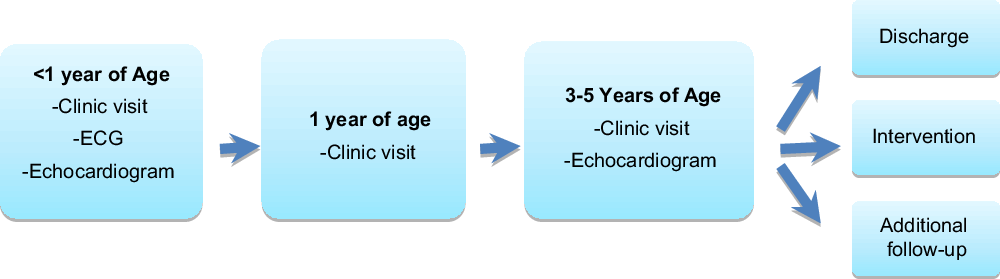
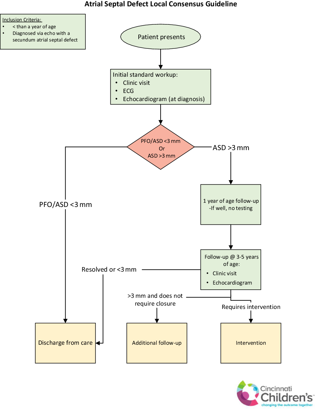
In order to derive our optimal use criteria, we examined data from natural history studies and prior published guidelines in addition to querying providers at our institution about their current clinical practice. Through this, we were able to create a universal consensus, among our group, for an ideal follow-up regimen for patients with isolated atrial septal defects, without symptoms (Figs 1 and 2). For patients with patent foramen ovale (or atrial septal defect <3 mm), optimal utilisation was defined as an initial clinic visit evaluation with no further follow-up recommended, as these lesions typically close spontaneously and are not hemodynamically significant.Reference Radzik, Davignon, van Doesburg, Fournier, Marchand and Ducharme7 For patients with an atrial septal defect, optimal utilisation was defined as an initial clinic visit, electrocardiogram and echocardiogram, a follow-up visit at 1 year without imaging to assess for symptoms and a follow-up at 3–5 years of age with an echocardiogram to assess for spontaneous closure or need for surgical/catheterisation intervention.
Optimal utilisation for patent foramen ovale.

Optimal utilisation for atrial septal defect.

Given the association between atrial septal defects and conduction abnormalities, especially with associated mutations in myocardial transcription factors NKX2.6, GATA5, and TBX5, we deemed a baseline electrocardiogram an important component of the initial assessment.Reference Williams and Perry13 Further electrocardiograms would be indicated based upon clinical concern for conduction abnormalities and are not included in our algorithm. Consistent with prior guidelines, we also deemed advanced imaging, specifically CT and MRI, as unnecessary in the analysis of atrial septal defects for these patients and unlikely to change the clinical course.Reference Haas and Schirmer2
Charge analysis
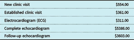
Charge data were obtained from the Cincinnati Children’s Hospital Medical Center financial department (Table 1). These charges were based upon estimates for financial year 2017. Each patient’s total charge was calculated by summing the charge for each clinic visit, total number of electrocardiograms and, finally, the initial and subsequent follow-up echocardiogram(s). Optimal charges were defined at $6250 for a patent foramen ovale and $10,567 for an atrial septal defect.
Charge values (in dollars)

Statistical analysis
Descriptive statistics were performed using means and standard deviations. Statistical analysis was performed using analysis of variance (ANOVA).
Results
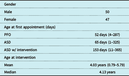
Ninety-seven patients were included in this analysis: 40 had patent foramen ovale (or atrial septal defect <3 mm), 43 had atrial septal defects not requiring intervention, and 14 had atrial septal defects that required intervention. The mean age at intervention was 4.03 years old (Table 2). After analyzing the clinic notes for each patient encounter, no patients had documented symptoms localised to the cardiovascular system during any clinic encounters (shortness of breath, feeding or exercise intolerance, failure to thrive, or oedema) and no patients required urgent intervention.
Patient demographics (range)

ASD = atrial septal defect; PFO = patent foramen ovale.
One patient had surgery performed at 9.5 months of age due to very large ASD (17 mm × 18 mm) with significant right heart dilation at initial diagnosis around 6 months of age. Interestingly, in the OR, this patient was found to have anomalous pulmonary vein draining to the right atrium, which was not noted on prior echocardiograms and likely contributed to the severe right heart dilation necessitating early surgery. This was clearly an outlier from the rest of the patients in this cohort who all had their intervention between 3.5 and 5.3 years of age.
Comparison of lesion types
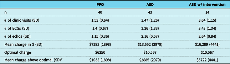
The utilisation data are shown in Table 3. For all lesion types, the mean utilisation for clinic visits, electrocardiograms, and echocardiograms were above optimal. The mean charges for patent foramen ovale (or atrial septal defect <3 mm), atrial septal defect (>3 mm), and atrial septal defect requiring intervention were $7283, $13,552, and $16,289, respectively. Optimal charges for these groups were $6250 for patent foramen ovale (or atrial septal defect <3 mm) and $10,567 for atrial septal defect (with or without intervention). While comparing these charges to optimal, there was statistically significant difference in mean charge above optimal among the three groups of $1033, $2885, and $5722 (p < 0.02; Table 3). While comparing these retrospective charges to proposed optimal charges, estimated savings would have been $242,472 for the years 2010–2013 for all lesion types.
Comparison of lesion types

ASD = atrial septal defect; PFO = patent foramen ovale; SD = standard deviation
* p < 0.02
Comparison by cardiology sub-specialists
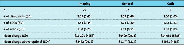
There was also statistically significant variation of mean charges between cardiologist sub-specialists irrespective of lesion type. Catheterisation providers’ mean charge per patient was $14,089, imaging providers’ mean charge was $11,322, and general cardiologists’ mean charge was $9429 (p < 0.02; Table 4). While comparing these charges to optimal for the specific lesion types, the data show that catheterisation providers on average charged around $4916 above optimal, with imaging providers at $2482 above optimal and general cardiologist at $1147 above optimal. Patient distribution was skewed, which is likely reflective of the composition of the cardiology department, with six patients being seen by a catheterisation specialist, 70 by an imaging specialist, and 17 by a general cardiologist. This variability in mean charge was seen with a relatively even distribution of patients that required intervention (one cath, 12 imaging, and two general) and patients with patent foramen ovale (atrial septal defects <3 mm) (three cath, 30 imaging, and eight general).
Comparison of provider types

SD = standard deviation
* p < 0.02
Comparison by years in practice
Provider experience was also assessed as a potential factor for variability in resource utilisation. Providers were stratified based on years of practice since graduating fellowship. Two groups were created, providers with less than 10 years of practice experience or greater than 10 years of practice experience. There was no significant variability among the groups. The median charge above optimal for providers with less than 10 years of practice experience was $2258 with an interquartile range from $672 to $3086. The median charge above optimal for providers with greater than 10 years of practice experience was $1504 with an interquartile range of $0 to $3086. The p-value was not statistically significant at 0.25.
Discussion
Our study found wide variability in outpatient follow-up for secundum atrial septal defects at our institution. Our data show that providers are seeing patient more frequently in clinic and performing more imaging, both electrocardiograms and echocardiograms than our locally agreed upon guide for optimal utilisation. Overall, from 2010 to 2013, total patient charges could have been reduced by $242,472 at our institution, had optimal utilisation been achieved.
Our data also demonstrated variability among paediatric cardiac sub-specialists in how often routine testing was performed with catheterisation providers being more likely to perform interval imaging than general cardiologists and imaging specialists. There was also no statistically significant difference in practice patterns based on provider years of experience. This variability likely comes from differences in clinical assessment, but ultimately did not change the outcome for these patients as no patients presented with pulmonary hypertension or heart failure requiring urgent intervention. The majority of our patients did not require surgical intervention, which is consistent with previous studies, and when interventions were performed, the mean age was around 4 years old, with one outlier.Reference McMahon, Feltes and Fraley1,Reference Radzik, Davignon, van Doesburg, Fournier, Marchand and Ducharme7
As it is rare for patients with isolated secundum atrial septal defect to be clinically symptomatic in the early years of life, we believe that a thoughtful, cost-conscious approach to outpatient, pre-intervention follow may be effective.Reference Goetschmann, Dibernardo, Steinmann, Pavlovic, Sekarski and Pfammatter3 This assumption is limited to a very specific patient population, that is, asymptomatic patients with isolated secundum atrial septal defects. However, given the relatively high prevalence of atrial septal defects, reduction of unnecessary testing could lead to significant savings in healthcare dollars. Verghese et al showed that standardisation can decrease practice variation and resource utilisation in paediatric patients presenting with chest pain. Through the implementation of a Standardized Clinical Assessment and Management Plan, they were able to see a significant reduction of exercise stress tests, Holter monitors, and long-term event monitors in this patient population, which ultimately would lead to charge and cost savings.Reference Verghese, Friedman and Rathod14 We hope our data will help guide our institution to make similar savings and provide more streamlined, cost-effective care.
Limitations
Given the retrospective nature of this study, we were unable to obtain provider rationale for their frequency of follow-up and imaging. Our study was also limited by the efficacy of our query of the electronic medical record as we may have missed patients who did not meet our search criteria but who in fact would have qualified for our study. As our study only looked at charges, based upon charge data for fiscal year 2017, we did not assess the total cost and therefore true saving of healthcare dollars. However, we can extrapolate that if we were able to reduce the amount of tests (electrocardiograms and echocardiograms) and clinic visits, those savings would be passed along to our patients and the healthcare system as a whole.
Future directions
With this data, we will be able to implement a pathway to help guide our clinicians at our institution regarding when to follow up these patients in clinic and when to perform routine testing (Fig 3). Prospectively, we will be able to gather data and assess whether having this pathway in place will lead to a reduction in routine testing and decrease resource utilisation in our clinics.
Atrial septal defect local consensus guideline.

Acknowledgements
We would like to acknowledge all members of Cincinnati Children’s Hospital and the Heart Institute who helped with this study.
Financial Support
This research received no specific grant from any funding agency, commercial or not-for-profit sectors.
Conflicts of Interest
None.