Hypertrophic cardiomyopathy is the commonest medical cause of sudden unexpected death in older children,1 and in athletes.2 The prevalence of the disease in young adults is approximately 1 in 500,3 but although overt disease is uncommon in childhood,4, 5 the disease then has a higher mortality than in adulthood,6, 7 with an annual fatality rate of 6.6% in untreated patients.8 Studies in adults have identified some risk factors for sudden death, such as non-sustained ventricular tachycardia as identified on the 24 hour Holter electrocardiogram,9, 10 left ventricular hypertrophy, in particular a maximal mural thickness exceeding 3 centimetres,11 malignant family history,12 a hypotensive response of blood pressure on exercise testing,13 and presence of dynamic obstruction within the left ventricular outflow tract.14 These risk factors have a low positive predictive accuracy.15–17 All studies, furthermore, have contained no, or few children. As it is affected children who have the highest annual rate of death, we carried out our present study in order to determine which clinical measures were of value in identifying those children at high-risk.
Materials and methods
Patients studied
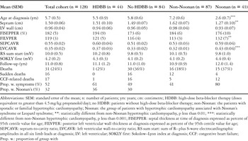
We included all patients in whom a diagnosis of hypertrophic cardiomyopathy had been made before 19 years of age, and who had attended the Divisions of Paediatric Cardiology at the John Radcliffe Hospital, Oxford, and Southampton General Hospital in Great Britain, the University Hospital, Lund, the Academic Hospital, Uppsala, the Queen Silvia Hospital for Children, Gothenburg, and the Astrid Lindgren Hospital, Stockholm, in Sweden. For those patients still surviving, we required a follow-up of at least one year by the end of 2003. Hypertrophic cardiomyopathy was defined as primary and inappropriate hypertrophy in a non-dilated heart, with normal or exaggerated systolic function, in the absence of valvar outflow obstruction or underlying systemic disease. We did not include any infants with hypertrophic cardiomyopathy occurring secondary to gestational diabetes in the mother, or children with hypertrophic cardiomyopathy secondary to mitochondrial disorders, Friedreich's ataxia or other myopathies. Of the subjects identified, 50 (39%) had familial hypertrophic cardiomyopathy, and 37 had sporadic disease. The familial and sporadic cases are analysed together as the non-Noonan group. Based on dysmorphic features, we categorised 39 patients as having Noonan's syndrome (30%), and 2 as Leopard syndrome. These patients were analysed as the Noonan group, as the syndromes are closely related. The characteristics of the patients at diagnosis, and the duration of follow-up in the various groups, are shown in Table 1. The majority had severe hypertrophy, bringing them substantially above the limit of plus 2 standard deviations, with an average septal thickness 82% above the upper limit of normal for age.
Table 1. Characteristics of important predictors and outcomes in children with hypertrophic cardiomyopathy.

In each hospital, patients were recruited from the time of introduction of complete diagnostic registers and availability of diagnostic ultrasound, going back up to 33 years. All hospitals are regional centres, with a geographical base for referrals. In all, we identified 128 patients with hypertrophic cardiomyopathy diagnosed between the ages of birth and 18 years. The average follow-up for the patients was 11.0 years, plus or minus the standard error of the mean of 0.8, giving us 1406 patient-years for analysis. Patients who presented because of sudden death, and those diagnosed at post mortem, are not included. The mean age at diagnosis was 5.7 years, with a median age of 3.6 years. There were a total of 31 disease-related deaths, at a median age of 10.6 years. Out of these deaths 16 were sudden and unexpected, these patients dying at a median age of 13.3 years.
We reviewed the original records in all cases. All but 1 of the patients dying had post mortem records. In the exceptional patient, we had access to recent electrocardiographic and echocardiographic data. We classified the therapeutic regimes at the latest assessment in four categories:
- “no specific therapy” – no therapy or diuretics only. Those in this group had followed the same regime prior to the latest assessment on average 8.1 plus or minus 1.2 years
- “conventional doses of beta-blockers”, at 0.8 to 4 milligrams per kilogram of propranolol per day, or other equivalent beta-blockade. These treatments had continued on average for 6.3 plus or minus 0.7 years
- “high-dose beta-blocker therapy”, at 5 to 23 milligrams per kilogram per day of propranolol, or equivalent doses of metoprolol, atenolol or bisoprolol, with an average duration of 7.9 plus or minus 0.9 years, the dose titrated by 24 hour Holter-recordings as previously described8
- “calcium-blocker therapy”, mainly verapamil in doses of 2.3 to 12 milligrams per kilogram per day, given for an average duration of 5.1 plus or minus 1.1 years.
There is a difference in the earliest year of recruitment into the four groups analysed in respect of treatment, since in the period from 1965 through 1980 only two types of treatment were used, namely no treatment or beta-blockade at conventional doses. Beta-blockade at increased dosage, and calcium-channel blockers, started to be used in 1981 and 1984, respectively. Patients have continued to be recruited into all of the groups, nonetheless, from 1984 onwards to date. Of the two groups with most recent start date, those treated with calcium channel blockers have the worst annual mortality, at 8.8%, whilst those treated with high doses of beta-blockers have the lowest annual mortality, at 0.3%. The annual mortality for the other groups is 4.2% for those receiving no specific therapy, and 4.1% for those treated with beta-blockers in conventional doses. The median year for commencement of treatment is 1994 for beta-blockade at conventional doses, 1993 for high-dose beta-blockade, and 1989 for calcium channel blockers, so there is no era difference between the three groups receiving active treatment. Since the group of patients receiving no specific therapy, with a median year of inclusion of 1983, has the same annual mortality as the group undergoing treatment with beta-blockers at conventional dosage with the latest median year of recruitment, there is no evidence to suggest a chronological era effect on deaths of the patients. In addition to medical therapy, 12 patients were referred for myectomy, 1 patient having two procedures.
Electrocardiographic measurements
Original electrocardiograms were available for re-measurements according to standardised criterions in 126 of the 128 patients, and in all 16 patients dying suddenly. From the electrocardiograms, we calculated the Sokolow–Lyon index,18 albeit that this proved impossible in infants and in patients with dominant S-waves across the praecordium. Accordingly, we used an additional electrocardiographic measure, namely the sum of the total deflections in the R and S waves, including Q-waves if deeper than S waves, in all six limb leads.8 Normal values for this measure were obtained in 55 children with echocardiographically normal hearts aged from 0.1 to 18 years.
Echocardiographic M-mode measurements
Echocardiographic M-mode measurements were available in 119 of the 128 patients. In 8 of the remaining patients, severe hypertrophy had been documented during angiography, and in addition there was histological confirmation of hypertrophic cardiomyopathy from post mortem or myomectomy specimens. In the other, diagnosis had been made by repeated cross-sectional echocardiography, with Doppler-confirmation of a significant gradient across the left ventricular outflow tract. Original M-mode tracings were re-measured using long-axis measurements according to current conventions,19, 20 but in 5 of the patients where no tracings had been archived, we accepted the M-mode measurements as recorded in the notes. All 5 had been seen at a time when current conventions for measurement had been adopted. To estimate the severity of hypertrophy in the growing child, we calculated two types of ratios. First, we related mural thickness to cavity dimensions by dividing the thicknesses of the septum and posterior wall by the diastolic diameter of the cavity, referred to as the septum-to-cavity ratio, and the left ventricular wall-to-cavity ratio, respectively, as well as calculating the ratio of the posterior left ventricular wall thickness to the diameter of the cavity in systole, referred to as the systolic wall-to-cavity ratio.8, 20 Second, the observed values of both septal and left ventricular posterior mural thicknesses were expressed as a percentage of the respective predicted 95th centile values for age,8 measurements referred to as the age-corrected mural thickness.
Outflow obstruction had been observed in 66 patients, in 64 documented at cardiac catheterisation with a gradient exceeding 16 millimetres of mercury, or with Doppler measurements showing velocities across the left ventricular outflow tract over 2 metres per second. In the other 2 patients, obstruction was inferred from the co-existence of systolic murmurs and M-mode features of systolic anterior movement of the mitral valvar apparatus. Because of the difficulty in comparing catheter and Doppler gradients, we have simply classified patients as with or without obstruction in the left ventricular outflow tract.
Mode of death
We analysed all 31 disease-related deaths. Of these, 16 patients had died suddenly, 2 died perioperatively associated with septal myectomy, and 17 were classed as associated with congestive heart failure. The latter include 1 of the myectomy deaths, an infant with severe heart failure symptoms, and 3 patients with failure and a dilated end-stage. Two patients with dilated end-stage and heart failure died suddenly. These patients are included in both groups for analysis of risk factors.
Statistics
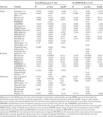
Statistical analysis was carried out using commercial software (Statgraphics Plus v5.2, SPSS 11.5 and GraphPad Prism 4). Between group comparisons of measurements used the Mann–Whitney U-test. Candidate risk factors for death were identified by multivariate correlation analysis, and substantiated with Cox proportional hazards regression analysis. Cox regression was carried out first on categorical variables and continuous variables with the complete data set, secondly on 98.4 percent complete electrocardiographic data set, and thirdly on the 90.6 percent complete echocardiographic data set, with forward stepwise method based on likelihood ratios, but forced entry method to obtain regression coefficient (B) and change in odds resulting from a unit change in the predictor (Exp(B)) for risk factors with a p-value between 0.05 and 0.10. These data are showed in Tables 2 and 3. This was done in order to have maximal data for each estimation of regression coefficients.
Table 2. Cox hazard regression analysis of risk factors.

Table 3. Comparisons between the effect of significant predictors of risk in patients with hypertrophic cardiomyopathy with and without associated Noonan's syndrome.

Lastly, fully to assess the relative weight of various risk factors in relation to each other, a forward stepwise analysis was carried out with the predictors identified to that stage only in patients with a complete data set. These data are given in the text.
The distributions of values of different risk factor measures were compared in survivors and non-survivors with box-and-whisker plots and receiver-operator characteristic curves to identify suitable cut-off values. The Fisher exact test was used for categorical data analysis, and odds ratios were used to compare groups above and below cut-off values. The survival of patients in groups with different risks was compared with Kaplan–Meier survival curves and log-rank t-tests. For those risk factor measures that were independent predictors on Cox regression analysis, we calculated the specificity, sensitivity, positive predictive value and negative predictive values.
Results
Population of patients
Patients with Noonan or Leopard syndromes were diagnosed at a younger age than those categorised as non-Noonan, but age-corrected measures of septal hypertrophy, such as septum-to-cavity ratio and percent age-corrected septal thickness, did not differ (Table 1). Hypertrophy of the free left ventricular wall, however, was more pronounced, with significantly higher left-ventricular wall-to-cavity ratios and age-corrected left ventricular wall thickness in those with Noonan's and Leopard syndrome as compared with the non-Noonan group.
Risk factors for disease-related death
Both electrocardiographic and echocardiographic measures show a positive correlation of risk with degree of hypertrophy, and on Cox regression analysis, risk also correlates with rate of progression of the disease (Table 2). Values of the sum of the R and S waves, and the rate of increase over 12 months, showed significant positive associations with death. We used the measurements of relative mural thickness, that is the septum-to-cavity ratio, the left ventricular wall-to-cavity ratio, and age-corrected septal thickness, in the Cox regression analysis, as they had higher correlation coefficients with death on univariate correlation analysis than did absolute mural thickness. On Cox regression, they showed highly significant correlation with risk of death. It was also evident that predictors of sudden death are different from predictors for death related to cardiac failure (Table 2).
Risk factors for sudden death
The median age at sudden death was 13.3 years, with many such deaths occurring in asymptomatic patients aged from 8 to 12 years (see Fig. 1).

Figure 1. A frequency histogram of the age of the patients dying suddenly.
Sudden death shows highly significant positive correlation with electrocardiographic voltages, both the sum of the R and S waves and the Sokolow–Lyon index, and with septal wall thickness expressed as age-corrected septal thickness. When these three strong predictors were analysed together in the patients with a complete data set, forward stepwise analysis left the sum of the R and S wave at diagnosis (p equal to 0.001), and latest age-corrected septal thickness (p equal to 0.036), as independent predictors for sudden death. The Sokolow–Lyon index is removed from the equation. The presence of a gradient across the left ventricular outflow tract showed no separate predictive power.
Risk factors for death related to cardiac failure
These deaths occurred in two distinct groups, 1 in infants with vigorous systolic function, and 1 where cardiac failure was a late result of progressive disease, commonly with a dilated end-stage. Deaths in infants with vigorous systolic function occurred during the first 2 years of life (56%), and in those with progressive disease after the age of 13. Absolute electrocardiographic voltages showed no significant correlation with risk, and neither did absolute mural thickness. Young age at diagnosis, the left ventricular wall-to-cavity ratio, and the presence of Noonan's syndrome were significant risk factors (see Table 2), but on forward stepwise analysis on patients with complete data, the age at diagnosis is removed from the equation, leaving the left ventricular wall-to-cavity ratio (p equal to 0.005) and Noonan's syndrome (p equal to 0.043) as significant independent predictors. The left ventricular wall-to-cavity ratio shows higher levels of significance early in the course of the disease, as infants dying from failure invariably have high values, whereas patients progressing to a dilated end-stage get a late fall in the ratios. An elevated left ventricular wall-to-cavity ratio is an indicator of the presence of generalised hypertrophy, as no child in this cohort had hypertrophy restricted to the free left ventricular wall.
Effect of therapy
Neither myectomy (p equal to 0.96; not shown in Table), nor treatment with verapamil (see Table 2), had any significant effect on subsequent survival. Beta-blocker therapy, on the other hand, showed a strong negative correlation, the higher the dose, the lower being the risk, to all disease-related deaths (p equal to 0.003) as well as sudden deaths (p equal to 0.008). This mode of treatment was also approaching significance with those dying in cardiac failure (p equal to 0.087). When beta-blockade is divided into regimes using conventional and high doses, only the regime using the high doses showed significant reduction of risk, this finding being in keeping with our previous study.8 The reduction was significant both for those dying suddenly, and for those dying with cardiac failure (see Table 2). Because of this, we repeated the Cox regression analysis having excluded those patients receiving high doses of beta-blockers. Our repeated analysis confirmed the significant predictive powers already at the time of diagnosis for the sum of the R and S waves, the septum-to-cavity ratio, the left ventricular wall-to-cavity ratio, and the age-corrected septal thickness (Table 2). In Figure 2, we illustrate the striking differences in the values of the sum of the R and S waves at diagnosis of children with hypertrophic cardiomyopathy who do or do not subsequently die suddenly, comparing these values also with those obtained in our normal children.

Figure 2. A box-and-whisker plot of the electrocardiographic amplitudes at the time of initial diagnosis, quantified as the sum of the amplitudes of the R and S waves in millivolts. The Q waves are included if deeper than the S waves. Patients are divided into those with hypertrophic cardiomyopathy and dying suddenly (HCM SD), those with hypertrophic cardiomyopathy not dying suddenly or in cardiac failure (HCM Surv), and 55 normal children aged from 0.1 to 18 years (Normals).
The patients treated with beta-blockers at high doses were fully comparable with the remainder of the cohort in their clinical features (see Table 1). As any protective effect of treatment would distort the evaluation of risk factors, particularly the assessment of the size of the risk, we also removed the 44 patients treated with beta-blockers in high doses from the analysis of cut-offs for the risk factors, leaving 84 subjects. All but one of the 31 patients dying were included among these 84 subjects, giving 36% mortality in this remaining cohort.
Are the risk factors the same in hypertrophic cardiomyopathy associated with Noonan's syndrome?
As the risk of dying in heart failure is higher in children with hypertrophic cardiomyopathy in the setting of Noonan's syndrome, we studied whether some risk factors are specific for this syndrome by separate Cox-regression analysis of the two subgroups (see Table 3). As only 5 patients without Noonan's or Leopard syndrome died in cardiac failure, and only 4 patients with these syndromes died suddenly, the statistical power is limited. Nevertheless, the last measured sum of the R and S waves was statistically significantly correlated with sudden death in both groups. The last age-corrected measurement of septal thickness just failed to reach statistically significant correlation with sudden death in the patients with Noonan's or Leopard syndrome, but the change in odds resulting from a unit change in age corrected septal thickness was almost identical to the patients who were not syndromic (see Table 3). Thus, there is no evidence to suggest that risk factors for sudden death are different in the two groups. Likewise, the regression between left ventricular wall-to-cavity ratio at diagnosis and death in cardiac failure has very similar regression coefficients, and changes in odds resulting from a unit change, in both groups (see Table 3).
Selection of cut-off values
We compared the measures that proved to be independent predictors of different types of disease-related death on Cox regression analysis between survivors and non-survivors so as to identify suitable cut-off values for further evaluation of their predictive efficacy. The cut-off levels that appeared to give optimal separation with box-and-whisker plots (for an example see Fig. 2) and receiver-operator characteristic curves proved to be:
- The sum of the R and S waves exceeding 10 millivolts, with an odds ratio of 8.4 at diagnosis comparing patients above and below the cut off, and 95% confidence intervals from 2.2 to 33.7. The p value is equal to 0.0012 as a risk factor for sudden death.
- Initial septal thickness exceeding 190% of the 95th centile value for age, with an odds ratio of 6.2, and confidence intervals from 1.5 to 25.1. The p value is equal to 0.011 as a risk factor for sudden death.
- An initial left ventricular wall-to-cavity ratio greater than 0.30, with an odds ratio of 36.0, confidence intervals from 4.2 to 311, and a p value of less than 0.0001, for death related to cardiac failure.
With increased duration of follow-up, the predictive strength of the measures predicting sudden death tends to increase further, with the latest measurement of the sum of the R and S waves greater than 10 millivolts giving an odds ratio of 34.3, confidence intervals from 2.0 to 600, and a p value of less than 0.0001. Similarly, when the last age-corrected ratio of septal thickness exceeds 190%, the odds ratio is 11.1, with confidence intervals from 1.3 to 95.2, and a p value equal to 0.013. In Figure 3, we have compared the difference in survival between patients who are positive for at least one risk factor at the time of diagnosis, these patients having a median survival of 16.0 years, with that of patients without risk factors at diagnosis. The latter patients have a superior survival (p equal to 0.0002), with a 96.7 percent survival proportion at the point where 5 patients remain at risk after 20 years of follow-up. The 1 patient who died without any risk factors at diagnosis was a child who subsequently fulfilled two criterions for risk 2.8 years before her sudden death. The annual mortality in patients lacking risk factors at diagnosis was 0.27 percent, whereas annual mortality in patients with two risk factors at diagnosis is 4.7 percent, reaching 6.6% in those patients with three risk factors.

Figure 3. Kaplan–Meier survival curve comparing the survival of patients with values for the sum of the elctrocardiographic amplitudes, the age-corrected septal thickness, and left ventricular wall-to-cavity ratios below the cut-offs established as configuring high risk at diagnosis (○) with patients with at least one of these measures above the cut-offs ([squf ]). The survival is significantly different on log-rank test (p equal to 0.0002), and the hazard ratio is 16.0. Only the 84 patients not receiving protective high doses of beta-blockers are included in these survival curves.
Performance of the indicators of increased risk as measures for screening
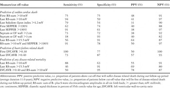
The measures highlighted above as particularly important indicators of early risk, and a few more measures suggested by previous investigations of adults with hypertrophic cardiomyopathy, were evaluated for their efficacy in screening by calculating their sensitivity and specificity, and their positive and negative predictive values (Table 4). The best measures in predicting sudden death are the sum of the R and S waves in the electrocardiogram exceeding 10 millivolts, and a septal thickness exceeding 190% of the 95th centile for age. Each of these measures has much higher sensitivity, and positive and negative predictive values, than do absolute measures of mural thickness, regardless of whether a cut off of greater than 3 or 2 centimetres is employed (see Table 4). It is also possible to give a good early prediction of the likelihood of dying in cardiac failure using a cut-off for the ratio of left ventricular wall-to-cavity of more than 0.3 (Table 4). Values substantially above the lowest cut off, and being positive on more than one criterion, give an incremental risk, as shown by increasing positive predictive values (Table 4). The highest risk for death is seen in patients with the summed R and S waves exceeding 10 millivolts combined with a left ventricular wall-to-cavity ratio over 0.40, where a positive finding indicates a risk of 78% of disease-related death.
Table 4. Performance of selected indicators of high risk as measures for screening.

Discussion
Risk factors for children with primary hypertrophic cardiomyopathy, excluding cases of secondary cardiomyopathy, have been analysed in four previous studies, albeit with considerably fewer cases. The first two studies, using groups of 35 and 46 patients, failed to identify any predictors.21, 22 A later study from a supra-regional referral centre, analysing 97 children, identified increased QTc-dispersion, ventricular tachycardia on ambulatory 24 hour electrocardiograms, and myocardial bridging on coronary angiography, as predictors of sudden death. This investigation clearly dealt with selected cases, since two-thirds of their subjects had undergone coronary angiography.23 The smaller precursor to the present study, also on a complete geographical cohort, showed that large electrocardiographic voltages, both as the Sokolow–Lyon index and the sum of the R and S waves in the limb leads, correlated with mortality on correlation analysis, as did echocardiographic measures of ventricular mural thickness expressed as a proportion of the 95th centile for age.8 Cox proportional regression to determine which risk factors were independent, and estimates of screening efficacy, were not performed. Furthermore, our current study is the first to show the differences between the risk factors for dying suddenly, as opposed to dying in cardiac failure.
What are the special aspects of risk-stratification for children with hypertrophic cardiomyopathy?
In adults with hypertrophic cardiomyopathy, stratification is generally based on family history, exercise testing, ambulatory electrocardiographic monitoring, and echocardiography. Although identification of particularly malignant mutations is feasible, the laboratory techniques for screening for an unknown mutation are only available in selected research laboratories. Hence, genotyping is not yet a usable tool in routine clinical practice. Furthermore, only 1 percent of patients with hypertrophic cardiomyopathy have known “malignant” mutations.24 A malignant family history remains a significant risk factor for children with hypertrophic cardiomyopathy,8 but is not often helpful, as three-fifths lack a family history. With a median age at presentation of 3.6 years, a formal exercise test is also not often an option. Thus, in the child with hypertrophic cardiomyopathy, the prediction of risk must rest largely on 12 lead electrocardiography, echocardiography, and ambulatory electrocardiographic monitoring. We have demonstrated four facts of major importance.
First, there is a strong correlation between electrocardiographic amplitudes and the risk for sudden death. This is not simply a general reflection of degree of cardiac hypertrophy, as we have also shown that electrocardiographic amplitudes are independent predictors from measures of mural thickness. We have observed that patients belonging to the same family with familial hypertrophic cardiomyopathy tended to have similar electrocardiographic amplitudes. This latter observation has been substantiated by evidence that electrocardiographic phenotypes, such as the Sokolow–Lyon voltage, show heritability.25 There is little published data about the association between electrocardiographic voltages and particular causative mutations for hypertrophic cardiomyopathy. In families with eight different cardiac beta-myosin heavy chain mutations, the proportion with a raised Sokolow–Lyon index ranged from zero in a family without sudden deaths, to two-fifths in a family with two sudden deaths.26 Patients with mutations in the gamma-2 subunit of AMP-activated protein kinase have strikingly large electrocardiographic amplitudes.27 In this study, we found that the sum of the amplitudes of the R and S waves in the limb leads proves a better predictor than the Sokolow–Lyon index, with a sensitivity of 94% as compared to 79%. Indeed, the sum of the amplitudes is the only electrocardiographic measure that can always be quantified in children, regardless of their age. The odds ratios for this measure when used as a risk factor are considerably larger, 32.3 using the latest follow-up measurement, than other risk factors hitherto described in adults with hypertrophic cardiomyopathy, where relative risks range between 1.8 and 5.3.28
Second, although the risk for sudden death is correlated with maximal ventricular mural thickness, the degree of hypertrophy has to be related to what is normal for age. In this respect, children have an increased risk of sudden death at much lower absolute thicknesses than do adults. Mural thickness exceeding 3 centimetres has been advocated as a cut-off for adults deemed at high risk.11 This is far too high a cut-off level for children, where it would give a sensitivity of only 18%. In childhood, the best predictor is to divide the septal thickness with the 95th centile value arrived at by the equation:

expressing the result as a percentage.8 A value exceeding 190% implies a state of high risk, with a sensitivity of 91%, and odds ratios of 6.2 to 11.1 for sudden death, compared with a relative risk of 2.07 described for absolute mural thickness exceeding 3 centimetres in adults with hypertrophic cardiomyopathy.17
Third, a large proportion of children with hypertrophic cardiomyopathy dying suddenly are asymptomatic, and they die before, or in early, puberty between the ages of 8 and 13 years. This underlines the importance of considering early electrocardiographic and echocardiographic screening, ideally no later than the age of 6 or 7 years, of children with one parent with hypertrophic cardiomyopathy. Although the hypertrophy in many individuals may not be severe at this age, this test would identify those subjects at high risk. Probable gene carriers can also be identified for further monitoring by using both diastolic and systolic wall-to-cavity ratios.20, 29
Fourth, although the risk factors for dying suddenly are different from those for dying in cardiac failure, the same measures prove to be predictive in those children having hypertrophic cardiomyopathy associated with Noonan's syndrome, as well as in those with hypertrophic cardiomyopathy unrelated to Noonan's syndrome. This is interesting from the mechanistic point of view. In particular, the evidence for inheritability of electrocardiographic characteristics suggests the possibility that risk associated with a mutation causing hypertrophic cardiomyopathy might be modified by other mutations or gene polymorphisms.25
Limitations of our study
Use of retrospective complete cohorts is ideal for identifying risk factors for adverse outcomes, but is not ideal for assessing the effects of treatment, as the allocation of treatment is not randomised. The advantage of using such data obtained retrospectively in rare conditions such as hypertrophic cardiomyopathy seen in children is that a large number of patient years can be analysed. As the patients treated with high doses of beta-blockers in our cohort were indistinguishable from the rest of the patients with hypertrophic cardiomyopathy in respect of all identified risk factors (see Table 1), it would therefore seem appropriate to note that the results of Cox Hazard analysis supports the finding of an improved survival on Kaplan–Meier survival analysis in patients treated in this fashion as reported in our previous study.8 In the present study, all 9 patients in the groups of patients not receiving beta-blockers in high doses died when they fulfilled the 3 criterions for high risk, whereas death occurred in only one of the 9 patients fulfilling the same three criterions in the group of patients receiving beta-blockers in high doses. This difference is highly significant, with a p value of 0.0004 as calculated using Fisher's exact test. Furthermore, none of these patients has required an implantable cardiac defibrillator. That lipophilic beta-blocker therapy may give significant protection against both sudden arrhythmic death, and death in cardiac failure, is given credence by the findings in large and randomized prospective studies in adults of beta-blocker therapy in dilated cardiomyopathy, and in ischaemic heart disease,30 which report significant protection using metoprolol and bisoprolol. The mechanisms by which such treatments may work in hypertrophic cardiomyopathy have been discussed earlier.8
Conclusions
Stratification using measures obtained from unfiltered 12-lead electrocardiograms and echocardiography, investigations available in most district hospitals, allows separation of patients at low risk, with an annual mortality of 0.27%, from those at high risk, with a subsequent annual mortality of up to 6.6%, with high sensitivity and negative predictive value. This allows selection of children not only for appropriate medical therapy, but also for consideration of other therapeutic options.
Acknowledgements
The uniquely long follow-up in patients with echocardiographic data would not have been possible without the pioneering efforts of Nils-Rune Lundström in the application of echocardiography for children in Lund, and the meticulous diagnostic registers and record-keeping by him and his Swedish contemporary colleagues, in particular, Bengt Eriksson in Gothenburg, Magnus Michaëlsson in Uppsala, and Claes Thorén in Stockholm. Financial support: Grants from the Lund-Oxford Biomedical Exchange programme, Lund, Sweden, The Childrens Echocardiography Fund, Oxford Radcliffe Hospital Trust Fund, United Kingdom and a LUA Project Grant from the Gothenburg University, Sweden.