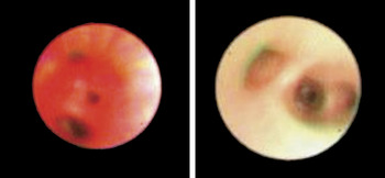
A male infant, born at term with trisomy 21, underwent surgical closure of a perimembranous ventricular septal defect at 4 months of age. He subsequently developed pulmonary hypertension secondary to severe stenosis of a common left pulmonary vein, which was treated by dilation with a cutting balloon. In view of recurrent infections of the lower respiratory tract, flexible bronchoscopy was performed at 1 year of age. This revealed normal bronchial anatomy, but marked left-sided hyperaemic changes (
Figure 1). We interpreted the bronchoscopic findings as indicative of venous congestion, due to unilateral pulmonary venous obstruction. In cases where such differential hyperaemia is noted, the possibility of an underlying cardiac lesion always should be considered.
Figure 1

Acknowledgment
We would like to thank Dr Aruna Ranasinghe for his contributions to this publication.