Introduction
Acanthocephalans are helminths that may be pathogenic to birds (Taraschewski, 2005; Atkinson et al., Reference Atkinson, Thomas, Hunter, Atkinson, Thomas and Hunter2008), and they have been associated with varying levels of mortality, mainly in coastal birds. For example, mass mortalities have been reported in common eiders, Somateria mollissima, in the Dutch Wadden Sea (Camphuysen et al., Reference Camphuysen, Berrevoetsc, Cremersd, Dekinga, Dekker, Ens, van der Have, Kats, Kuiken, Leopold, van der Meer and Piersma2002) and mute swans, Cygnus olor, in Canada and Scotland (Sanford, Reference Sanford1978; Pennycott, Reference Pennycott1998).
Olrog's gull is a species listed as vulnerable to extinction (IUCN, 2010) and is endemic to the Atlantic Coast of Argentina, Uruguay and southern Brazil (Yorio et al., Reference Yorio, Bertellotti and García Borboroglu2005). Variable mortality has been reported among Olrog's gull prefledged chicks since 2003 in the largest known breeding colony for this species, and the acanthocephalan Profilicollis chasmagnathi was postulated as one of the possible causes of death (La Sala & Martorelli, Reference La Sala and Martorelli2007). Although this first study provided baseline information on disease pathology, there is still an important gap in knowledge related to pathological and epidemiological aspects of acanthocephalan infections in this species.
The objective of this work is to extend previous findings in acanthocephalan-infected Olrog's gulls by: (1) assessing the role of chicks' size/age as risk factors for higher intensity of infection and increased pathology; (2) studying the association between intensity of infection and pathology; and (3) further characterizing pathological features of infection.
Materials and methods
Source of the data
The study incorporated data from necropsies of freshly dead, prefledging chicks salvaged during four breeding seasons of Olrog's gull (2003: 6 birds; 2005: 66 birds; 2006: 37 birds; 2007: 1 bird) at the Isla del Puerto breeding colony (38°48′S, 62°15′W) in the Bahía Blanca Estuary, Argentina.
The total head–beak (hereafter head–beak) and tarsus–metatarsus (hereafter TMT) lengths of each dead chick were measured to the nearest 0.01 mm using digital callipers, and this metric was used as a proxy for each chick's age. Chicks were necropsied within 4 h of collection, and each gastrointestinal tract (hereafter GI) was removed and fixed in 10% formalin until it could be examined. At the laboratory, each GI was opened and specimens of P. chasmagnathi present in the GI were counted. Complete, acanthocephalan-associated perforations were recorded and their intensity was used as an estimate of severity of infection. Acanthocephalans found in the coelomic cavity, which had perforated the GI, were quantified. Affected regions of the gut were prepared for histopathological examination; samples were dehydrated in ethanol, embedded in paraffin, sectioned at 5 μm, stained with haematoxylin/eosin, periodic acid–Schiff (PAS) or Masson's trichrome, and examined under light microscopy.
Statistical analyses
The analyses included combined sets of data from 2005 and 2006. Prevalence of infection and its 95% confidence interval (95% CI) and mean intensity (number of parasites/number of infected hosts) were computed and interpreted as measures of frequency and intensity of infection, respectively. The degree of clustering of parasites in the host population (dead prefledging chicks) was calculated using the variance-to-mean ratio (Elliot, Reference Elliot1977).
Because the dependent variables ‘intensity of infection’ and ‘number of perforations’ approximated a theoretical negative binomial distribution, their association with the explanatory variable ‘chick size’ (approximated by head–beak and TMT) in the subpopulation studied here (dead chicks) was assessed using generalized linear models (GLM) with a negative binomial response using the function ‘glm.nb’ in the package MASS (Vernables & Ripley, Reference Venables and Ripley2002) of the statistical program R (R Development Core Team, 2010). Because head–beak and TMT were highly correlated (>70%), they were examined separately to avoid collinearity, and the Akaike information criterion (AIC; Akaike, Reference Akaike1974) was used to select the most parsimonious, final model, as suggested by the lowest AIC value. The association between ‘intensity of infection’ and ‘number of perforations’ was assessed using the same approach described above.
For continuous independent variables, such as head–beak, the predicted percentage of change in the dependent variable for every one-unit increase in the value of the independent variable was computed as [exp(β) − 1] x 100, where β is the regression coefficient of the dependent variable. Simulations were used to fit curves of the evolution of infection intensity and associated intestinal perforations as a function of head–beak. The relative goodness of fit of the models was assessed based on the AIC criterion. Terms (variables) were considered to improve the model fit if their exclusion increased AIC by more than two units (Burnham & Anderson, Reference Burnham, Anderson, Burnham and Anderson2002).
Results
Parasite identification
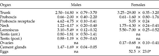
Based on morphological characteristics and measurements of 10 males and 10 females (table 1), the parasite was identified as P. chasmagnathi (Holcman-Spector et al., Reference Holcman-Spector, Mañé-Garzón and Dei-Cas1977). The body consisted of an elongated trunk with abundant spines on its anterior third, a slender neck and a large spherical proboscis. Proboscis hooks were arranged in 16–20 longitudinal rows with 8–9 hooks each. The proboscis receptacle was cylindrical, double-walled, extending between the proboscis base and the first third of the trunk. Lemnisci were larger than the receptacle. Males had spherical or oval testes, four paired cement glands and a prominent copulatory bursa with numerous cuticular rays and two muscular sphincters. Females had a uterine bell, uterine duct, uterus, muscular vagina, vulva and subterminal genital pore. Eggs were ovoid without filaments.
Measurements of P. chasmagnathi from Olrog's gull, Larus atlanticus.
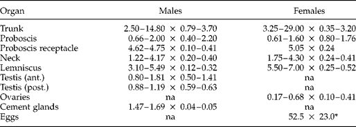
* Measurements in μm; na, not applicable.
Parasite aggregation, distribution and accumulation over time
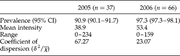
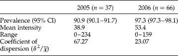
The degree of parasite aggregation in the studied subpopulation (i.e. the dead chicks) is presented along with frequency estimates in table 2. The intensity of infection in different regions of the GI was consistent between years (2005: jejunum = 51.4%, ileum = 39.9%, caeca = 1.2%, rectum = 0.8%, coelom = 6.7%; 2006: jejunum = 36.6%, ileum = 50.7%, caeca = 1.3%, rectum = 0.7%, coelom = 10.8%), with the exception that the proportions were inverted between the jejunum and ileum between years. Complete perforation of the intestines was observed in 85% (34/40) and 97.3% (36/37) of the chicks studied in 2005 and 2006, respectively.
Infection levels of P. chasmagnathi in dead chicks of Olrog's gull.
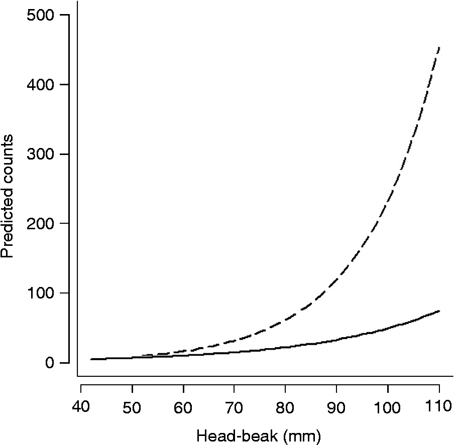
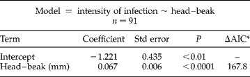
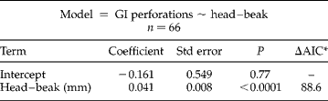
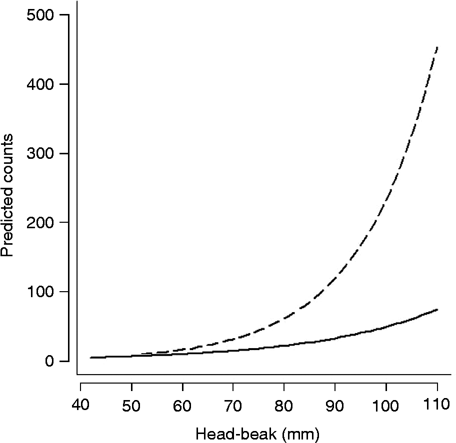
Based on the AIC, the probabilities of infection intensity and number of GI perforations were better described by head–beak than by TMT, so only the former variable was included in the models. Head–beak was positively associated with intensity of infection (Z= 11.29, df = 90, P< 0.001), which increased by 6.9% for each millimetre of increment in head–beak (table 3, fig. 1). Similarly, head–beak was positively associated with the number of intestinal perforations (Z= 5.29, df = 65, P< 0.001), which increased by 4.1% for each millimetre of increment in head–beak (table 4, fig. 1). The number of perforations increased by 2.2% (Z= 9.82, df = 76, P< 0.001) for each unit of increment in intensity.
Simulation of P. chasmagnathi intensity (dashed line) and associated intestinal perforations (solid line) as a function of chick size (head–beak length) in dead Olrog's gull chicks.
Negative binomial model describing the association between head–beak and intensity of infection by P. chasmagnathi in dead chicks of Olrog's gull.
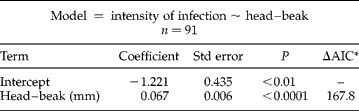
* AIC value increment if the single term is dropped.
Negative binomial model describing the association between head–beak and the number of GI perforations associated with P. chasmagnathi in dead chicks of Olrog's gull.
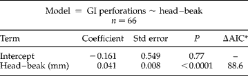
* AIC value increment if the single term is dropped.
Gross pathology
Many of the worms perforated the intestinal wall with their trunk and attached with their proboscis to the serosa of adjacent intestinal loops, causing intense peritoneal reactions observed as strong fibrous adhesions. Some extraintestinal worms perforated from the serosal towards mucosal surfaces of the intestinal wall (both completely and incompletely). Intestinal rupture was observed in one bird where the entire metasomae of five adult acanthocephalans protruded into the peritoneal cavity. Many of the worms found free in the coelom were encapsulated by a thick layer of fibrotic tissue which often contained the decomposed parasite. Some worms were found with their proboscis embedded into the parenchyma of the pancreas. The majority of the large, intra-intestinal female worms found in the largest chicks were gravid.
Histopathology
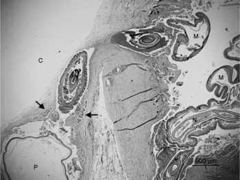
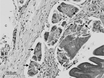
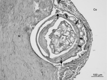
In the earliest stages of parasite penetration into the GI wall, a band of necrotic debris was observed around the leading edge of the presoma. An intense proliferation of fibroblasts was present throughout the thickness of the muscularis externa, and a mixed inflammatory infiltrate composed of macrophages, lymphocytes and eosinophils surrounded the parasites' presomae. With deeper invasion into the muscularis externa, abundant multinucleated giant cells were observed surrounding the parasite. In the most advanced stage of penetration, the presoma was surrounded by a granulomatous reaction composed of giant cells, macrophages, eosinophils and lymphocytes (figs 2, 3 and 4). Grossly, these appeared on the serosal surface of the GI as prominent nodules containing the parasite's proboscis. Occasionally, P. chasmagnathi perforated the intestinal wall with minimal evidence of granuloma formation.
Profilicollis chasmagnathi perforating the intestinal wall to the serosa level. Two adults of P. chasmagnathi can be observed with their metasoma (M) in the intestinal lumen, and their presoma (neck = N; proboscis = P) penetrating the intestinal wall and contained in a granuloma with abundant giant cells (arrows).

Granuloma surrounding P. chasmagnathi. Multinucleated giant cell (solid arrows), macrophage (dashed arrow), lymphocytes (arrowheads), and host necrotic tissue (NT) are observed adjacent to P. chasmagnathi (P). Abnormal fibroblast proliferation and fibrosis (F) surrounds the parasite-containing granuloma isolating it from the coelomic cavity (C).

Interface between P. chasmagnathi (P) and the thickened GI serosa (S). Intense mononuclear cell infiltration with lymphocytes (solid arrow) and macrophages (dashed arrow) are observed at the interface between P. chasmagnathi and host tissue. Co, coelomic cavity; M, muscularis externa; H, proboscis hooks.

Profilicollis chasmagnathi was also present in the caecum and tended to occupy most of its lumen. The degree of penetration was variable and ranged from superficial to complete perforations of the organ's blind end. The intensity of infection was limited to one worm per caecum. There were profound architectural changes of the crypts, acute and chronic cellular infiltrates in the lamina propria, granuloma formation and intense hyperplasia of mucosa-associated lymphoid tissue.
Discussion
This study shows: (1) the presence of advanced pathology associated with P. chasmagnathi infections in Olrog's gull chicks from very early in the post-hatching period; (2) that the intensity and severity of infection increase significantly as a function of chick size during that period; and (3) that the severity of infection increases with intensity of infection. Also, the present work elaborates on previous findings describing the pathological features of acanthocephalan infections in this vulnerable species.
In general, the pathological findings presented here agree with descriptions made in domestic and wild birds (Nath & Pande, Reference Nath and Pande1963; Bishop & Threlfall, Reference Bishop and Threlfall1974; Taraschewski & Hofmann, Reference Taraschewski and Hofmann1991), mammals (Nelson & Nickol, Reference Nelson and Nickol1986; Richardson & Barnawell, Reference Richardson and Barnawell1995) and fish (de Buron & Nickol, Reference de Buron and Nickol1994; Sanil et al. Reference Sanil, Asokan, John and Vijayan2011) infected with different species of acanthocephalans. Fibrous nodules similar to those observed in Olrog's gull chicks have been reported in other avian species, such as Melanerpes carolinus (Picidae) infected by Mediorhynchus centurorum (Nickol, Reference Nickol1969), domestic ducks infected by Polymorphus minutus and Filicollis anatis (Nicholas & Hynes, Reference Nicholas and Hynes1958; Taraschewski & Hofmann, Reference Taraschewski and Hofmann1991), and Somateria mollissima (Anatidae) infected by Profilicollis botulus (Bishop & Threlfall, Reference Bishop and Threlfall1974). Also, the composition of inflammatory cells surrounding the parasites and comprising the granulomas resembles those described by Gonzales-Viera et al. (Reference Gonzales-Viera, Luján-Vega, Chavera-Castillo, Cárdenas-Callirgos and Tantaleán2009) in Leucocephalus modestus (Laridae) infected by Profilicollis altmani, as well as by Taraschewski & Hofmann (Reference Taraschewski and Hofmann1991) in domestic ducks infected by Filicollis anatis.
Granulomas are an important protective innate response which becomes stronger as the bird matures (Juul-Madsen et al., Reference Juul-Madsen, Viertlboeck, Smith, Göbel, Davidson, Kaspers and Schat2008). Complete perforations by small, immature P. chasmagnathi were observed in some very small chicks (c. 5 days of age) without the nodule formation typically observed in larger birds, and may thus be explained by a lack of competent innate responses to P. chasmagnathi in the smallest chicks that died.
Here, P. chasmagnathi was identified at very high prevalence and high intensities in the subpopulation of dead chicks, with full-thickness intestinal perforations being observed in most of the birds. The intensity and severity of infection (number of GI perforations) increased sharply as a function of chick's size/age, whereas infection became more severe with increasing intensity.
During their breeding season, Olrog's gull parents feed chicks from day one of hatching and themselves almost exclusively on Cyrtograpsus angulatus (pers. obs.) and Neohelice granulata (Delhey et al., Reference Delhey, Carrete and Martínez2001) crabs, which are intermediate hosts of P. chasmagnathi larvae (Martorelli, Reference Martorelli1989). This would be expected to increase infection intensity over time through constant re-infections, with chicks in good condition being capable of fighting off infection but feeble birds succumbing to it.
Our study population was composed only of dead chicks, whereas no data were available from live, healthy birds. Therefore, the chicks studied here were likely to have been in poor condition prior to death and are expected to have had weakened immune responses. This could explain, at least in part, the significant increase in infection intensity and severity as a function of chick size/age, as well as the increase in severity associated with parasite intensity.
At this early stage there is no evidence to support a causal association between P. chasmagnathi and death. Then, one may speculate that poor condition may have increased infection-associated pathology, which further reduced condition and so on, or, alternatively, that infection and the subsequent pathology may have led to poor condition. Regardless of the directionality of events leading to the death of these birds, and despite the lack of data from live chicks, our results: (1) show that before death, intensity and severity of infection build up gradually over time as a function of size; and (2) further support previous findings suggesting that the pathology associated with P. chasmagnathi may be regarded as sufficient to cause death.
Acknowledgements
We especially thank Club Náutico Bahía Blanca, N. Acosta, J. Cereguetti, M. Sotelo and L. Diaz for their unwavering support. This study was funded in part by the Agencia Nacional de Promoción Científica y Tecnológica (PICT 34412/05) and by the Wildlife Health Fund of the Field Veterinary Program (Wildlife Conservation Society).