Implications
A high molecular weight protein (HMWP) was isolated from sow milk, and some of its biochemical characteristics, origin and biological functions were identified. HMWP could be a soluble and globular monomeric glycoprotein, which had no homology with sequences of other protein and was deduced to be synthesized and secreted by mammary epithelial cells. HMWP concentrations in mature milks were higher in colostrums. HMWP displayed anti-bacterium activity in vitro. This research is intended to lay the foundation for further investigating the biological functions of HMWP for both the mammary gland and the piglet.
Introduction
Proteins are one of the major components of milk. They exist in various forms and have numerous functions. There is ample information on the main milk proteins especially in humans and dairy cattle. However, studies on sow milk proteins are relatively few. In recent years the importance of sow milk proteins has received increasing attention (Hansen et al., Reference Hansen, Strathe, Kebreab, France and Theil2012; Krogh et al., Reference Krogh, Flummer, Jensen and Theil2012) and a number of new milk proteins have been reported (Chen et al., Reference Chen, Frankshun, Wiley, Miller, Welch, Ho, Bartol and Bagnell2011). A polymorphic high molecular weight protein (HMWP) in sow milk was reported by Zou et al. (Reference Zou, McLaren and Hurley1992), Zhang et al. (Reference Zhang, Zou, Qin and Chen2004) and Zhang et al. (Reference Zhang, Zong, Zou, Chen and Qin2006). There are four variants of HMWP namely A, B, C and D among sow populations (Qin et al., Reference Qin, Xu, Zou and Hurley2003; Zhang et al., Reference Zhang, Zou, Qin and Chen2004) by the analysis of SDS-PAGE with the B variant being the most common in sows milk (Qin et al., Reference Qin, Xu, Zou and Hurley2003). We studied the relationship between HMWP polymorphism, gene expression and the reproductive performance of sows (Qin et al., Reference Qin, Xu, Zou and Hurley2003; Qin et al., Reference Qin, Dong, Zou and Xu2004a, Reference Qin, Xu, Zou and Hurley2004b). The results showed that the effects of HMWP genotypes as discrete variables were significant for some aspects of lactation and reproduction. HMWP gene expressive quantity was also related to some lactation and reproduction traits. These findings suggested HMWP may be useful as a marker assisted selection tool to improve the reproductive and lactational performances of sows. The results also aroused our attention as to the biochemical characteristic and biological function of HMWP. However, there is yet not any report known about biochemical characteristics and biological functions of HMWP. In this research, we isolated B variant of HMWP from sows milk and identified some of its biochemical characteristics. We also studied the specificity, content change and origin of HMWP in sow milk and its antibacterial activity in vitro. This research intended to elucidate further the biochemical characteristics of HMWP, and lay the foundation for further studying its molecular structure, gene clone and biological function for both the mammary gland and the piglet.
Material and methods
Experimental animal and milk preparation
Lactating Erhualian sows were provided in this study by Wuxi Xishan pig breed farm, Jiangsu province of China. These sows have only B type HMWP in milk (determined by SDS-PAGE). All experimental sows were raised with the same feeding and management in barns. Milk samples of 10 lactating Erhualian sows were collected by hand milking on the 1st, 2nd, 4th, 7th, tenth, 13th, 17th and 20th day after farrowing. HMWP was isolated from milk collected during day’s 10 to 20 of lactation during which milk samples were pooled to 150 ml. Then milk sample containing sodium azide (0.02%) was carried by ice bottle, frozen at −70°C until analyzed. Samples of bovine, rabbit and goat milk were provided by the experimental pasture of Nanjing agricultural university, and some human milk was provided by obstetric and gynecological department of Nanjing general hospital of Nanjing military command. All above were mature milk also preserved in −70°C refrigerator freezer. Four adult male New Zealand white rabbits weighing (4.0±0.4) kg, as experimental animals to prepare antiserum of HMWP, were purchased from Anhui Provincial Center for Medical Experimental Animals. Some blood (~40 ml) of Erhualian sows during day’s 10 to 20 of lactation was collected with the inferior vena cava in several times, serum of which was made at 4°C and frozen at −70°C until analyzed.
Escherichia coli ATCC 25922, which was used for the study as bacterial reference strain, was originally purchased from ATCC.
All animal experiments were carried out under the protocol approved by the Institutional Animal Care and Use Committee of the Anhui Medical University. During animal experiments, as far as possible animal suffering was ameliorated.
Materials and reagents
Mono Q column (alkaline anion exchange column, Φ10 mm×10 cm), nitrocellulose (NC) blot membranes, PhastGel IEF 3 to 9 gels and Calibration kits for isoelectric point (pI) determination using isoelectric focusing were the product of GE Healthcare (Uppsala, Sweden). Lectins tagged with biotin were the products of Sigma (Saint Louis, Missouri, USA). These lectins included peanut lectin, wheat germ agglutinin (WGA), soybean lectin, concanavalin A (ConA), Phaseolus vulgaris P (PHA-P) and Ulex europaeus 1 (UEA-1). Alkaline phosphatase signed with avidin (Avidin-AP), alkaline phosphatase signed with immunoglobulin G (IgG) of goat antibody again rabbit, 5-bromo-4-chloro-3-indolyl-phosphate (BCIP)/nitro blue tetrazolium (NBT) stain kit and marker proteins of SDS molecular weight were purchased from Sigma. All chemicals were of analytical or chromatographic grade.
Electrophoresis of proteins
SDS-PAGE was used in all experiments (except for giving specific electrophoresis) for detecting HMWP according to the method described by Sambrook and Russell (Reference Sambrook and Russell2001). In SDS-PAGE, the samples were treated with 4% β-mercaptoethanol to keep reductive state. Protein bands in the gel were stained with a silver-based or coomassie brilliant blue method. The proteins of the gel were analyzed with Quantity-One software version 4.62 (Bio-Rad, Hercules, USA) to assay HMWP purity.
Determination of protein and HMWP concentrations
The protein concentrations in the samples were measured by the technique of Bradford (Reference Bradford1976) with bovine serum albumin (BSA) as the reference. HMWP concentrations in sow milks were measured by ELISA method described by Crowther (Reference Crowther2008), taking antiserum of HMWP (rabbit polyclonal antibody against HMWP) as the first antibody.
Removal of cream and caseins from sow milk
The 100 ml sow milk was centrifuged at 6000× g for 20 min. Then the cream and cellular debris were removed, the skim milk was obtained. According to previously described methods (Liang et al., Reference Liang, Chen, Chen and Chen2006; Hernandez and Harte, Reference Hernandez and Harte2009), whey was made after removal of caseins. Afterwards, whey pH was adjusted to 7.0 with 0.5 M sodium hydroxide.
Fractional precipitation of whey with ammonium sulfate
On the basis of Erat’s method (Erat et al., Reference Erat, Demir and Sakiroglu2005), whey proteins were fractionated by different saturations of ammonium sulfate. The precipitated whey proteins at 10%, 20%, 30%, 40%, 50%, 70% and 100% saturations of ammonium sulfate were obtained, respectively. The resulting protein pellets were dissolved in a minimum volume of distilled water. The dissolved pellets were dialyzed in a 6000 to 8000 MW cutoff dialysis membrane against 3000 ml distilled water. The distilled water was changed once every 4 h until no precipitation of BaSO4 in distilled water with BaCl2 checkout. The dialyzed solutions were condensed by lyophilization, and then analyzed by SDS-PAGE.
Sample purification by ion-exchange chromatography on protein purification system
The dissolved protein pellet at 40% saturation of ammonium sulfate, namely the crude HMWP sample, was chromatographed by Mono Q column (anion-exchanger) on AKTA explorer 100 protein purification system (GE Healthcare, Uppsala, Sweden). There were three steps as follows. First, the sample and chromatography column were pretreated. The sample was filtered through filter membrane to remove other insoluble materials, then dialyzed in a 6000 to 8000-MW cutoff dialysis membrane against 500 ml buffer A (20 mM Tris-HCl, pH 8.0, TBSA) for 5 h. Mono Q column was equilibrated with buffer A. Second, HMWP was primarily purified by first ion-exchange chromatography. The crude samle containing 6 mg protein was applied onto Mono Q column equilibrated with buffer A. The column was washed with one and half bed volume of buffer A to remove unbounding proteins and other impurity. The column was further eluted with a linear gradient of buffer B (buffer A+0.5 M sodium chloride, TBSB) at a flow rate of 0.5 ml/min. The fractions with HMWP were pooled, concentrated and kept at −70°C for further purification. Third, HMWP was further purified by second ion-exchange chromatography in different elution requirement. The concentrated eluent in which HMWP was main component was rehandled according to above procedure and purified again at different elution requirement. The column was eluted with a linear gradient of (0% to 40%) buffer B, the ion intensity of which was lowered. Finally, the pure HMWP sample was collected, concentrated and kept at −70°C for next experiment.
Determination of HMWP molecular weight
The molecular weight of HMWP was determined by SDS-PAGE and gel filtration (Sephadex G-150, Pharmacia, Uppsala, Sweden) respectively. The method of SDS-PAGE was the same as the above. The molecular weight was estimated from marker proteins of SDS molecular weight with analysis of Quantity-One software version 4.62 (Bio-Rad). Sephadex G-150 was packaged in a Φ1.6×60 cm column and pre-equilibrated with 20 mM Tris-HCl buffer (pH 7.0). About 0.5 ml concentrated HMWP was applied to the column and eluted with the same buffer at a flow rate of 0.3 ml/min. Protein profile was monitored by measuring A280nm. The molecular weight was estimated from a standard curve obtained from the proteins with their molecular weights known.
N-terminal amino acid sequencing and mass spectrometry identifying
The N-terminal amino acid sequence of HMWP was determined with automatic protein sequencer in Shanghai institute of biochemistry and cell biology, Chinese academy of science, China. HMWP was also identified by one dimensional-PAGE liquid chromatography mass spectrometry (1D-LC-MS). Electrospray ionization mass spectrometry (ESI-MS) measurement was performed on a Thermo LCQ ion trap instrument equipped with a nanoelectrospray (Thermo, San Jose, USA). The hydrolysates of HMWP by trypsin digestion were separated by C18 reversed phase capillary column in LC, and identified by data-dependent MS/MS.
Amino acid assay of HMWP
The purified HMWP (1 ml containing 200 μg) was hydrolyzed by 6 M HCl at 110°C for 18 h. After removal of HCl by vacuum dryness, the hydrolyzate was dissolved in ddH2O. The 50 μL hydrolyzate was chromatographed with HP-1050 HPLC (Agilent, Santa Clara, USA) for assaying amino acids of HMWP. Tryptophan (Trp) content was determined in the following. The HMWP sample was hydrolyzed with 5 mol/l NaOH at 110°C for 22 h. The fluorescence intensities of hydrolysate at pH 11 were determined by Shimadzu RF-540 fluorescence spectrophotometer (Shimadzu, Japan), with the excitation wavelength of 280 nm and emission wavelength 360 nm. Then Trp concentration of the sample was calculated by the standard curve.
Identification of HMWP glycosyls
Western blot of lectin was used to characterize the carbohydrate residues attached to HMWP. Lectins used in this study included peanut lectin (from Arachis hypogaea, specific for galactose, Gal), WGA (from Triticum vulgaris, specific for N-acetylneuraminic acid, NeuNAc), soybean lectin (from Glycine max, specific for N-acetylgalactosamine, GalNAc), ConA (from Canavalia ensiformis, specific for mannose, Man and/or N-acetylglucosamine, GlcNAc), PHA-P (specific for oligosaccharide) and UEA-1 (specific for fucose, Fuc) (He et al., Reference He, Liu, Zhu, Xie, Costello, Talsma, Flack, Crowley, Dimeco, Vescovi, Fan and Lubman2011). With SDS-PAGE and transfer electrophoresis, HMWP was transferred to NC on the base of the method described by Sambrook and Russell (Reference Sambrook and Russell2001) and Egito et al. (Reference Egito, Miclo, Lopez, Adam, Girardet and Gaillard2002). The rest of the operating procedures were undertaken in biochemical incubator at room temperature (22°C) according to the method described by Chan et al. (Reference Chan, Poon, Huang and Choi1999).
Determination of HMWP pI
HMWP pI was determined by electrophoresis of isoelectric focusing. PhastGel IEF 3 to 9 gels and Calibration kits for pI determination were used in isoelectric focusing with 3.5 W, 15°C and 410 Vhr. Protein in the gel was stained with a silver-based method. Detailed manipulation referred to the handbook of Phastsystem apparatus provided by GE Healthcare.
Isolation of milk fat globule membrane protein (MFGMP)
MFGMP were isolated from sow milk as described previously (Pallesen et al., Reference Pallesen, Andersen, Nielsen, Berglund, Petersen, Rasmussen and Rasmussen2001; Wilcox et al., Reference Wilcox, Janolino and Swaisgood2002; Le et al., Reference Le, van Camp, Rombaut, van Leeckwyck and Dewettinck2009). The following procedures were undertaken at 4°C.
Preparation of rabbit polyclonal antibody against HMWP, identification of HMWP origin by Western blot
The purified HMWP was used to immunize adult male New Zealand white rabbits by intradermal injection. At first, the vaccine dose for each rabbit was 50 µg with Freund complete adjuvant for emulsification. Next, the rabbits were immunized with HMWP for strengthening for three times, every 2 weeks for one time. The vaccine dose for each rabbit was 20 µg with Freund incomplete adjuvant for emulsification. The antiserum of HMWP (rabbit polyclonal antibody against HMWP) was obtained, the titer of which was detected by agar gel precipitation and ELISA. The rabbit polyclonal antibody against HMWP was used to identify the origin and species specificity of HMWP, and assay HMWP concentrations in sow milks by ELISA. Western blot was operated as follows. With SDS-PAGE and transfer electrophoresis, the proteins of sow milk and serum were transferred to NC membrane on the base of the method described by Sambrook and Russell (Reference Sambrook and Russell2001) and Egito et al. (Reference Egito, Miclo, Lopez, Adam, Girardet and Gaillard2002). After blocking the nonspecific binding sites with BSA, the NC membrane was incubated with antiserum of HMWP (rabbit polyclonal antibody against HMWP) and then incubated with alkaline phosphatase conjugated secondary antibody (goat anti-rabbit IgG). The proteins were detected with BCIP/NBT system. Detailed processing referred to Sambrook and Russell (Reference Sambrook and Russell2001).
Assay of HMWP inhibiting bacterium growth
Escherichia coli were chosen to test the microorganism growth inhibitory activities of HMWP. Briefly, the log-growing phase was reached by incubating each strain in Luria-Bertani (LB) broth. The chosen strain was seeded into 96-well plates in sextuplicate at a starting density of 5×10⁵ colony-forming unit/ml in LB. HMWP proteins were added at four levels of final concentration (0, 50, 100 and 200 μg/ml, 0 as control). Each sample was incubated with shaking at 37°C, and the OD600 was measured every 3 h. Each experiment was triplicated independently.
Statistical analysis
All measured data were presented as mean±s.d. The differences among groups were analyzed using the one-way ANOVA by SPSS13.0 statistical software. Statistical significance was defined as P<0.05.
Results
Isolation of HMWP
The sow milk was defatted to obtain skim milk, and then whey was made after removing caseins (main milk proteins) from skim milk. Whey proteins were fractionated by different saturations of ammonium sulfate. The maximal precipitation of HMWP appeared at 40% saturation of ammonium sulfate. The resulting protein pellets were dissolved in a minimum volume of distilled water. After dialysis, the crude HMWP sample was obtained. By quantity-One software analysis of the electrophoresis gel, HMWP purity had achieved (25.52±8.13%) in the crude.
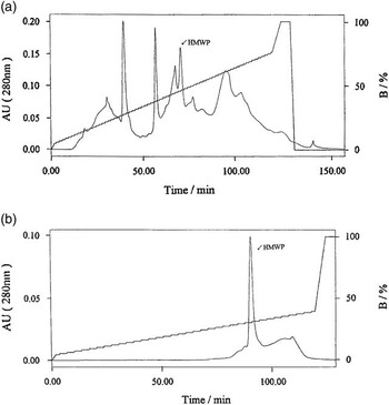
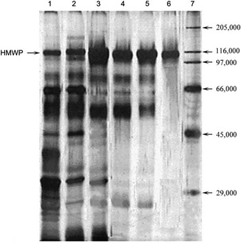
HMWP crude sample was chromatographed by Mono Q column (anion-exchanger) on AKTA explorer 100 protein purification system (GE Healthcare). The chromatogram is shown in Figure 1a. By SDS-PAGE identification, HMWP was detected in the sixth peak, retention time of which was 70.95 min. The HMWP fraction was collected, pooled and concentrated to obtain the primary purified HMWP sample for further purification. With different elution condition, the primary purified HMWP sample was further purified by Mono Q column again on AKTA explorer 100 protein purification system. The high single peak appeared when retention time was 90.10 min as shown in Figure 1b. By SDS-PAGE identification, HMWP was detected in the peak. The HMWP fraction was collected, pooled and concentrated to obtain the purified HMWP sample. The purified HMWP sample exhibited a single band on SDS-PAGE, the purity of which reached (95.60±7.40%) (Figure 2).
The anion exchange chromatogram of a high molecular weight protein (HMWP) sample from sows milk on Mono Q column. (a) The 40% saturation ammonium sulfate pellet (the crude HMWP sample) on Mono Q column. buffer A: 20 mM Tris-HCl, pH 8.0; buffer B: buffer A+0.5 M NaCl, gradient elution. (b) The primary purified HMWP sample on Mono Q column. buffer A: 20 mM Tris-HCl, pH 8.0; buffer B: buffer A+0.5 M NaCl (0% to 40%), buffer B gradient elution.

SDS-PAGE profile of a high molecular weight protein (HMWP) from sows milk at different steps of purification process (stacking gel: 50 g/l; lower gel: 100 g/l; silver stained). 1: milk; 2: whey; 3: protein pellet at 40% saturation ammonium sulfate; 4 to 5: the primary purified HMWP sample by Mono Q column chromatographing for the first time; 6: the purified HMWP by Mono Q column chromatographing further; 7: molecular weight protein standard.

N-terminal amino acid sequencing and LC-MS identifying of HMWP
The first 10 N-terminal amino acid sequence of HMWP was Ala-Leu-Val-Gln-Ser- Cys or Gly-Leu-Asn-Leu-Val. The sequence was blasted against GenBank. No protein showed significant similarity with this sequence suggesting the HMWP may be novel. The result of ESI-MS (see Supplementary Figure S1) also proved HMWP could be a novel protein.
Amino acid compositions of HMWP
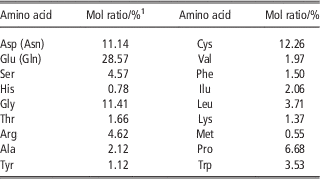
The amino acid assay of HMWP with HP-1050 HPLC is shown in Table 1. HMWP was rich in glutamate (including glutamine), cysteine, glycine, aspartic acid (including asparagines) and proline. The richest residue was glutamate (including glutamine), consisting of 28.57% of the total amino acids (mol). The content of hydrophobic amino acids (Ala, Val, Leu, Ile, Met, Phe and Pro) was lower at 18.59% of the total amino acids suggesting HMWP has high solubility in water. Rich in glycine and proline, HMWP easily created β-turn on its polypeptide chain. The β-turn is a necessary secondary structure of protein, and globulin has a large number β-turns. Moreover, HMWP easily folded because of abounding in cysteine to form disulfur bonds. These results suggest the HMWP possesses the characteristic of globulin.
Amino acid compositions of high molecular weight protein (HMWP) isolated from sows milk

1 Mol ratio: percentages of amino acid taking up the total amino acid (mol).
Biochemical properties of HMWP
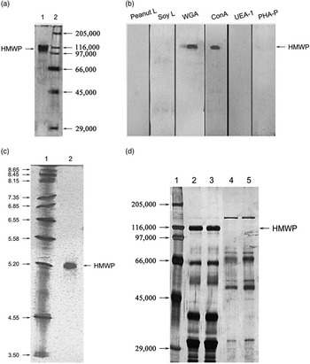
The molecular weight of HMWP on SDS-PAGE was determined to be 115 000 in reductive state (the sample with 4% β-mercaptoethanol, Figure 3a). On gel filtration chromatography, the molecular weight of HMWP was determined to be to 114 800. The results suggested HMWP was a monomer, not dimer or polymer.
Biochemical properties analysis of a high molecular weight protein (HMWP) isolated from sows milk. (a) SDS-PAGE of HMWP (stacking gel: 50 g/l; lower gel: 100 g/l; silver stained). 1: HMWP; 2: molecular weight protein standard. (b) Western blot of HMWP with lectins. (c) Isoelectric focusing electrophoresis of HMWP. 1: pI marker; 2: HMWP. (d) HMWP was found not to be a milk fat globule membrane protein (MFGMP) by SDS-PAGE profile of milk protein and MFGMP (stacking gel: 50 g/l; lower gel: 100 g/l; silver stained). 1: molecular weight protein standard; 2 to 3: sow milk; 4 to 5: MFGMP of sow milk. Peanut L=peanut lectin; Soy L=soybean lectin; WGA=wheat germ agglutinin; ConA=concanavalin A; UEA-1=Ulex europaeus 1; PHA-P=Phaseolus vulgaris P.

Various lectins can combine specific glycosyls in glycoprotein (Hurley et al., Reference Hurley, Grieve, Magura, Hegarty and Zou1993; Patton et al., Reference Patton, Gendler and Spicer1995). On base of this principle, Western blots of lectins were used to identify the kinds of carbohydrate residues attached to HMWP qualitatively. The results are shown in Figure 3a and Figure 3b. HMWP was bound by WGA and ConA, implying that HMWP was a kind of glycoprotein containing NeuNAc, Man and/or GlcNAc.
With isoelectric focusing analysis, HMWP pI was estimated to be 5.1 from a standard curve obtained from the proteins with their pI, as shown in Figure 3c.
MFGMP were isolated from sow milk. The SDS-PAGE of MFGMP was conducted to determine if MFGMP include HMWP. As shown in Figure 3d, MFGMP did not contain HMWP. HMWP is proposed to be a secretory milk protein.
HMWP being unique to sows milk
The sow milk proteins were compared with bovine, goat, rabbit and human milk proteins by SDS-PAGE (Supplementary Figure S2). HMWP did not exist in bovine, goat, rabbit and human milk in SDS-PAGE gel and NC membrane of western blot (data not shown). We speculate that HMWP may be unique to sows milk.
HMWP in sow milk originating in the breast
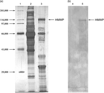
By western blot, HMWP could be detected in sow milk, not in sow serum, as shown in Figure 4. It was deduced to be synthesized and secreted by the mammary gland.
A high molecular weight protein (HMWP) isolated from sows milk originated in the breast, and not from blood. (a) SDS-PAGE of HMWP (stacking gel: 50 g/l; lower gel: 100 g/l; silver stained). 1: molecular weight protein standard; 2: sow serum; 3: sow milk; (b) Western blot identification of HMWP. 4: sow serum; 5: sow milk.

Changes of HMWP content in sow milk
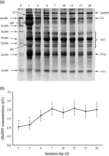
Figure 5a shows the content changes of main milk proteins in a sow during lactating 1 to 21 days by SDS-PAGE. Figure 5b shows the general variation of HMWP concentration in 1 to 21 days of lactation in 10 sows. HMWP concentrations in sows milk were the lowest (1.03±0.20 g/l) in the 1st day of lactation after parturition, rose significantly during lactation 1 to 7 days, in which the concentrations of lactation 1 to 2 days were significantly lower than that on the 20th day (P<0.05). The HMWP content of sows milk remained relatively constant (1.95±0.13 g/l) during lactation 7 to 20 days (P>0.05).
Changes in a high molecular weight protein (HMWP) content in the milk of 10 sows over 20 day lactation. (a) SDS-PAGE of milk protein in a sow (stacking gel: 50 g/l; lower gel: 150 g/l; coomassie brilliant blue stained). (b) Changes of HMWP content in milk of 10 sows. Data are presented as mean±s.d. of 10 sows, *P<0.05 compared with HMWP concentration on the twentieth day. S=molecular weight protein standard; 1, 2, 4, 7, 10, 13, 17 and 20 represent lactation day after parturition respectively; Lf=lactoferrin; IgH=immunoglobulin heavy chain; IgL=immunoglobulin light chain; SA=serum albumin; tCN=total caseins; β-Lg=β-lactoglobulin; α-La=α-lactalbumin.

The effect of HMWP on bacterium growth inhibition
To determine HMWP function, its inhibition effect on the growth of bacterium was examined. HMWP significantly inhibited E. coli in a dose related manner (Supplementary Figure S3).
Discussion
Different variants of HMWP have analogical protein structure and biochemical characteristics (Zhang et al., Reference Zhang, Zou, Qin and Chen2004). It is very difficult to separate HMWP variants. B variant of HMWP was the most common in sows milk. For this reason the milk from sows which have only B pattern HMWP was used in our studies and the results are reflective of the structure and function of HMWP-B.
In our study, whey protein was fractionated by different saturations of ammonium sulfate. The maximal precipitation of HMWP appeared at 40% saturations of ammonium sulfate. This result is similar to Ig (Bergmann-Leitner et al., Reference Bergmann-Leitner, Mease, Duncan, Khan, Waitumbi and Angov2008). It is not understood if HMWP has the immunity function as Ig and further research is required.
Hydrochloric acid hydrolysis taken in this study destroyed tryptophan for amino acid assay of HMWP. Thus, tryptophan content in HMWP was alone determined by fluorescence spectrophotometer after hydrolyzing the sample with sodium hydrate. Because of deamidation, hydrochloric acid hydrolysis cannot distinguish asparagines from aspartic acid and glutamine from glutamic acid. Therefore, HMWP should contain all 20 amino acids. By SDS-PAGE, gel filtration chromatography, pI determination, amino acid assay and references (Kontopidis et al., Reference Kontopidis, Holt and Sawyer2004; Andrews et al., Reference Andrews, Schmidt and Asenjo2005), HMWP appears to be a soluble, acidic and monomeric globulin. However, in order to more fully understand the biochemical properties and biological function of HMWP, further study is required.
Big molecular proteins in milk are often glycoprotein. Our results showed HMWP is a glycoprotein but has fewer kinds of glycosyl. Many other big proteins in milk have many kinds of glycosyl. According to the general linking rule and our results the linking between glycan and protein in HMWP was deduced to be N-link though this is yet to be proven. Because the glycosyl of HMWP was merely qualitative identification by Western blot of lectins, other means must be used to understand more accurately the degree of glycosylation and glycosyl content of HMWP. At present, a good method of studying protein glycosylation is glycanase treatment and mass spectrometry analysis (Gusakov et al., Reference Gusakov, Antonov and Ustinov2008). From the result of identification, HMWP glycosyls contained NeuNAc, which could combine and inhibit bacteria and virus. For example, Patton et al. (Reference Patton, Gendler and Spicer1995) reported that NeuNAc of mucin in milk could combine rotavirus that resulted in infant gastroenteritis. Our results showed HMWP also has anti-bacterial activity.
There are many kinds of proteins in milk, the most being secreted proteins including casein proteins and whey proteins. In addition, there is a small amount of membrane-bound proteins in milk, such as MFGMP. MUC1, a kind of MFGMP, is currently the most actively researched (Wilson et al., Reference Wilson, Robinson, Donnet, Bovetto, Packer and Karlsson2008). We previously thought HMWP was similar to MUC1. However the current results do not support this assumption.
The present results suggest HMWP may be unique to sows milk. However the milk from species other than those investigated here will need to be tested to confirm this.
There are two sources of secretory proteins in milk. Many milk proteins are synthesized and secreted by breast epithelial cells, such as total caseins (tCN), α-lactalbumin (α-La), β-lactoglobulin (β-Lg), lactoferrin (Lf) and bioactive peptide (Demeter et al., Reference Demeter, Markiewicz, van Arendonk and Bovenhuis2010). But serum albumin, Ig, enzyme, hormone and growth factor are derived from the blood (Qin and Zou, Reference Qin and Zou2003). In the colostrum, hematogenous proteins are the most abundant. While in the mature milk, proteins are mainly the proteins synthesized and secreted by breast epithelial cells (Qin and Zou, Reference Qin and Zou2003). By Western blot, HMWP was deduced to be synthesized and secreted by mammary cells.
HMWP was low in colostrum and averaged only 1.03 g/l in milk collected the 1st day of lactation. The concentration of HMWP in milk increased as lactation progressed and averaged 1.95 g/l in mature milk. According to secretory characteristics of other proteins synthesized by mammary epithelial cells, change pattern of HMWP suggests that it may be synthesized and secreted by mammary epithelial cells, such as tCN, α-La and β-Lg.
The inhibition of E. coli by HMWP suggests it may have antibacterial function. Compared with colostrum, Ig, Lf and other immune-active substances decline sharply in mature milk. By contrast, HMWP content in mature milk is higher than that in colostrum, which could increase breast defense capabilities in mature milk. However, we do not know whether HMWP has a wide antimicrobial, immune, anti-inflammatory, mammary epithelial repair and other effects on the mammary gland and/or piglet. Further research is clearly required to fully elucidate the role and functions of HMWP.
Conclusions
HMWP was isolated and purified from sow milk, purity of which was (95.60±7.40%). HMWP had no homology with sequences of other protein suggesting it may be novel. HMWP has a molecular weight of ~115 000 and is a kind of glycoprotein containing NeuNAc, Man and/or GlcNAc. HMWP appears to be synthesized and secreted by mammary epithelial cells and may be unique to sows milk. HMWP concentration in sow milk rose significantly during lactation 1 to 7 days, and was relatively constant during lactation 7 to 20 days. HMWP also significantly inhibited E. coli in vitro.
Acknowledgments
The work reported in this paper, and the preparation of the paper, was funded was funded by National Natural Science Foundation of China (30872992) and the National Natural Science Foundation of China (81472448). The authors of this paper would like to thank Mr Pingrong Wang in Wuxi Xishan pig breeds farm, Wuxi, China, for help in sampling sow milk. The authors also thank Research Centre for Life Sciences, University of Science and Technology of China, for help HMWP identification by 1D-LC-MS.
Supplementary material
To view supplementary material for this article, please visit http://dx.doi.org/10.1017/S1751731114003280