Introduction
Malaysia is categorised by the World Health Organization as an intermediate tuberculosis burden country (i.e. less than 100 tuberculosis cases per 100 000 population per year) within the Western Pacific region.1 Approximately 15 per cent of reported tuberculosis cases involve only extrapulmonary sites, or both pulmonary and extrapulmonary sites.2 Tuberculous otitis media is rare, and accounts for 0.05–0.9 per cent of chronic infections of the middle ear.Reference Bhalla, Jones, Rothburn and Swift3, Reference Vaamonde, Castro, Garcia-Soto, Labella and Lozano4 The condition remains a significant diagnostic challenge for the otorhinolaryngologist, as the clinical symptoms and signs are non-specific and standard microbiological and histological tests for tuberculosis often give false negative results. Therefore, a high index of clinical suspicion of tuberculous otitis media is required in patients who do not respond to standard therapy for (nontuberculous) chronic middle-ear infection, in order to avoid irreversible complications, surgical intervention and propagation of the disease.
We report four cases of tuberculous otitis media with different clinical presentations, encountered between 1998 and 2002. None of the cases showed improvement with local or systemic antibiotics. The diagnosis, complications and management of these cases are discussed.
Case reports
Case one
A 26-year-old woman presented with left ear discharge mixed with blood, together with a two-year history of progressive hearing loss. She had previously been treated by general practitioners and ENT surgeons with courses of Sofradex®, ciprofloxacin and clotrimazole eardrops. She had also been treated with courses of cefuroxime, amoxicillin with clavulanate, and erythromycin systemic antibiotics. She had failed to respond to any of these treatments.
Otoscopic examination revealed mucopurulent discharge from the left middle ear, with subtotal tympanic membrane perforation and granulation tissue in the middle-ear cleft. The patient's lungs were normal, and this was confirmed on chest X-ray.
Pure tone audiography revealed a moderate to severe, mixed hearing loss in the left ear. A plain film radiograph of the mastoid showed a sclerotic mastoid antrum.
A modified radical mastoidectomy was performed. Intra-operatively, the cortex of the mastoid bone was found to be eroded and the antrum to be full of granulation tissue. The malleus, incus and stapes were found to be necrotic.
Histopathological examination of surgical material confirmed tuberculous otitis media.
The patient was treated with antituberculous medication and recovered uneventfully.
Case two
A five-year-old boy presented with right ear discharge together with a four-month history of decreased hearing, otalgia and multiple, bilateral neck swellings. He was not responding to multiple local and systemic antibiotics.
Clinical examination revealed multiple, bilateral cervical lymphadenitis. Otoscopic examination revealed a thin, watery, clear discharge with swelling of the right external ear canal. There was central perforation of the right tympanic membrane, but the right middle-ear space was unable to be visualised fully due to narrowing of the external ear canal.
A mastoid X-ray revealed a sclerotic mastoid antrum.
Cervical lymph node biopsy indicated tuberculous lymphadenitis.
The patient underwent right cortical mastoidectomy. Intra-operatively, granulation tissue under pressure was discovered in the antrum. All the ossicles were intact.
Histopathological examination of the granulations confirmed tuberculosis infection.
The patient's lungs were normal, and this was confirmed by chest X-ray.
The patient was treated with antituberculous medication. His cervical lymphadenitis subsided and he recovered well.
Case three
A 23-year-old woman presented with chronic, foul-smelling right ear discharge together with a two-year history of otalgia and gradual hearing loss. She was treated with multiple local and systemic antibiotics but her symptoms persisted. There were no other associated symptoms, specifically related to the chest.
Otoscopic examination revealed a central perforation of the right tympanic membrane, with foul-smelling discharge. Granulation tissue was noted in the middle-ear cleft. Both lungs appeared normal.
Pure tone audiography revealed right, moderate to severe, mixed hearing loss. A mastoid X-ray revealed a sclerotic right mastoid antrum with a soft tissue shadow in the middle-ear cleft.
A right-sided modified radical mastoidectomy was performed. Intra-operatively, cholesteatoma was noted in the antrum, along with granulation tissue.
However, the histopathology report on the granulation tissue described classical tuberculous granulomas consisting of lymphocytes, epithelioid cells and Langhans multinucleated giant cells with areas of caseous necrosis. Culture of granulation tissue grew Mycobacterium tuberculosis.
The patient was commenced on antituberculous treatment and made a full recovery.
Case four
A 23-year-old man presented with a two-week history of right facial muscle weakness, together with a one-year history of right-sided otorrhoea which had failed to respond to local or systemic antibiotics. He had initially been treated by his general practitioner with a two-week course of Sofradex ear drops but the discharge persisted. He was referred to an ENT surgeon who prescribed a two- to three-weeks' course of ciprofloxacin and clotrimazole eardrops together with systemic ciprofloxacin for two weeks, the latter then being replaced by amoxicillin with clavulanate (Augmentin). There were no associated chest symptoms.
Clinical examination revealed a right, House–Brackmann grade IV facial nerve palsy. Granulation tissue was observed within the middle-ear cleft. Both lungs were normal.
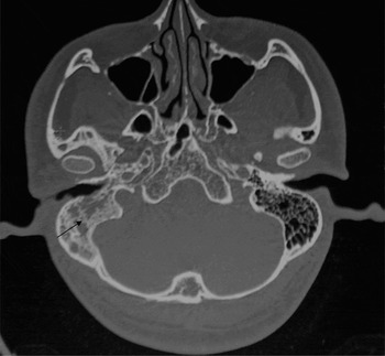
Computed tomography scanning of the temporal bone revealed right mastoiditis and middle-ear infection (Figure 1).
Axial computed tomography scan showing opacification of the right mastoid air cells (arrow), which appears as a soft tissue mass in the middle-ear cleft. No bony erosion is seen around the right facial canal. The right ossicles, cochlea and semicircular canals appear normal.

The patient underwent right modified radical mastoidectomy and facial nerve exploration. Intra-operatively, pale granulation tissue was noted in the antrum and middle ear. The facial nerve was found to be dehiscent in the region of the second genu.
Histopathological examination of granulations showed tuberculosis infection.
The patient was commenced on antituberculous treatment. His right ear discharge resolved. However, his facial nerve paralysis showed only partial improvement, from grade IV to grade II, after a year of follow up.
Discussion
Tuberculous otitis media is now rarely seen. The diagnosis is made difficult by a low index of clinical suspicion, variable clinical signs, infrequent associations with other systemic involvements, and false negative cultures owing to the fastidious nature of M tuberculosis.Reference Cho, Lee, Kim, Chung, Lee and Koh5 With tuberculosis and human immunodeficiency virus (HIV) infections both growing threats worldwide, the number of cases involving both tuberculosis and HIV has risen over the past 12 years. In 2004 in Malaysia, 15 429 new cases of tuberculosis were reported, 8.2 per cent of which involved co-infection with HIV.1 Co-infection of HIV with tuberculosis accounted for 51 per cent of the tuberculosis deaths notified for the year 2001 in Selangor state, Malaysia.Reference Venugopalan6
Most patients with tuberculous otitis media are adolescent or younger.Reference Nishiike, Irifune, Doi, Osaki and Kiuchi7 This was borne out by our cases: three out of our four patients were in their 20s, and the fourth was five years old. It has been postulated that tuberculosis bacilli spread to the middle ear via the eustachian tube, via haematogenous spread from other tuberculous foci, or via direct implantation from the external auditory canal via perforation of the tympanic membrane.Reference Mongkolrattanothai, Oram, Redleaf, Bova and Englund8
The classical features of tuberculous otitis media have been described as: painless otorrhoea with multiple tympanic membrane perforations; exuberant granulations; early, severe hearing loss; and bone necrosis.Reference Cho, Lee, Kim, Chung, Lee and Koh5 However, it is clear that the actual clinical manifestations of tuberculous otitis media do not always comply with this classical description. Otorrhoea was present in all of our cases, refractory to standard antibiotic treatment; this should raise suspicion of tuberculous otitis media. Two of our patients had otalgia. This agrees with previous studies which have reported otalgia to occur relatively frequently, in approximately 30 per cent of tuberculous otitis media patients in association with acute infection or super-infection and mastoid involvement.Reference Vaamonde, Castro, Garcia-Soto, Labella and Lozano4, Reference Lee and Drysdale9 One of our cases had facial nerve palsy. None of our patients had concomitant or previous pulmonary tuberculosis. Tuberculous otitis media should be suspected in a patient with chronic middle-ear infection who presents with facial nerve palsy. Three out of our four patients had progressive hearing loss. Tuberculous otitis media gradually erodes the ossicles and bony structures of the inner ear as the disease progresses, causing progressive hearing loss.
Tuberculosis infection may be suggested by an otoscopic examination revealing necrotic components, abundant granulation tissue, multiple perforations or whitish exudates not readily aspirated. All of our patients had otorrhoea and showed granulation tissue in the middle-ear cleft. However, none of our patients showed multiple tympanic membrane perforations, as per the classical description of tuberculous otitis media. It has been presumed that multiple perforations are a feature only of early tuberculous infection, and that at a later stage they fuse to form a single, large perforation.Reference Cho, Lee, Kim, Chung, Lee and Koh5
Definitive confirmation of a diagnosis of tuberculous otitis media comes from culture of tissue specimens or ear discharge, as the low number of mycobacteria present may not be detectable on Ziehl–Neelsen staining of a histological section or ear discharge swab. Serological tests based on recognition of serum immunoglobulin G antibodies to selected mycobacterial antigens have been developed, and these are useful in the diagnosis of extrapulmonary tuberculosis.Reference Meher, Singh, Yadav and Gathwala10 Polymerase chain reaction will be the next important alternative to culture to enable the definite diagnosis of extrapulmonary tuberculosis infection in cases in which only a small number of organisms is present. Smear tests, microbiological culture, serological tests, polymerase chain reaction and histological analysis have their respective advantages and disadvantages, and must be repeated to achieve a definite diagnosis. Radiological examination is useful in assessing the extent of the lesion.
The standard treatment of tuberculous otitis media is antituberculous medication. Patients should be treated for at least six months, except in cases of disseminated tuberculosis and tuberculous meningitis.Reference Nicolau, Northrop and Eavey11 In Malaysia, extrapulmonary tuberculosis is treated for at least nine to 12 months. The duration of treatment may be extended depending on the clinical or radiological response.1 Treatment is divided into two phases: an intensive phase for two months, and a maintenance phase for at least seven months or longer. Four drugs are given daily during the intensive phase: isoniazid, rifampicin, pyrazinamide and ethambutol. This leads to rapid conversion, achieving a negative result in term of microbial culture of TB, and amelioration of clinical symptoms. Two drugs are given during the maintenance phase: ethambutol and rifampicin. The sterilising effect of treatment eliminates remaining bacilli and reduces drastically the chances of subsequent relapse. Second line antituberculous treatment should be initiated in cases of microbial resistance; the patient's compliance is important in reducing the risk of such resistance.1 All of our patients were treated with antituberculous medication for nine months, and recovered uneventfully.
• A high index of clinical suspicion of tuberculous otitis media is required in patients who do not respond to standard antibiotic therapy for chronic middle-ear infection
• The diagnosis is hampered by variable clinical signs, infrequent association with other systemic involvements and false negative cultures
• Middle-ear granulation tissue is a consistent finding
• Standard treatment for tuberculous otitis media is antituberculous medication for at least six months
The role of surgery in tuberculous otitis media remains controversial. Although surgery is indicated for the removal of sequestrum, it is not always easy to establish whether sequestrum is present. Mastoidectomy was indicated and undertaken in all our patients. One patient presented with facial nerve palsy, and three patients presented with chronic, unresolved otitis media refractory to standard antibiotic treatment, with sclerotic mastoiditis, indicating the need for surgery. Cho et al. reported that tuberculous otitis media patients who underwent chemotherapy after surgery appeared to achieve a dry ear earlier than those not receiving surgery, and that the surgical group tended to have better outcomes in terms of tympanic membrane closure.Reference Cho, Lee, Kim, Chung, Lee and Koh5 However, in cases of facial nerve palsy, the indication for mastoidectomy and decompression of the facial nerve has been debated.Reference Singh12
Conclusion
Tuberculous otitis media remains a significant diagnostic challenge, because clinical signs are nonspecific and standard microbiological and histological tuberculosis tests often give false negative results. A high index of clinical suspicion of tuberculous otitis media is required in patients who do not respond to standard antibiotic therapy for (nontuberculous) chronic middle-ear infection. The usefulness of smear tests, microbial culture, serological tests, polymerase chain reaction and histological analysis depend on the availability of the test within a hospital's laboratory services, and the tests must be repeated to enable definite diagnosis in highly suspicious cases. Early diagnosis and treatment of tuberculous otitis media is important to avoid irreversible complications, surgical intervention and propagation of the disease.