Introduction
Major depressive disorder (MDD) is a serious illness with severe impact on daily functioning. Because of its chronic course, associated impaired functioning and high health-care costs, MDD accounts for a considerable part of the population burden of disease (Mathers & Loncar, Reference Mathers and Loncar2006). Many studies in younger populations indicate that, compared to healthy controls, patients with MDD have structural brain abnormalities on magnetic resonance imaging (MRI) (Campbell & MacQueen, Reference Campbell and MacQueen2004, Reference Campbell and MacQueen2006; Videbech & Ravnkilde, Reference Videbech and Ravnkilde2004; Geuze et al. Reference Geuze, Vermetten and Bremner2005; Konarski et al. Reference Konarski, McIntyre, Kennedy, Rafi-Tari, Soczynska and Ketter2008; Koolschijn et al. Reference Koolschijn, van Haren, Lensvelt-Mulders, Hulshoff Pol and Kahn2009; Lorenzetti et al. Reference Lorenzetti, Allen, Fornito and Yucel2009). Studies characterizing MDD in detail suggest that smaller volumes of brain regions may occur especially in patients with longer duration of depression and greater severity, and in those with repeated episodes (Konarski et al. Reference Konarski, McIntyre, Kennedy, Rafi-Tari, Soczynska and Ketter2008; Lorenzetti et al. Reference Lorenzetti, Allen, Fornito and Yucel2009; McKinnon et al. Reference McKinnon, Yucel, Nazarov and MacQueen2009); some studies did not find an association between depression severity and brain volumes (Koolschijn et al. Reference Koolschijn, van Haren, Lensvelt-Mulders, Hulshoff Pol and Kahn2009; van Tol et al. Reference van Tol, van der Wee, van den Heuvel, Nielen, Demenescu, Aleman, Renken, van Buchem, Zitman and Veltman2010).
The majority of extant studies on MDD and structural brain changes have examined specific brain regions, in particular the hippocampus, and have been conducted in relatively young populations. Less is known of the association between MDD and brain volumes in older people (Konarski et al. Reference Konarski, McIntyre, Kennedy, Rafi-Tari, Soczynska and Ketter2008). Increased understanding of the relationship between MDD and brain abnormalities in older people is important, because older people may be especially vulnerable to the adverse consequences of brain abnormalities, in particular adverse cognitive outcomes. Indeed, loss of total brain volume in older people is a risk factor for cognitive decline and dementia (Jack et al. Reference Jack, Shiung, Weigand, O'Brien, Gunter, Boeve, Knopman, Smith, Ivnik, Tangalos and Petersen2005; Ikram et al. Reference Ikram, Vrooman, Vernooij, den Heijer, Hofman, Niessen, van der Lugt, Koudstaal and Breteler2010), and older people with a history of depression may be at increased risk for Alzheimer's disease (Ownby et al. Reference Ownby, Crocco, Acevedo, John and Loewenstein2006; Geerlings et al. Reference Geerlings, den Heijer, Koudstaal, Hofman and Breteler2008; Byers & Yaffe, Reference Byers and Yaffe2011). From this, it has been hypothesized that depression is a causal risk factor for Alzheimer's disease (Jorm, Reference Jorm2001; Byers & Yaffe, Reference Byers and Yaffe2011). However, because of the long preclinical phase of Alzheimer's disease, the direction of association is still not well understood; several other hypotheses may explain the association between depression, brain atrophy and dementia, including depression being a prodomal phase of dementia (Jorm, Reference Jorm2001). The results from two recent population-based studies with long follow-up periods showed that depressive symptoms increased the risk for dementia many years before dementia diagnosis (Saczynski et al. Reference Saczynski, Beiser, Seshadri, Auerbach, Wolf and Au2010) and that increasing numbers of depressive episodes increased dementia risk (Dotson et al. Reference Dotson, Beydoun and Zonderman2010), suggesting that depression may contribute to the development of dementia. However, these studies did not use brain MRI and it is unclear whether brain volume loss was underlying these associations. In another population-based study, history of depressive symptoms increased risk for dementia but this was not explained by smaller hippocampal or amygdalar volumes (Geerlings et al. Reference Geerlings, den Heijer, Koudstaal, Hofman and Breteler2008).
Clarification is needed regarding several issues related to depression and brain volumes in older populations. Because the majority of older people with MDD will also have a history of MDD, it is important to make a distinction between first onset and a history with early onset or multiple episodes so that the distinction can be made between late-life MDD resulting from, or contributing to, brain atrophy. Furthermore, it is unclear whether MDD leads to smaller brain volume only in the acute state and reverses in remission (Hsieh et al. Reference Hsieh, McQuoid, Levy, Payne, MacFall and Steffens2002; MacQueen et al. Reference MacQueen, Yucel, Taylor, Macdonald and Joffe2008; Ahdidan et al. Reference Ahdidan, Hviid, Chakravarty, Ravnkilde, Rosenberg, Rodell, Stodkilde-Jorgensen and Videbech2011; Geerlings et al. Reference Geerlings, Brickman, Schupf, Devanand, Luchsinger, Mayeux and Small2012).
Previous studies that examined the association of MDD later in life with total brain volume had a case–control design and a relatively small sample size (Sheline et al. Reference Sheline, Wang, Gado, Csernansky and Vannier1996; Kumar et al. Reference Kumar, Jin, Bilker, Udupa and Gottlieb1998; Ashtari et al. Reference Ashtari, Greenwald, Kramer-Ginsberg, Hu, Wu, Patel, Aupperle and Pollack1999; Ballmaier et al. Reference Ballmaier, Sowell, Thompson, Kumar, Narr, Lavretsky, Welcome, DeLuca and Toga2004; Konarski et al. Reference Konarski, McIntyre, Kennedy, Rafi-Tari, Soczynska and Ketter2008). Relatively few studies have examined this relationship in the general elderly population and these studies used depressive symptoms as opposed to a formal diagnosis of MDD to assess depression (Dotson et al. Reference Dotson, Davatzikos, Kraut and Resnick2009; Goveas et al. Reference Goveas, Espeland, Hogan, Dotson, Tarima, Coker, Ockene, Brunner, Woods, Wassertheil-Smoller, Kotchen and Resnick2011; Geerlings et al. Reference Geerlings, Brickman, Schupf, Devanand, Luchsinger, Mayeux and Small2012). To our knowledge there are no studies that had lifetime DSM-IV diagnoses of MDD and MRI measures on a population-based cohort of older people.
In the current study we investigated the associations of lifetime DSM-IV diagnoses of MDD with total brain volume on MRI in a large population-based study of older people without dementia. We hypothesized that if MDD contributes to brain atrophy, an early onset and repeated episodes of MDD will be associated with a smaller brain volume, whereas if MDD is a sequela of brain atrophy, current MDD will be associated with a smaller brain volume.
Method
Participants
Study participants were from the Age, Gene/Environment Susceptibility (AGES)–Reykjavik Study, a population-based cohort study originating from the Reykjavik study, as described fully elsewhere (Harris et al. Reference Harris, Launer, Eiriksdottir, Kjartansson, Jonsson, Sigurdsson, Thorgeirsson, Aspelund, Garcia, Cotch, Hoffman and Gudnason2007). In brief, from 2002 to 2006, 5764 persons, randomly chosen from survivors of the Reykjavik Study, were examined for the AGES–Reykjavik Study. As part of comprehensive assessments at the Reykjavik research center, participants answered questionnaires and underwent clinical examinations, had blood drawn, cognitive testing, and brain MRI. The AGES–Reykjavik Study was approved by the Icelandic National Bioethics Committee (VSN: 00-063), the Icelandic Data Protection Authority, Iceland, and the Institutional Review Board for the National Institute on Aging (NIA), National Institutes of Health (NIH), USA. Written informed consent was obtained from all participants.
MRI protocol
All participants without contraindications were eligible for a brain MRI scan on a study-dedicated 1.5-T Signa Twinspeed system (General Electric Medical Systems, USA). The image protocol included an axial T1-weighted three-dimensional spoiled gradient echo sequence [time to echo (TE) 8 ms, repetition time (TR) 21 ms, flip angle (FA) 30°, field of view (FOV) 240 mm, matrix 256 × 256, slice thickness 1.5 mm]; a fluid attenuated inversion recovery (FLAIR) sequence (TE 100 ms, TR 8000 ms, inversion time 2000 ms, FA 90°, FOV 220 mm, matrix 256 × 256); a proton density (PD)/T2-weighted fast spin echo (FSE) sequence (TE1 22 ms, TE2 90 ms, TR 3220 ms, echo train length 8, FA 90°, FOV 220 mm, matrix 256 × 256); and a T2*-weighted gradient-echo echo planar imaging (GRE-EPI) sequence (TE 50 ms, TR 3050 ms, FA 90°, FOV 220 mm, matrix 256 × 256). The FLAIR, PD/T2 and T2* sequences were acquired with 3-mm-thick interleaved slices. All images were acquired to give full brain coverage and slices were angled parallel to the anterior commissure–posterior commissure line.
Brain segmentation
The intracranial volume (ICV) and the brain parenchyma compartments were segmented automatically with an AGES–Reykjavik Study modified algorithm described previously (Sigurdsson et al. Reference Sigurdsson, Aspelund, Forsberg, Fredriksson, Kjartansson, Oskarsdottir, Jonsson, Eiriksdottir, Harris, Zijdenbos, van Buchem, Launer and Gudnason2012). The pipeline is based on a multispectral tissue segmentation method that estimated volumes for four tissue classes: gray- and white-matter regions, white-matter lesions (WMLs) and cerebrospinal fluid (CSF). These four classes were summed to obtain the total ICV. Total brain volume was defined as the sum of gray-matter, normal white-matter and WML volumes and was expressed relative to ICV as the brain parenchymal fraction (BPF), an indicator of global brain atrophy. Calculation of regional tissue volumes was based on a regional probabilistic atlas, created from a large sample of the AGES cohort (n = 314), that was warped non-linearly to the T1-weighted images of each study participant.
Cerebral infarcts, identified by trained radiographers, were defined as defects in the brain parenchyma with associated hyperintensity on T2 and FLAIR images with a maximum diameter of at least 4 mm. For infarcts in the cerebellum and brain stem or infarcts with cortical involvement, no size criterion was required.
Dementia diagnosis
Dementia ascertainment was a three-step protocol as described previously (Harris et al. Reference Harris, Launer, Eiriksdottir, Kjartansson, Jonsson, Sigurdsson, Thorgeirsson, Aspelund, Garcia, Cotch, Hoffman and Gudnason2007). All participants were screened using the Mini-Mental State Examination (MMSE; Folstein et al. Reference Folstein, Folstein and McHugh1975) and the Digit Symbol Substitution test. Those with positive screen results were administered a diagnostic battery of neuropsychological tests and, among them, those with positive screen results were examined by a neurologist and a proxy interview was administered regarding medical history, social, cognitive and daily functioning changes of the participant. A consensus diagnosis, according to DSM-IV criteria (APA, 1994), was made by a panel that included a geriatrician, neurologist, neuropsychologist and neuroradiologist.
Diagnosis of MDD
The presence of MDD in the preceding 2 weeks and in the past was assessed according to DSM-IV criteria (APA, 1994) using the Mini-International Neuropsychiatric Interview (MINI; Sheehan et al. Reference Sheehan, Lecrubier, Sheehan, Amorim, Janavs, Weiller, Hergueta, Baker and Dunbar1998). The MINI was administered by five trained and standardized health professionals. To rule out possibly unreliable answers to history of depression questions, only participants with no diagnosis of dementia and an MMSE (Folstein et al. Reference Folstein, Folstein and McHugh1975) score ⩾21 were eligible to receive the MINI. In this sample, we applied the following screening criteria to identify persons who may have had past or current episodes of depression: if they had a score ⩾6 on the 15-item Geriatric Depression Scale (GDS-15; Yesavage et al. Reference Yesavage, Brink, Rose, Lum, Huang, Adey and Leirer1982) or a GDS-15 score of 4 or 5 and a positive response to three out of four of the following anxiety questions: ‘In the past month, have you felt anxious or frightened?’; ‘Were there times lately that you felt anxious?’; ‘Are there special situations that make you anxious?’; ‘Have you ever had attacks of fear or panic?’, or if they reported ever having had a doctor diagnosis of depression, or if they reported ever having used antidepressant medications, or if they used antidepressant medication at the time of interview as assessed from medication bottles brought to the clinic.
To evaluate the screening properties of our algorithm, 358 consecutive non-demented participants (mean age 76 years, range 66–91 years) were evaluated with the MINI from June 2002 until May 2003. For current MDD the sensitivity and specificity were 100% and 64% respectively; for past MDD the sensitivity and specificity were 93% and 66% respectively.
The MINI includes questions on age at onset of first MDD episode and number of episodes. In the analyses, participants were first classified as ever versus never diagnosed with MDD. Second, the ‘ever MDD’ group was divided into past (and not current) MDD and current MDD (irrespective of past); into persons with an age of first onset before age 60 (early onset) and at ⩾60 years (late onset); and into persons with 1–2 previous episodes and ⩾3 episodes. Third, explorative analyses were performed by further subdividing the current and past MDD groups according to their history and age at onset (Fig. 1).
Definition of different depression groups: (a) onset of depression; (b) number of episodes.

Other variables
Age, sex, education (categorized into primary, secondary and college/university education), smoking history (ever versus never), current alcohol intake (yes versus no) and subjective memory complaints (yes versus no) were assessed by questionnaires. Body mass index (BMI) was calculated as measured weight (kg) divided by height (m) squared. Systolic and diastolic blood pressure was measured with a standard mercury sphygmomanometer and the mean of two measurements was calculated. Diabetes mellitus was defined as a self-reported doctor's diagnosis of diabetes, use of blood glucose-lowering drugs, or fasting blood glucose level ⩾7.0 mmol/l. History of stroke was based on a self-reported doctor's diagnosis of stroke.
Analytical sample
Of the 5764 persons included, 4614 participants had complete data after post-processing for brain volume analysis (Sigurdsson et al. Reference Sigurdsson, Aspelund, Forsberg, Fredriksson, Kjartansson, Oskarsdottir, Jonsson, Eiriksdottir, Harris, Zijdenbos, van Buchem, Launer and Gudnason2012). The majority of the 1150 persons without successful brain segmentation did not have an MRI (e.g. contraindications, refusal, scheduling conflicts, home visit) or the MRI had artifacts or did not have all the sequences necessary for brain segmentation. In addition, 260 (5.6%) had a diagnosis of dementia and were excluded from the study sample, leaving 4354 participants for analysis.
Data analyses
We used multiple imputation (Rubin & Schenker, Reference Rubin and Schenker1991) with 10 datasets to address the missing values in the study sample of 4354 persons, using the statistical program S-PLUS version 6.0 (Insightful Corp., USA). Data were analyzed using PASW version 17.0 (USA), by pooling the 10 imputed datasets. The percentage of missings on variables varied from 0% to 4.8%.
First, characteristics were calculated according to the MDD group (never, past, current). Second, linear regression analyses were used to estimate the associations of ever MDD, and current and past MDD, with BPF. We also examined the associations of early-onset and late-onset depression with BPF. Similar analyses were performed for the association of number of episodes of MDD (1–2 episodes and ⩾3 episodes) with BPF. In all analyses those with never MDD comprised the reference group. Analyses were adjusted for age, sex and education (model 1), and additionally for MMSE score, subjective memory complaints, smoking history, alcohol intake, BMI, systolic and diastolic blood pressure, diabetes, history of stroke, WML volume, and presence of infarcts on MRI (model 2). In model 3, additional adjustments were made for current antidepressant use. All analyses were repeated with gray-matter fraction (GMF) and white-matter fraction (WMF) as outcome variables.
To explore in more detail the relative influence of history and age at onset within participants with current and past MDD, we compared the following depression groups to those never depressed: current MDD and no history (first onset), current MDD and early-onset history, current MDD and late-onset history, past MDD and early-onset history, and past MDD and late-onset history.
Finally, we estimated associations of never, past and current MDD with z score transformed of regional brain volumes, adjusted for age, sex, education and ICV.
Results
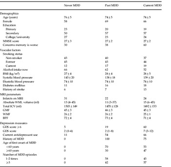
The mean age of the study population was 76 (s.d. = 5) years and 58% were female. Of the total sample, 95.5% persons did not have a lifetime diagnosis of MDD, 3.0% had a past but no current diagnosis of MDD and 1.5% had a current diagnosis of MDD (i.e. in the past 2 weeks). Of the persons with current MDD, 75% also had a past history of MDD. Compared to those never depressed and those with past MDD, persons with current MDD had higher depressive symptom levels, more often used antidepressants and had more previous depressive episodes (Table 1).
Characteristics of the study samplea

MDD, Major depressive disorder; MMSE, Mini-Mental State Examination; MRI, magnetic resonance imaging; BMI, body mass index; WML, white-matter lesion; ICV, intracranial volume; GMF, gray-matter fraction; WMF, normal white-matter fraction; BPF, brain parenchymal fraction; GDS, Geriatric Depression Scale.
Values presented as percentage, mean ± standard deviation or median (10–90 th percentile).
a Based on the sample before imputation (never MDD n = 4050; past MDD n = 125; current MM n = 62).
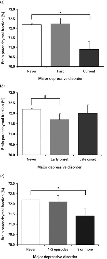
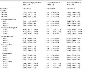
Compared to those never depressed, participants with ever MDD had borderline significantly smaller relative brain volumes, adjusted for age, sex and education, which attenuated in models 2 and 3. When we differentiated between current and past MDD, participants with current MDD had statistically significantly smaller relative brain volume, indicating more global brain atrophy, adjusted for age, sex and education [B = − 1.44%, 95% confidence interval (CI) −2.26 to −0.63; p = 0.001]. After additional adjustment in models 2 and 3, the estimate attenuated somewhat but remained statistically significant. Current MDD was associated with more atrophy in the gray matter and normal white matter (Table 2). Persons with past/not current MDD did not have more brain atrophy than those never depressed (Fig. 2a, Table 2).
Mean brain parenchymal fraction (BPF) according to (a) past and current major depressive disorder (MDD); (b) early-onset (first MDD at age <60 years) and late-onset (first MDD at age ⩾60 years) MDD; the groups include persons with past and current MDD; and (c) number of episodes of MDD. Means are adjusted for age, sex, education, Mini-Mental State Examination (MMSE) score, subjective memory complaints, smoking habits, alcohol intake, body mass index (BMI), systolic and diastolic blood pressure, diabetes, history of stroke, white-matter lesion (WML) volume and presence of infarcts on magnetic resonance imaging (MRI). Error bars represent standard errors. * p < 0.05, #p = 0.095.

Results of the linear regression models for the associations of depression groups with relative brain volumes

MDD, Major depressive disorder; CI, confidence interval.
B (regression coefficient) represents the difference in percentage brain volume between the respective depression group and participants without a lifetime MDD diagnosis.
Model 1: adjusted for age, sex and education.
Model 2: model 1 with additional adjustment for Mini-Mental State Examination (MMSE) score, subjective memory complaints, smoking habits, alcohol intake, body mass index (BMI), systolic and diastolic blood pressure, diabetes, history of stroke, white-matter lesion (WML) volume and presence of infarcts on magnetic resonance imaging (MRI).
Model 3: model 2 with additional adjustment for current antidepressant use.
* p < 0.05.
Participants with an early-onset MDD (<60 years) had borderline significantly smaller relative brain volume than those never depressed in models 1 and 2, which attenuated after adjusting for antidepressant use; those with a late onset did not have smaller relative brain volume (Fig. 2b, Table 2). Additional analysis within only the persons with a history of MDD showed no significant differences in total brain volume between late-onset and early-onset MDD (mean difference in relative total brain volume adjusted for age, sex, education was 0.15%; 95% CI −0.90 to 1.19).
Participants with 1–2 episodes did not have smaller relative brain volume than those never depressed; those with ⩾3 episodes had statistically significantly smaller relative brain volume in model 2, which attenuated after adjusting for antidepressant use (Fig. 2c, Table 2). Additional analysis within only the persons with a history of MDD showed no significant differences in total brain volume between ⩾3 episodes and 1–2 episodes (mean difference in relative total brain volume adjusted for age, sex, education was −0.58%; 95% CI −1.58 to 0.42).
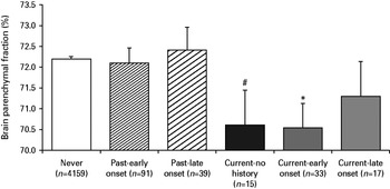
When exploring the relative influence of age at onset within participants with current and past MDD, participants with current MDD with early onset had more brain atrophy than those never depressed in model 1 (B = − 1.66%, 95% CI −2.82 to −0.49, p = 0.005) whereas this association was less strong and not significant for participants with current MDD/late onset (B = − 0.87%, 95% CI −2.50 to 0.76, p = 0.29). However, participants with current MDD without a history had moderately more brain atrophy (B = − 1.62%, 95% CI −3.30 to 0.05, p = 0.058 (Fig. 3). Additional analyses of presence of depressive symptoms as measured with the GDS score (⩾6 v. <6) within the group without a lifetime diagnosis of MDD were consistent with this latter finding, where presence of depressive symptoms was associated with more brain atrophy [B(model 2) = −1.03%, 95% CI −1.50 to −0.55, p < 0.0001]. Finally, participants with past MDD/early onset (B = − 0.09%, 95% CI −0.81 to 0.63, p = 0.81) or those with past MDD/late onset (B = 0.18%, 95% CI −0.94 to 1.30, p = 0.75) (Fig. 3) did not have smaller brain volumes than those who were never depressed.
Mean brain parenchymal fraction (BPF) according to past, current and age at onset of major depressive disorder (MDD). Means are adjusted for age, sex and education. Error bars represent standard errors. * p < 0.05, #p = 0.058.

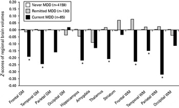
Figure 4 shows the z scores adjusted for age, sex, education and ICV in different brain tissue regions for persons never depressed, those with past MDD and those with current MDD. As can be seen, compared to participants never depressed, those with current MDD had significantly more atrophy in most of the brain regions, including frontal and temporal gray and white matter, parietal white matter, and the hippocampus and thalamus. When we additionally adjusted for other covariates (model 2), estimates attenuated somewhat and some regions lost statistical significance (frontal gray matter p = 0.06, hippocampus p = 0.08, and thalamus p = 0.11).
Z scores of regional brain volumes according to past and current major depressive disorder (MDD). Z scores are adjusted for age, sex, education and intracranial volume (ICV). * p < 0.05 compared to never MDD. Unadjusted mean volume in ml (s.d.) of brain regions: frontal gray matter (GM) 213 (22), temporal GM 128 (13), parietal GM 86 (10), occipital GM 87 (11), hippocampus 5.6 (0.6), amygdala 4.8 (0.6), thalamus 15 (1), striatum 20 (2), frontal white matter (WM) 137 (18), temporal WM 63 (9), parietal WM 71 (10), and occipital WM 53 (8).

Discussion
In a community-based cohort of older people without dementia, we observed that persons with current MDD, but not those in remission, had more global brain atrophy, including more gray- and white-matter atrophy in the majority of lobes, and also more atrophy of the hippocampus and thalamus. Within those with current MDD, persons with a first onset and those with an early onset had more global brain atrophy than those never depressed. Multiple previous episodes were also associated with more global brain atrophy.
To our knowledge, this is the first study in a community-based sample of older people to examine the associations of lifetime DSM-IV diagnoses of MDD with brain volumes on MRI. A major strength of this study lies in the combination of characteristics that reduced selection bias, information bias and confounding, which include the population-based design, the exclusion of participants with dementia, the use of structured diagnostic interviews to obtain DSM-IV lifetime depression diagnoses, and the large number of confounders taken into account. In addition, the large sample size and the volumetric brain measures increased the precision and power to detect small differences. We were also able to examine several depression characteristics, including current and past MDD, age at onset and number of episodes.
A limitation is the cross-sectional design. We tried to distinguish the temporal relationship by differentiating between history of MDD and MDD at time of MRI. We also excluded participants with dementia and adjusted for cognitive functioning to decrease the possibility that the association was explained by subclinical dementia.
Of note is the low prevalence of lifetime MDD compared to other studies (Kessler et al. Reference Kessler, Berglund, Demler, Jin, Merikangas and Walters2005, Reference Kessler, Birnbaum, Bromet, Hwang, Sampson and Shahly2010). Whereas the low prevalence of current MDD can be explained by the short time period used (i.e. MDD in the past 2 weeks as opposed to the more commonly used 12-month prevalence), there are several factors, pertinent in general to studies of older persons, that may account for the low prevalence of past MDD. For example, the prevalence may be lower because depression is a risk factor for non-response and for mortality (Penninx et al. Reference Penninx, Geerlings, Deeg, van Eijk, Van Tilburg and Beekman1999; Schoevers et al. Reference Schoevers, Geerlings, Beekman, Penninx, Deeg, Jonker and van Tilburg2000), so it is possible that persons with MDD earlier in life died before the start of the AGES Reykjavik Study. Furthermore, depression and dementia/mild cognitive impairment are highly co-morbid (Thomas & O'Brien, Reference Thomas and O'Brien2008); as we excluded the participants with dementia and those with MMSE scores <21, we also probably excluded a proportion of persons with depression. Finally, although the sensitivity of our screening algorithm was very high, most of the depression questions used in the screening algorithm were based on current complaints and to a lesser extent on history of complaints. This may have resulted in identifying a lower proportion of persons with past MDD receiving the MINI. As a result, the reference group (never MDD) will include a proportion of persons who may have had a history of MDD or who had current depressive symptoms or subthreshold depression. Nevertheless, those persons receiving a diagnosis of current or past MDD are likely to be correctly classified. Although the comparison between the ever MDD and the never MDD group may be diluted, this will not have affected the comparison to a great extent, given the very large size of the reference group; and we do not consider this can explain the difference in brain volume between the current MDD and remitted MDD group.
When we adjusted for current use of antidepressants, associations attenuated. This might suggest that medication users had more severe depression and therefore the greatest risk for brain atrophy. It might also suggest that antidepressant use is associated with brain atrophy independent of MDD. Antidepressants and, in particular, selective serotonin reuptake inhibitors (SSRIs) are frequently prescribed for other indications than depression, such as anxiety or sleeping problems. A recent population-based study in old persons without dementia showed that use of antidepressants was associated with more brain atrophy, independent of depressive symptom level (Geerlings et al. Reference Geerlings, Brickman, Schupf, Devanand, Luchsinger, Mayeux and Small2012). More studies are needed with detailed data on type, dose, duration and prescription indication to determine whether or not antidepressants are harmful for the brain.
Many studies in younger populations found volume reductions of the hippocampus (MacQueen & Frodl, Reference MacQueen and Frodl2011) and other specific brain regions thought to be involved in emotion regulation (Lorenzetti et al. Reference Lorenzetti, Allen, Fornito and Yucel2009; MacQueen & Frodl, Reference MacQueen and Frodl2011; Kupfer et al. Reference Kupfer, Frank and Phillips2012) in patients with MDD compared to healthy controls. Our findings are not consistent with these previous reports because we observed gray- and white-matter atrophy in the majority of lobes and also in the hippocampus and thalamus, suggesting that, at older age, atrophy associated with MDD is widespread. From our data we cannot know, however, whether the volume reduction started in specific brain regions at younger age and expanded with older age, or whether a general neurodegenerative process underlies the association with MDD later in life.
Previous population-based studies did not have data on lifetime diagnoses of MDD and findings relied on depressive symptom scales and one or two questions to determine history of depression (Geerlings et al. Reference Geerlings, den Heijer, Koudstaal, Hofman and Breteler2008, Reference Geerlings, Brickman, Schupf, Devanand, Luchsinger, Mayeux and Small2012; Dotson et al. Reference Dotson, Davatzikos, Kraut and Resnick2009; Goveas et al. Reference Goveas, Espeland, Hogan, Dotson, Tarima, Coker, Ockene, Brunner, Woods, Wassertheil-Smoller, Kotchen and Resnick2011). As a result, it is difficult to differentiate between depressive symptoms indicating MDD and depressive symptoms associated with disease and disability, or between a first onset and depression as part of a lifelong history of depressive episodes. If depression is a causal risk factor for brain atrophy and dementia, then a history of depression, and in particular an early onset and repeated episodes, might be expected to be associated with more brain atrophy. However, if depression is a consequence of brain volume loss or a prodrome of dementia, then depression closest in time of the MRI might be expected to be associated with more brain atrophy. When we differentiated history within those with current MDD, we observed that those with an early onset had smaller brain volumes, suggesting depression preceded or promoted brain tissue loss. Consistent with this, we also found that multiple episodes were associated with smaller brain volumes. One population-based study examining the bidirectional relationship between depression and hippocampal volume loss found that depressive symptoms at baseline predicted faster hippocampal volume loss, but hippocampal volume at baseline was not associated with incident depression, which supports our findings (den Heijer et al. Reference den Heijer, Tiemeier, Luijendijk, van der Lijn, Koudstaal, Hofman and Breteler2011). However, we also found that persons with first-onset MDD had smaller brain volumes and, consistent with this, we also found that a high GDS-15 score in the absence of a lifetime diagnosis of MDD was associated with smaller brain volumes. Although numbers in the subgroups were small, this suggests that depression can be both a contributor to and a consequence of smaller brain volume.
Older persons with MDD in remission did not have smaller brain volumes than those never depressed. Although this finding is somewhat counterintuitive, one explanation could be that MDD is associated with smaller brain volume only in the acute state. Several studies have shown that patients with MDD who showed remission at follow-up had larger baseline hippocampal volume than patients with MDD who did not show remission at follow-up (Hsieh et al. Reference Hsieh, McQuoid, Levy, Payne, MacFall and Steffens2002; MacQueen et al. Reference MacQueen, Yucel, Taylor, Macdonald and Joffe2008; Ahdidan et al. Reference Ahdidan, Hviid, Chakravarty, Ravnkilde, Rosenberg, Rodell, Stodkilde-Jorgensen and Videbech2011). In addition, one study found that patients with current depression had a smaller hippocampal volume than patients in remission at the time of the MRI (Caetano et al. Reference Caetano, Hatch, Brambilla, Sassi, Nicoletti, Mallinger, Frank, Kupfer, Keshavan and Soares2004). It should be noted that these studies examined hippocampal volume instead of total brain volume and hence the findings may not be fully comparable. Two studies that examined both hippocampal and total brain volume observed that patients with a history of MDD but not current major depression had a smaller hippocampal volume, but not a smaller total brain volume, when compared to healthy controls (Sheline et al. Reference Sheline, Wang, Gado, Csernansky and Vannier1996; Neumeister et al. Reference Neumeister, Wood, Bonne, Nugent, Luckenbaugh, Young, Bain, Charney and Drevets2005). However, we did not find an association between past MDD and hippocampal volume. Possibly current depression is associated with increased cortisol levels, which may be neurotoxic, and in remitted depression cortisol levels return to normal and atrophy is reversed (Caetano et al. Reference Caetano, Hatch, Brambilla, Sassi, Nicoletti, Mallinger, Frank, Kupfer, Keshavan and Soares2004). Few studies in humans, however, have investigated the relationship between depression, cortisol and brain volumes within one study. Although higher basal cortisol levels may be associated with smaller hippocampal volume (Knoops et al. Reference Knoops, Gerritsen, van der Graaf, Mali and Geerlings2010), they may not explain the relationship between MDD and hippocampal volume (Gerritsen et al. Reference Gerritsen, Comijs, van der Graaf, Knoops, Penninx and Geerlings2011). It should be noted that the findings on depression in the study by Gerritsen et al. (Reference Gerritsen, Comijs, van der Graaf, Knoops, Penninx and Geerlings2011) are inconsistent with ours because in their study current MDD was not associated with hippocampal volume whereas remitted depression was associated with a smaller entorhinal cortex volume and we did not measure entorhinal cortex volume. Furthermore, a history of depression was based on the two core symptoms of MDD and not on a clinical diagnosis (Gerritsen et al. Reference Gerritsen, Comijs, van der Graaf, Knoops, Penninx and Geerlings2011). Clearly, more studies are needed to examine the role of hypothalamic–pituitary–adrenal (HPA) axis dysregulation in the relationship between MDD, brain atrophy and the development of dementia.
In conclusion, in this population-based study of older persons without dementia, current MDD, irrespective of prior history, was associated with widespread gray- and white-matter brain atrophy whereas MDD in remission was not associated with more brain atrophy. Prospective studies should examine whether MDD is a consequence of, or contributes to, brain volume loss and development of dementia.
Acknowledgements
The AGES–Reykjavik Study is funded by the NIH contract N01-AG-12100, the NIA Intramural Research Program, Hjartavernd (the Icelandic Heart Association), and the Althingi (the Icelandic Parliament). Dr M. I. Geerlings is funded by a grant from the Netherlands Organization for Scientific Research (NWO: project no. 917-66-311) and the University Medical Center Utrecht (program Internationalization). The funding sources had no involvement in the design and conduct of the study; collection, management, analysis and interpretation of the data; and preparation, review or approval of the manuscript. Dr L. J. Launer (PI) had full access to all of the data in the study and takes responsibility for the integrity of the data and the accuracy of the data analysis.
Declaration of Interest
None.