Introduction
Pulmonary arteriovenous fistulas are abnormal direct connections between pulmonary arteries and veins, bypassing the capillary bed. These can arise idiopathically, be part of hereditary haemorrhagic telangiectasia, or develop following surgical interventions such as the Kawashima procedure. This procedure, performed in patients with single-ventricle anatomy and interrupted inferior vena cava with azygos continuation, reroutes venous blood flow to the pulmonary arteries while excluding hepatic blood flow from the lungs. This exclusion has been associated with the development of pulmonary arteriovenous fistulas due to the absence of certain “hepatic factors” believed to maintain pulmonary vascular health. Reference Alsoufi, Rosenblum and Travers1,Reference Alibrahim, Mohammed and Kabbani2 Additionally, a recent comprehensive review on pulmonary arteriovenous fistulas in children has emphasised the importance of early detection and surveillance and reinforced the central role of embolisation in preventing severe complications. Reference Lim and Ratjen3 Embolisation using devices like the Medtronic® Micro Vascular Plug has proven to be an effective treatment option for pulmonary arteriovenous fistulas, offering immediate vessel occlusion and low recanalisation rates. Reference Latif, Bailey and Motaghi4–Reference Duvnjak, Di Ciesco and Andersen6
Case description
We describe the case of a 15-year-old patient (52 kg) with a double-outlet right ventricle (S, D, D) and complete atrioventricular septal defect, left isomerism, and interrupted inferior vena cava with azygos continuation, who had previously undergone a Kawashima procedure (in a different institution) with banding of the pulmonary artery trunk. The patient’s oxygen saturation was 82%.
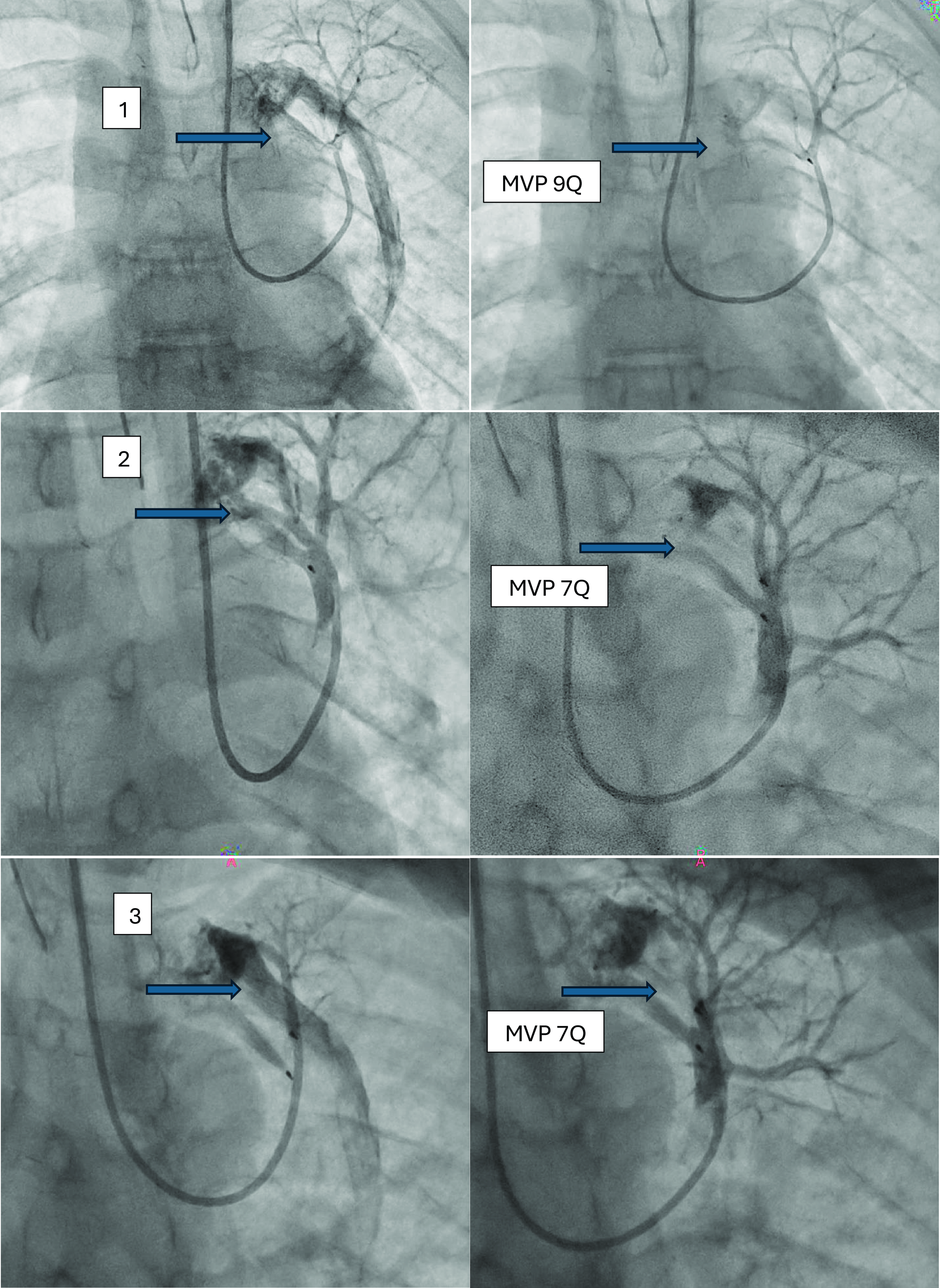
Pre-catheterisation evaluation aimed to assess pulmonary arterial pressures and vascular resistance for a potential total cavopulmonary connection was performed. Cardiac catheterisation revealed a mean pulmonary pressure of 14 mmHg. Angiography indicated oxygen enrichment in the right pulmonary artery (72%), compared to 50–60% in the superior vena cava and azygos system showing a preserved antegrade flow through the pulmonary artery, mostly directed to the right lung. A haemodynamically significant pulmonary arteriovenous fistula, not described in the imaging prior to the Kawashima procedure, originating from the left upper lobe and draining into the left upper pulmonary vein was identified, with bubble contrast studies confirming significant shunting to the left side (Figure 1). Through a short 5 French sheath placed in the left internal jugular vein an antegrade approach for embolisation was chosen. A 5 French Judkins right catheter was advanced in the main feeding vessel (Figure 2), and a Micro Vascular Plug 9Q device was implanted distally prior to the convolute part of the fistula (Figure 2, Panel 1). Control angiography showed two remaining residual feeding vessels into which a Judkins right catheter with a telescopic system of a PROGREAT® Microcatheter on a 0.014 HI-TORQUE PILOT™ guidewire was advanced to implant a Micro Vascular Plug 7Q (Figure 2, panels 2 and 3). Control angiography showed improved perfusion of the upper left pulmonary lobe and minimal residual flow through the pulmonary arteriovenous fistulas. Post-intervention mean pulmonary pressure raised to 16 mmHg and oxygen saturation to 86%. The embolisation procedure was completed in 90 minutes, with a fluoroscopy time of 30 minutes and a total radiation dose of 8.02 mGy.
Pre-interventional imaging showing a haemodynamically significant pulmonary arteriovenous fistula in the left upper lobe. The fistula is marked with a blue arrow.

Sequential embolisation of the three feeding vessels (3.9 mm, 3.3 mm, 2.7 mm) with a Micro Vascular Plug (MVP) with progressive reduction of the flow into the fistula and augmentation of the flow in the pulmonary branches.

Discussion
Pulmonary arteriovenous fistulas are a recognised complication following the Kawashima procedure, attributed to the lack of hepatic venous return to the lungs. Reference McElhinney, Kreutzer, Lang, Mayer, Del Nido and Lock7,Reference Mahmoud, Zahrani, Bahaidarah, Kouatli and Baslaim8 The “hepatic factor hypothesis” supports this, indicating that hepatic venous effluent is essential for maintaining pulmonary vascular integrity. Reference Alsoufi, Rosenblum and Travers1,Reference Alibrahim, Mohammed and Kabbani2,Reference Latif, Bailey and Motaghi4 In this case, it is interesting to note, that, at least partially, a hepatic flow was preserved, but it was mostly directed to the right lung, since the upper cavopulmonary connection drained on the left pulmonary artery.
We decided to close this significant left-sided pulmonary arteriovenous fistula to evaluate the pulmonary pressure correctly to confirm the prerequisites for a total cavopulmonary connection and to avoid a chronic desaturation after total cavopulmonary connection. Furthermore, considering the size of this pulmonary arteriovenous fistulas a spontaneous closure after connection of the hepatic veins was unrealistic.
The Micro Vascular Plug was chosen for embolisation due to its proven ability to rapidly and effectively occlude vessels, with minimal risk of embolisation or late recanalization. Micro Vascular Plug devices offer advantages over traditional coils by providing precise placement and quick occlusion ideal for such an inconveniently located vascular structure. Reference Bailey, Arun and Towsley5,Reference Duvnjak, Di Ciesco and Andersen6,Reference Haddad, Bonnet and Malekzadeh-Milani9 Its low profile permitted us to accommodate the acute angle avoiding a long and stiff sheath to perform embolisation. Reference Bailey, Arun and Towsley5,Reference Duvnjak, Di Ciesco and Andersen6,Reference Haddad, Bonnet and Malekzadeh-Milani9 Furthermore, since the feeding vessels were long, we could accommodate the devices without obstruction of the pulmonary branches. We decided to embolise the feeding vessels Reference Kuczyńska, Pyra, Światłowski, Sobstyl, Kuklik and Jargiełło10 rather than the nidus to minimise the risk of distal device embolisation, considering the size of the downstream vessels. Moreover, as all three feeding vessels were embolised and the residual shunt observed immediately after implantation was minimal, we believe this approach provided an optimal balance between risks and benefits.
Conclusion
This case highlights the successful use of three Micro Vascular Plug for embolisation of a haemodynamically significant pulmonary arteriovenous fistula in a post-Kawashima surgery paediatric patient.
Financial support
All authors have no potential conflicts of funding or financial relationship to disclose.
Competing interests
All authors have no potential conflicts of interest to disclose.
Ethical standards
All subjects gave their informed consent. The study was conducted in accordance with the Declaration of Helsinki.