Introduction
While inspecting the collection of unidentified lichenicolous fungi of the late Rolf Santesson, preserved in the herbarium of the Museum of Evolution at Uppsala University (UPS), I came across an interesting specimen of a pyrenocarpous ascomycete from Chile annotated as ‘Polycoccum sp.’ that causes conspicuous galls on the thallus of the lichen Bunodophoron ramuliferum (I.M. Lamb) Wedin. A search for similar material among the Bunodophoron A. Massal. specimens stored in UPS uncovered another specimen of this fungus from New Zealand. Subsequent examination of these specimens showed that they represent a species from the lichenicolous genus Clypeococcum D. Hawksw. that is unknown to science. This paper aims to describe this new species and provide an updated key to the 15 accepted species of Clypeococcum.
Materials and Methods
Microscopy was carried out and images were captured using a Zeiss Axio Zoom V16 microscope and a Zeiss Axio Imager A1 microscope equipped with Nomarski differential interference contrast optics and fitted with a Zeiss AxioCam MRc5 digital camera. Microscopic characters were studied using sections hand-cut with a razor blade and mounted in water, 10% potassium hydroxide (K), Lugol's iodine directly (I) or after a K pretreatment (K/I), or concentrated nitric acid (N). Measurements were taken from water mounts and rounded to the nearest 0.5 μm. The length, width, length/width ratio (l/w) and upper cell length/lower cell length ratio of the ascospores, as well as the diameter of the ascomata are given as (min‒) (x̄ − SD) ‒ (x̄ + SD) (‒max), where ‘min’ and ‘max’ are the extreme values observed, x̄ the arithmetic mean and SD the corresponding standard deviation. Specimens examined are housed in LE and UPS.
Specimens examined for comparison
Clypeococcum cladonema (Wedd.) D. Hawksw. (type of Clypeococcum). Russia: Krasnodar Territory: on Flavoparmelia caperata (L.) Hale, 2014, M. P. Zhurbenko 14326a (LE 309410a).
Didymocyrtis consimilis Vain. (type of Didymocyrtis Vain.). Russia: Republic of Adygeya: on Parvoplaca tiroliensis (Zahlbr.) Arup et al., 2014, M. P. Zhurbenko 14431 (LE 309489).
Polycoccum trypethelioides (Th. Fr.) R. Sant. (type of Polycoccum Körb.). Norway: Troms County: on Stereocaulon glareosum (Savicz) H. Magn., 2003, M. P. Zhurbenko 0383 (LE 260505).
Verrucoccum spribillei V. Atienza et al. USA: Alaska: on Lobaria pulmonaria (L.) Hoffm., 2001, M. P. Zhurbenko 01197 (LE 233929).
The new species
Clypeococcum wedinii Zhurb. sp. nov.
MycoBank No.: MB 844870
Differs from Clypeococcum galloides Etayo mainly by its non-amyloid hymenium, 8-spored asci, larger ascospores (14–)18–22(–25) × (5–)6.5–8(–9) μm, and host preference (Bunodophoron).
Type: New Zealand, Southland, Fiordland National Park, Lake Haurako, Oblong Hill, along Look Out Track, open Nothofagus solandri-Weinmannia dominated forest, on a dead trunk of Podocarpus sp., 45°59ʹS, 167°22ʹE, elev. 360 m, on thallus of Bunodophoron notatum, 21 January 1990, M. Wedin 2947 (UPS 56614—holotype).
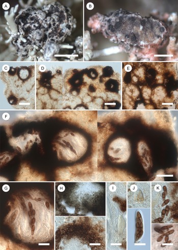
(Fig. 1)
Clypeococcum wedinii (A–C & K (below), holotype; D–J & K (above), UPS F-8925990). A, final stage of gall development. B, section through a gall showing immersed ascomata. C–E, galls with immersed ascomata and varying degrees of stromatic development in cross-section, in water. F, ascomata and clypeus-like stromatic growths in cross-section, in water. G, ascoma with asci, ascospores and interascal filaments in cross-section, in water. H, ascomatal wall in surface view, in K (above) and in cross-section, in water (below). I, interascal filaments, in I. J, asci, in I. Note apical beak above. K, ascospores in I (above) and water (below). Scales: A = 1 mm; B = 500 μm; C–E = 100 μm; F = 50 μm; G–K = 20 μm. In colour online.

Lichenicolous ascomycete growing on species of Bunodophoron and inducing conspicuous galls. Vegetative hyphae light to medium brown, 2–4.5 μm diam. Galls subglobose, 250–2600 μm diam., initially the same colour as the host thallus, later getting darker, becoming constricted at the base and strongly tuberculate, mottled with protruding black tops of the parasitic ascomata/clypei. The interior of the galls is filled with parasitic ascomata (up to 100 or possibly more) interspersed with loose to dense stromatic growths in the form of strands or more massive aggregations between/around the ascomata mixed with host tissues.
Ascomata perithecioid, black in the exposed parts, subglobose, not beaked, (40–)90–180(–210) μm diam. (n = 69), completely immersed in the interior of the galls or slightly protruding above their surface in the ostiolar area, with ostiole c. 20 μm diam., dispersed to aggregated, sometimes contiguous with a small number merged together, often linked by brown plurihyphal stromatic strands, 10–55 μm wide, or united in groups of up to 10 or more by more extensive and dense stromatic growths that sometimes form clypeus-like structures (Fig. 1F). Ascomatal wall in surface view composed of interwoven hyphae (textura intricata) or pseudoparenchymatous cells (textura angularis), in cross-section light brown (inside) to medium or dark brown (outside), with the pigment deposited in the hyphal cell walls, K+ partly greyish, N−, without warts on the outside, mainly 10–30 μm wide; composed of c. 8–12 layers of roughly isodiametric or slightly tangentially elongated cells 1.5–5.5 μm long with walls 0.5–3 μm wide, in places passing into stromatic growths and widening to 80 μm, especially between and above adjacent ascomata, forming in the latter case clypeus-like structures. Hymenium not inspersed, I−, K/I−. Hamathecium composed of distinct, persistent, hyaline, filiform, branched, occasionally anastomosing, septate, short-celled interascal filaments that are not swollen at the apices, 1.5–2.5 μm wide; ostiolar filaments absent. Asci bitunicate in structure, roughly subcylindrical, slightly widened in the middle or lower half (obclavate), rounded at the apex, sometimes with an apical beak, short-stalked, 65–80 × 15–18 μm (n = 8), 8-spored, I−, K/I− except for ascoplast turning orange. Ascospores light to occasionally medium brown when mature, K+ brownish grey, very narrowly obovate, rounded at the ends, 1-septate; upper cell wider, often longer and rarely slightly darker than the lower one, upper cell length/lower cell length = 1–1.4(–1.6) (n = 23), not or only slightly constricted at the septum, (14–)18–22(–25) × (5–)6.5–8(–9) μm, l/w = (2.1–)2.4–3.2(–4.0) (n = 144), occasionally with 1–2 conspicuous guttules in each cell, finely verruculose with verrucae c. 0.5 μm diam.; when immature rarely with a gelatinous sheath to 1.5 μm wide observed in K, biseriate and mostly overlapping in the ascus.
Conidiomata not observed.
Etymology
The new species is named after the eminent Swedish lichenologist Mats Wedin, monographer of the family Sphaerophoraceae in the Southern Hemisphere (Wedin Reference Wedin1995), who collected the holotype of the new species.
Distribution and hosts
The new species is known from two specimens collected in Chile and New Zealand, therefore belonging to the Neotropical and Australasian biogeographical realms. It was collected on thalli of Bunodophoron notatum (Tibell) Wedin and B. ramuliferum (Lecanorales, Sphaerophoraceae). The former species occurs in Australia, Tasmania and New Zealand, and the latter in Australia, Tasmania, New Zealand, southernmost South America, Juan Fernandes and the Falkland Islands (Wedin Reference Wedin1995). Apart from gall-formation, the harmful effects on the hosts are not noticeable, with their surrounding parts retaining their normal coloration.
Notes
With its lichenicolous lifestyle, black, subglobose, ostiolate perithecioid ascomata, often united by stromatic growths, ascomatal wall of both textura intricata and angularis in surface view, I−, K/I− hymenium, hamathecium composed of persistent, filiform, branched and anastomosing interascal filaments, bitunicate, roughly subcylindrical, 8-spored asci, and 1-septate, brown, verruculose ascospores, biseriate in the ascus, the examined fungus is most consistent with the protologue of the genus Clypeococcum (Polycoccaceae, Dothideomycetes) typified on C. cladonema (Hawksworth Reference Hawksworth1977). The only difference from this protologue, although significant, is the absence of a persistent distinct clypeus, but the fungus occasionally develops clypeus-like structures. Morphologically, the new species also strongly resembles pyrenocarpous lichenicolous taxa belonging to Didymocyrtis, Polycoccum and Verrucoccum V. Atienza et al. However, Didymocyrtis and Polycoccum differ in the absence of stromatic ascomata and the presence of exclusively textura angularis tissue in the ascomatal wall; the former is additionally distinct in its narrowly cylindrical asci and uniseriate ascospores (Hawksworth & Diederich Reference Hawksworth and Diederich1988; Ertz et al. Reference Ertz, Diederich, Lawrey, Berger, Freebury, Coppins, Gardiennet and Hafellner2015). Verrucoccum hymeniicola (Berk. & Broome) D. Hawksw. et al. and V. spribillei also form tightly packed ascomata surrounded by stromatic tissue. However, their ascomata are exposed in the upper two-thirds with age, and the ascomatal wall is made exclusively of textura angularis with a warted surface (Atienza et al. Reference Atienza, Hawksworth and Pérez-Ortega2021).
Considering other genera of ascomycetes characterized by a lichenicolous lifestyle, closed ascomata and stromatic growths, it is appropriate to compare Clypeococcum wedinii with Homostegia Fuckel, Lasiosphaeriopsis D. Hawksw. & Sivan, Lichenostigma Hafellner, Macrographa Etayo, Perigrapha Hafellner, Plectocarpon Fée, Plowrightia Sacc. and Saania Zhurb. They are all characterized by their well-developed, usually superficial stromata. Lichenostigma is also distinguished by its asci arising directly within the stromatic pseudoparenchyma (Calatayud et al. Reference Calatayud, Hafellner, Navarro-Rosinés, Nash, Ryan, Diederich, Gries and Bungartz2004). Other genera have perithecial locules that are completely enclosed by stromatic tissue, whereas in Clypeococcum wedinii only some of such locules are surrounded by a stroma. Additionally, Homostegia differs in the wall structure of the perithecial locules (textura angularis) and its 3-septate ascospores (Hawksworth et al. Reference Hawksworth, Atienza and Cole2004); Lasiosphaeriopsis is distinguished by the presence of periphyses, the absence of interascal filaments, unitunicate asci and perithecial locule walls with Munk pores (Hawksworth Reference Hawksworth1980b); Macrographa is characterized by abundant periphysoids and hyaline ascospores (Etayo & Sancho Reference Etayo and Sancho2008); Perigrapha and Plectocarpon are well distinguished by their K/I+ blue hymenial gel and asci with a K/I+ blue apical ring (Hafellner Reference Hafellner1996; Ertz et al. Reference Ertz, Christnach, Wedin and Diederich2005; Zhurbenko & Ohmura Reference Zhurbenko and Ohmura2018); Plowrightia differs in the absence of interascal filaments and in its hyaline ascospores (Clauzade et al. Reference Clauzade, Diederich and Roux1989; Thambugala et al. Reference Thambugala, Ariyawansa, Li, Boonmee, Hongsanan, Tian, Singtripop, Bhat, Camporesi and Jayawardena2014); and Saania is distinguished by its N+ red ascomatal wall and hamathecium, consisting only of periphysoids (Motiejūnaitė et al. Reference Motiejūnaitė, Zhurbenko, Suija and Kantvilas2019).
In the genus Clypeococcum, 15 species are accepted to date; all are lichen-inhabiting and rather highly selective concerning their hosts, 12 of them being confined to one lichen genus, two confined to two genera in the same family, and one to three genera in the same family (see key below). Eight of the 15 known species of Clypeococcum, including C. wedinii, are restricted to lichens of the order Lecanorales, while C. wedinii is the first species of this genus found on members of the family Sphaerophoraceae, to which the genus Bunodophoron belongs.
Additional specimen examined
Chile: on Bunodophoron ramuliferum (thallus), leg. Poeppig, det. Kunze 1842, hb. Tuck, sheet no. 3746 (UPS F-8925990).
An updated key to the species of Clypeococcum
The previous key for Clypeococcum included nine species (Pirogov Reference Pirogov2015). Subsequently, six further species have been described, making it appropriate to present an updated key for the 15 currently known in this genus. This key is based on the literature cited below under ‘Lit.’.
1 Ascospores muriform; on Solorina Ach. Lit.: Zhurbenko (Reference Zhurbenko2020) C. lenae Zhurb.
Ascospores 1-septate 2
2(1) Galls present 3
Galls absent 5
3(2) Galls up to 2.6 mm diam., hymenium I−, asci 8-spored, ascospores (14–)18–22(–25) × (5–)6.5–8(–9) μm; on Bunodophoron. Lit.: present paper C. wedinii
Galls up to 1 mm diam., hymenium I+ violet, ascospores shorter 4
4(3) Asci (2–)4-spored, ascospores 11.5–14(–16) × 5.5–7(–8) μm; on saxicolous Lecidea Ach. s. lat. Lit.: Etayo (Reference Etayo2010) C. galloides
Asci 4–8-spored, ascospores 16.5–20 × 6.5–7.5 μm; on Umbilicaria Hoffm. Lit.: Körber (Reference Körber1865); Hawksworth (Reference Hawksworth1982); Alstrup & Hawksworth (Reference Alstrup and Hawksworth1990); Etayo (Reference Etayo2010)C. grossum (Körb.) D. Hawksw.
5(2) Hymenium predominantly I+ red; on Verrucaria latebrosa Körb. Lit.: Shivarov (Reference Shivarov2019) C. hemiamyloideum Shivarov
Hymenium not I+ red 6
6(5) Hymenium I+ violet; on Parmotrema A. Massal. Lit.: Etayo (Reference Etayo2017) C. amylaceum Etayo
Hymenium I− 7
7(6) Ascospores longitudinally striate by wrinkles, (16–)17–20(–21) × (7–)8–9(–10) μm; on Parmotrema. Lit.: Etayo (Reference Etayo2017) C. rugosisporum Etayo & Zhurb.
Ascospores not longitudinally striate 8
8(7) Asci 2-spored, ascospores (17–)20–27(–35) × (4–)5–5.5(–6) μm; on Cetraria Ach. and Nephromopsis Müll. Arg. Lit.: Zhurbenko (Reference Zhurbenko2009) C. bisporum Zhurb.
Asci with more than 2 spores, ascospores of a different size 9
9(8) Asci 4-spored 10
Asci 4–8-spored 11
10(9) Ascospores 8–10 × 5–7 μm; on Buellia melanostola (Hue) Darb. Lit.: Øvstedal & Hawksworth (Reference Øvstedal and Hawksworth1986); Grube & Hafellner (Reference Grube and Hafellner1990) C. epimelanostolum (Øvstedal & D. Hawksw.) Grube & Hafellner
Ascospores (12–)14.5–17.5(–19.5) × (4.5–)5–6.5(–7.5) μm; on Cetraria, Nephromopsis and Vulpicida J.-E. Mattsson & M.J. Lai. Lit.: Hafellner (Reference Hafellner1994); Zhurbenko & Zhdanov (Reference Zhurbenko and Zhdanov2013); Zhurbenko & Kobzeva (Reference Zhurbenko and Kobzeva2014) C. cetrariae Hafellner
11(9) Ascospores (24–)29–32(–34) × 5.5–6.5 μm; on Hypotrachyna (Vain.) Hale. Lit.: Etayo (Reference Etayo2017)C. cajasense Etayo
Ascospores shorter 12
12(11) Ascomata 80–200 μm diam., ascospores (15–)16–21(–22.5) × 6–8.5(–9.5) μm; on Squamarina Poelt. Lit.: Navarro-Rosinés et al. (Reference Navarro-Rosinés, Roux and Llimona1994) C. psoromatis (A. Massal.) Etayo
Ascomata smaller, ascospores shorter …13
13(12) Ascomata 60–150 μm diam., ascospores (13.5–)14–16(–18) × (5.5–)6–7(–7.5) μm; on Cetrelia W.L. Culb. & C.F. Culb. and Xanthoparmelia (Vain.) Hale. Lit.: Hawksworth (Reference Hawksworth1977) C. cladonema
Ascomata smaller 14
14(13) Ascomata c. 100 μm diam., ascospores 15.5–18 × 8–10 μm; on Placopsis (Nyl.) Linds. Lit.: Øvstedal & Hawksworth (Reference Øvstedal and Hawksworth1986) C. placopsiiphilum Øvstedal & D. Hawksw.
Ascomata 50–100 μm diam., ascospores (9–)10–12(–13) × (4–)5–6(–6.5) μm; on Hypocenomyce M. Choisy. Lit.: Hawksworth (Reference Hawksworth1980a) C. hypocenomycis D. Hawksw.
Acknowledgements
This study was carried out within the framework of the research project of the V. L. Komarov Botanical Institute of the Russian Academy of Sciences ‘Biodiversity, ecology, structural and functional features of fungi and fungus-like protists’ (122011900033-4) using the equipment of its Core Facility Center ‘Cell and Molecular Technologies in Plant Science’. The author's visit to UPS herbarium was financially supported by the Rolf Santesson foundation. Roland Moberg, Stefan Ekman and Martin Westberg are thanked for various kinds of help during his stay in Uppsala. Paul Diederich and Javier Etayo provided valuable comments on the manuscript. Mats Wedin helped to identify Bunodophoron ramuliferum.
Author ORCID
Mikhail P. Zhurbenko, 0000-0002-9839-4698.