Introduction
The immune system of multicellular organisms, and the pathogens it is intended to deal with, have co-evolved for millions of years. The vertebrate immune system has developed an array of sophisticated mechanisms to counter the multitude of pathogens that establish an infection and cause disease. At the same time, several pathogens have developed equally sophisticated strategies to suppress or evade the host immune mechanisms. The pathogens of bovine respiratory disease complex (BRDC) are no exception to this phenomenon. The characteristics of the major pathogens of BRDC are reviewed in other reviews of this issue, and this review will focus on the immune evasion strategies developed by these pathogens. Although the immune evasion mechanisms developed by the viral and bacterial pathogens appear to be different, conceptual similarities do exist (Finlay and McFadden, Reference Finlay and McFadden2006). Therefore, we will discuss viral and bacterial immune evasion mechanisms together, under the respective categories. A brief overview of the immune system, and the key elements involved in the orchestration of the immune response would enhance the understanding of the ‘non-immunologist’ readers.
The immune response to a pathogen consists of the innate and adaptive arms. Although the skin, mucosal surfaces and other physiological barriers are considered part of the innate immune system, the key cellular components are the macrophages, dendritic cells, neutrophils and natural killer (NK) cells (Goldsby et al., Reference Goldsby, Kindt, Osborne and Kuby2003). Macrophages and neutrophils phagocytose the extracellular pathogens and help to eliminate them. γδ T cells have been associated with immune response to certain bacterial infections (Fu et al., Reference Fu, Roark, Kelly, Drevets, Campbell, O'Brien and Born1994; Nishmura et al., Reference Nishmura, Emoto, Hiromatsu, Yamamoto, Matsuura, Gomi, Ilkeda, Itohara and Yoshikai1995; Mathy et al., Reference Mathy, Mathy, Lee, Walker, Lofthouse and Meeusen2002). However their precise role in the immune response is not very clear. The NK cells help in the elimination of intracellular pathogens such as the viruses. The other key components of the innate immune system are the early cytokines such as the interferons (IFNs) and the complement system. Innate immunity is innately available or induced very rapidly. But these components are not specific for the antigen, and more importantly, there is no immunologic memory. On the other hand, adaptive immunity is mediated by B and T lymphocytes, and the soluble factors produced by them – the antibodies produced by the B lymphocytes and the cytokines produced by the T lymphocytes. In addition to producing cytokines which regulate both humoral and cell-mediated immune responses, T cells also function as effector cells. In contrast to innate immunity, adaptive immunity is antigen-specific, diverse and has immunologic memory. However, these two arms of the immune response do not function independently in isolated compartments. Rather, there is interplay between the two components. For example, macrophages which are considered as a component of innate immune system play a key role in antigen presentation in the adaptive arm of the immune response. On the other hand, some of the cytokines such as interferon γ (IFN-γ) and interleukin 10 (IL-10) produced during the adaptive response to a pathogen enhance the activity of macrophages.
The immune response, whether it is humoral or cell-mediated, begins with antigen recognition, processing and presentation (Goldsby et al., Reference Goldsby, Kindt, Osborne and Kuby2003). Antigens are generally large and complex entities, and the lymphocytes do not recognize them in their entirety, but rather they recognize small and discrete regions or segments on the antigen, known as epitopes. B cells can recognize an epitope alone. But T cells can recognize an epitope only when it is associated with a major histocompatibility complex (MHC) molecule on the surface of a self-cell – either an antigen presenting cell (APC) or an altered self-cell such as a virus-infected cell. T cells are functionally divided into T helper (Th) cells and T cytotoxic (Tc) cells that generally express specific cell surface molecules CD4 and CD8, respectively. The recognition unit of T cells consists of a heterodimeric T cell receptor (TCR). The unique interaction between TCRs and MHC molecules defines self, and distinguishes that from non-self or foreign. The rules of MHC restriction govern the cognitive specificity of CD8+ and CD4+ T lymphocytes towards MHC class I and II molecules, respectively (Rothbard and Gefter, Reference Rothbard and Gefter1991). The humoral and cell-mediated arms of the immune system have developed to recognize pathogens in different milieus. The humoral arm represented by the antibodies recognize a variety of epitopes displayed on the surfaces of bacteria or viruses, or on the proteins, polysaccharides or lipopolysaccharides (LPSs) that have been released from invading pathogens. The cell-mediated immune system represented by the T cells, recognize peptide epitopes presented by the MHC molecules on self-cells such as virus-infected cells or cancer cells. This arrangement helps the host to meet the different challenges presented to the immune system by different pathogens. Extracellular pathogens are eliminated mostly by antibodies, whereas intracellular pathogens such as viruses are most effectively eliminated by cytotoxic T lymphocytes (CTLs). Of course, neutralizing antibodies at the portal of entry of the virus can effectively neutralize the virus. However, once the virus enters into the cell, antibodies are not effective since they cannot enter the cells. Antibody-dependent cellular cytotoxicity (ADCC) can help to eliminate virus-infected cells, but ADCC is a relatively inefficient process because CTLs require far fewer relevant functional molecules than ADCC to trigger an immunological response (Oldstone, Reference Oldstone1994).
Induction of both humoral and cell-mediated immune response depends on the activation of Th cells and the ensuing production of cytokines. The exogenous antigen is picked up by specialized APCs which include macrophages, dendritic cells, B cells and others, processed and the peptides are presented on the class II molecules on the surface of APCs to the Th cells. On recognition of the class II–peptide complexes and the co-stimulatory signals, the Th cells get activated and become effector Th and memory Th cells. The activated Th cells secrete various cytokines, the predominant of which is IL-2 which acts in an autocrine fashion and expands the Th cell population. The Th cells secrete the cytokines that drive the differentiation of B cells into memory B cells and plasma cells which secrete the antibodies. The Th cells also produce the cytokines that drive the Tc cells that recognize antigen presented by class I molecules, to become memory Tc cells and effector Tc cells, the CTLs, which recognize and lyse the altered self-cells such as the virus-infected cells. Therefore, processing of antigen and presentation of the relevant peptides by the MHC class I and II molecules is a crucial event that is central to the successful induction of an immune response to any pathogen. The importance of antigen presentation by MHC class I and II molecules is underscored by the number of pathogens that have evolved strategies to interfere with this intricate and multi-step process (Rinaldo, Reference Rinaldo1994; Yewdell and Hill, Reference Yewdell and Hill2002; Hewitt, Reference Hewitt2003). Nevertheless, the pathogens do target other events that constitute the host immune response, as well.
The strategies employed by the pathogens fall into two broad categories: (1) strategies by which the pathogens modulate the host immune response and (2) strategies by which the pathogens modify themselves or their location in the host to evade the host immune response.
Strategies pertaining to the host
Evasion of innate immune response
Interference with the physical barrier
The respiratory epithelial surface constitutes the first line of defense against the pathogens of BRDC. It provides mechanical, chemical and microbiological barriers to infection (Janeway and Travers, Reference Janeway and Travers2005). The pathogens attempt to establish an infection by adhering to and colonizing these surfaces. The respiratory epithelium secretes mucus which coats the pathogens preventing their adherence. More importantly, the pathogens are expelled in the flow of mucus driven by the constant upward movement of the epithelial cilia. The surfactant proteins A and D that bathe the lung epithelial surfaces coat the surfaces of pathogens rendering them more susceptible to phagocytosis by macrophages and neutrophils. Therefore, damage to the epithelium would be decidedly advantageous for the pathogens.
Bovine herpesvirus 1 (BHV-1) infects the epithelium of the upper respiratory tract which results in rhinotracheitis. Lesions include loss of cilia and goblet cells leading to epithelial erosions, which could progress to necrosis of epithelium and adjacent lymphoid tissue (Schuh et al., Reference Schuh, Bielefeldt-Ohmann, Babiuk and Doige1992). This sequence of events favors the migration and colonization of the lower respiratory tract by bacterial pathogens such as Mannheimia haemolytica which occur as commensals of the nasopharynx of healthy animals.
Interference with phagocytosis
Phagocytes constitute the next line of defense. Phagocytosis is a critical innate defense mechanism, but it is also an event where innate and adaptive immune responses come together (Janeway and Travers, Reference Janeway and Travers2005). Macrophages are the first phagocytes to encounter the pathogens. Macrophages continuously mature from blood monocytes that leave the circulation and migrate into various tissues. They are found in large numbers in both the interstitium and alveoli of the lung. They express several receptors for many bacterial components which include receptors for bacterial carbohydrates (Mannose and glucan receptors), lipids (LPS receptor, CD14) and other pathogen-derived components (toll-like receptors). Recognition of these structural motifs known as pathogen-associated molecular patterns (PAMPs), that are uniquely present in microbial pathogens but not in higher eukaryotes, by pathogen recognition receptors (PRRs) such as toll-like receptors leads to a cascade of signaling events which culminate in the activation of innate and adaptive immune responses (Gordon, Reference Gordon2002). Thus, phagocytosis results in uptake and intracellular killing of the pathogens, as well as the secretion of pro-inflammatory cytokines including IL-1β, IL-8 and tumor necrosis factor-α (TNF-α). IL-8 is a chemoattractant for neutrophils which draws the latter in large numbers into the lung. Macrophages and neutrophils play a key role in innate immunity since they can recognize, ingest and destroy many pathogens without the help of the adaptive immune response. Nevertheless, the internalization and killing of pathogens is an essential pre-requisite for microbial antigen processing and presentation for the induction of a specific adaptive immune response against the pathogen (Henneke and Golenbock, Reference Henneke and Golenbock2004). Neutrophils, and specialized APCs such as macrophages and dendritic cells are critically important for these functions. Because of their importance in the induction of innate and adaptive immune responses, pathogens have evolved strategies to combat the phagocytes (Coombes et al., Reference Coombes, Valdez and Finlay2004).
M. haemolytica is commonly found as a commensal in the tonsillar crypts and the upper respiratory tract of healthy cattle (Frank, Reference Frank, Adlam and Rutter1989). In conjunction with active viral infection and stress factors, it migrates to the lungs where it multiplies rapidly (Rehmtulla and Thompson, Reference Rehmtulla and Thomson1981; Confer et al., Reference Confer, Panciera, Clinkenbeard and Mosier1990). As discussed earlier, the alveolar macrophages, in response to LPS stimulation, secrete the pro-inflammatory cytokines IL-8, IL-1β and TNF-α (Stevens and Czuprynski, Reference Stevens and Czuprynski1995; Yoo et al., Reference Yoo, Maheswaran, Lin, Townsend and Ames1995; Malazdrewich et al., Reference Malazdrewich, Ames, Abrahamsen and Maheswaran2001). The chemoattractant effects of IL-8 results in a massive influx of neutrophils from the blood into the alveolar spaces of the lung. Both the cytokines IL-1β and TNF-α enhance the expression of β2 integrins on the surface of macrophages, neutrophils, lymphocytes and other leukocyte subsets (Leite et al., Reference Leite, Brown, Sylte, Briggs and Czuprynski2000, Reference Leite, O'Brien, Sylte, Page, Atapattu and Czuprynski2002, Reference Leite, Kuckleburg, Atapattu and Czuprynski2004). This sequence of events result in a situation that is very conducive to the immunosuppressive effects of M. haemolytica.
M. haemolytica produces an exotoxin which is cytolytic to macrophages, neutrophils and all other leukocyte subsets (Berggren et al., Reference Berggren, Baluyut, Simonson, Bemrick and Maheswaran1981; Shewen and Wilkie, Reference Shewen and Wilkie1982), and hence referred to as leukotoxin. The molecular basis of leukotoxin-induced cytolysis has been elucidated. Leukotoxin binds to β2 integrins which are leukocyte-specific integrins (Wang et al., Reference Wang, Kieba, Korostoff, Guo, Yamaguchi, Rozmiarek, Billings, Shenker and Lally1998; Ambagala et al., Reference Ambagala, Ambagala and Srikumaran1999; Li et al.,Reference Li, Clinkenbeard and Ritchey1999; Jeyaseelan et al., Reference Jeyaseelan, Hsuan, Kannan, Walcheck, Wang, Kehrli, Lally, Sieck and Maheswaran2000). β2 integrins mediate several vital functions of leukocytes including homing into areas of inflammation, phagocytosis, antigen presentation and cytotoxicity (reviewed in Gahmberg et al., Reference Gahmberg, Valmu, Fagerholm, Kotovuori, Ihanus, Tian and Pessa-Morikawa1998). The β2 integrins have a common β subunit, CD18, that associates with three distinct α subunits, CD11a, CD11b and CD11c to give rise to three different β2 integrins lymphocyte function associated antigen-1 (LFA-1), Mac-1 and complement receptor 4 (CR4), respectively. LFA-1 is enriched on lymphocytes although it is expressed on all leukocytes to some extent. Mac-1 is enriched on neutrophils, but is also expressed on monocytes, macrophages, NK cells and certain subpopulations of B and T lymphocytes. CR4 is enriched on monocytes and macrophages, but is expressed also on neutrophils and a small subpopulation of B cells. Recent studies have revealed that leukotoxin utilizes CD18 as its receptor which enables it to target not only the phagocytes, but also the lymphocytes and every other leukocyte subset as well (Deshpande et al., Reference Deshpande, Ambagala, Ambagala, Kehrli and Srikumaran2002; Dassanayake et al., Reference Dassanayake, Maheswaran and Srikumaran2007), which explains the earlier finding that leukotoxin-deletion mutant of M. haemolytica does not kill the animals and does not induce severe lesions in the lungs as the wild-type does (Petras et al., Reference Petras, Chidambaram, Illyes, Froshauer, Weinstock and Reese1995; Tatum et al., Reference Tatum, Briggs, Sreevatsan, Zehr, Ling Hsuan, Whiteley, Ames and Maheswaran1998; Highlander et al., Reference Highlander, Fedorova, Dusek, Panciera, Alvarez and Rinehart2000). It is an irony that the pathogen encodes a toxin that utilizes the very same host cell proteins that the host utilizes to orchestrate the immune response against the pathogens.
Histophilus (Haemophilus) somni infection also results in a large influx of neutrophils into the lungs. H. somni is able to compromise the phagocytic ability of these cells. Even the organisms that are phagocytosed by the neutrophils and mononuclear phagocytes are not killed (Czuprynski and Hamilton, Reference Czuprynski and Hamilton1985). H. somni evades killing by the phagocytes by inducing apoptosis of these cells (Yang et al., Reference Yang, Sylte and Czuprynski1998).
A wide range of bacteria possess polysaccharide capsules which are believed to compromise the phagocytic ability of neutrophils and mononuclear phagocytes. In an in vitro study using encapsulated and decapsulated M. haemolytica, it was found that the decapsulated organisms were more susceptible to serum agglutination, complement-mediated serum killing and phagocytosis by neutrophils (Chae et al., Reference Chae, Gentry, Confer and Anderson1990). Similarly, in a mouse model, acapsular mutant Pasteurella multocida were readily taken up by macrophages while the wild-type bacteria were significantly resistant to phagocytosis (Boyce and Adler, Reference Boyce and Adler2000), suggesting that the polysaccharide capsule may serve as an immune evasion molecule. H. somni also produces an exopolysaccharide, but it does not remain associated with the bacterium (Inzana et al., Reference Inzana, Glindemann, Cox, Wakarchuk and Howard2002). The polysaccharide capsule of Mycoplasma dispar impairs macrophage activation (as measured by glucose uptake), and production of pro-inflammatory cytokines IL-1β and TNF-α by alveolar macrophages (Almeida et al., Reference Almeida, Wannemuehler and Rosenbusch1992). The in vivo relevance of these in vitro findings remains to be elucidated.
Interference with intracellular killing
Macrophages and neutrophils produce several toxic products that aid in the killing of the phagocytosed bacteria (Janeway and Travers, Reference Janeway and Travers2005). The superoxide anion, hydrogen peroxide and nitric oxide are the most potent toxic products against bacteria. Membrane-associated NADPH oxidase generates superoxide anion in a process referred to as respiratory burst. This process is accompanied by a transient increase in oxygen consumption and is converted by superoxide dismutase into hydrogen peroxide which in turn is converted into other toxic chemicals such as hydroxyl radical, hypochlorite and hypobromite ions.
Bovine parainfluenza 3 (PI-3) virus inhibits the generation of superoxide anion in alveolar macrophages without killing the cells (Dyer et al., Reference Dyer, Majumdar, Douglas and Korchak1994). This inhibition is associated with increase in membrane permeability to extracellular calcium and increase in intracellular calcium. It is believed that the inhibition of superoxide anion generation is brought about by the inhibition of a cytosolic, calcium-independent, phosphatidyl serine/diglyceride-dependent protein kinase C. It has been proposed that PI-3-induced inhibition of oxygen-dependent bactericidal function of alveolar macrophages and disturbances in signal transduction contributes to immunosuppression and secondary bacterial infection in BRDC (Dyer et al., Reference Dyer, Majumdar, Douglas and Korchak1994).
H. somni also inhibits the production of superoxide anion by alveolar macrophages and neutrophils as a means of evasion of intracellular killing (Howard et al., Reference Howard, Boone, Buechner-Maxwell, Schurig and Inzana2004). This effect is believed to be dependent on direct cell–cell contact. Furthermore, H. somni has been reported to be resistant to nitric oxide-mediated killing also. The precise mechanisms involved in such resistance are not clear (Gomis et al., Reference Gomis, Godson, Wobeser and Potter1997; Reference Gomis, Godson, Wobeser and Potter1998).
Mycoplasma bovis inhibits the capacity of neutrophils to mount a chemiluminescent response subsequent to ingestion of these organisms (Thomas et al., Reference Thomas, Van Ess, Wolfgram, Riebe, Sharp and Schltz1991). It has been suggested that M. bovis inhibits neutrophil respiratory burst at the point of activation of protein kinase C or distal to it. Although this effect is believed to be mediated by a direct interaction between M. bovis cells and the neutrophil membrane, the precise mechanism of inhibition is not clear.
Interference with IFN system
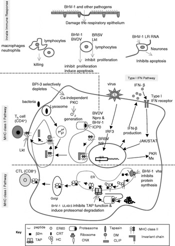
IFNs induce an anti-viral state and up-regulate immune functions by binding to type I or type II receptors on the cells, and activating the JAK/STAT signal transduction pathway (Darnell et al., Reference Darnell, Kerr and Stark1994). Type I IFNs (α and β) are produced by several cell types while type II IFN (γ) is produced primarily by T cells and NK cells. IFN-induced anti-viral state and up-regulation of immune functions are detrimental for the replication and dissemination of the viruses. Consequently, several viral pathogens have evolved strategies to interfere with IFN function (Fig. 1).
Immune evasion by pathogens of the bovine respiratory disease complex.

BHV-1 inhibits IFN-β-dependent transcription (Henderson et al., Reference Henderson, Zhang and Jones2005). The bovine infected cell protein 0 (bICP0) induces proteosome-dependent degradation of IFN response factor 3 (IRF3), a transcription factor essential for IFN promoter activation. The zinc RING finger and the sequences near the C-terminus of bICP0 are necessary for the inhibition of IRF-3-induced IFN-β promoter activity and IRF3 degradation (Saira et al., Reference Saira, Zhou and Jones2007). For more details, see the review on BHV-1 by Jones and Chowdhury in this issue).
Neither the noncytopathic (ncp) bovine viral diarrhea virus (BVDV) (Baigent et al., Reference Baigent, Zhang, Fray, Flick-Smith, Goodbourn and McCauley2002) nor the cytopathic (cp) BVDV (Gil et al., Reference Gil, Ansari, Vassilev, Liang, Lai, Zhong, Hong, Dubovi and Donis2006) infections induce detectable type I IFN responses in cell cultures to avoid stimulation of innate immune responses in cell culture (Chen et al., Reference Chen, Rijnbrand, Jangra, Devaraj, Qu, Ma, Lemon and Li2007). Antagonism of type I IFN induction in cells infected with BVDV is a function of the amino-terminal domain of BVDV NPRO (Gil et al., Reference Gil, Ansari, Vassilev, Liang, Lai, Zhong, Hong, Dubovi and Donis2006). BVDV NPRO inhibits production of type I IFN by specifically blocking the activity of IRF-3, preventing IRF-3 binding to DNA, enabling proteosomal degradation (Hilton et al., Reference Hilton, Moganeradj, Zhang, Chen, Randall, McCauley and Goodbourn2006).
Immunosuppression mediated by postnatal acute ncp BVDV infections is not associated with low IFN responses since experimental infections of calves with ncp BVDV results in type I IFN responses (Charleston et al., Reference Charleston, Fray, Baigent, Carr and Morrison2001). When type I IFN is released from virus-infected cells it transmits signals to remote cells via a receptor complex to induce an antiviral response by activating a number of genes. Among those genes activated are double-stranded RNA (dsRNA)-dependent protein kinase, 2′-5′-oligoadenylate synthetase and the Mx proteins. Mx proteins are induced specifically by type I IFN and exert an antiviral effect by targeting specific steps of the viral replication cycle. Mx proteins have been used as a marker for endogenous type I IFN in clinical studies (Muller-Doblies et al., Reference Muller-Doblies, Ackermann and Metzler2002). White blood cells of calves infected with ncp BVDV have elevated levels of Mx protein (Muller-Doblies et al., Reference Muller-Doblies, Arquaint, Schaller, Heegard, Hilbe, Albini, Abril, Tobler, Ehrensperger, Peterhans, Ackermann and Metzler2004). Conversely, fetal infections (60 days of gestation) with ncp or cp BVDV isolates result in elevated type I IFN production only in fetuses infected with cp BVDV. Foetuses infected with ncp BVDV produce only minimal Mx protein, suggesting that inhibition of type I IFN induction in infected fetal cells mediates establishment of persistent infection with ncp BVDV in cattle (Charleston et al., Reference Charleston, Fray, Baigent, Carr and Morrison2001).
Bovine respiratory syncytial virus (BRSV) is a weak inducer of type I IFN and is resistant to the antiviral effects of type I IFN. BRSV non-structural proteins NS1 and NS2 mediate virus escape from cellular antiviral mechanisms induced by type I IFN. Both proteins are required for this function; alone neither protein has substantial IFN antagonistic activity (Schlender et al., Reference Schlender, Bossert, Buchholz and Conzelmann2000). NS1 and NS2 also prevent induction of IFN-β after virus infection or dsRNA stimulation. NS protein inhibition of IFN induction is mediated by inhibition of IRF-3 activation (Bossert et al., Reference Bossert, Marozin and Conzelmann2003). Block of induction of type I IFN by NS proteins correlates with increased virulence of BRSV (Valarcher et al., Reference Valarcher, Furze, Wyld, Cook, Conzelmann and Taylor2003).
Regulation of apoptosis
Virus-infected cells trigger the programmed cell death or apoptotic pathways in order to prevent the replication and spread of the viral infection. In response, several viruses have evolved to encode viral proteins that inhibit apoptosis to keep their host cells long enough for replication and production of progeny virus. BHV-1 infects the epithelial cells of the upper respiratory tract of cattle. It induces apoptosis of infected epithelial cells near the end of productive infection in order to ensure efficient virus release (Devireddy and Jones, Reference Devireddy and Jones1999). However, BHV-1 undergoes latent infection in sensory neurons. In these cells, the latency-related (LR) RNA of BHV-1 is abundantly expressed (Geiser and Jones, Reference Geiser and Jones2005). The LR gene products inhibit apoptosis of latently infected cells in order for the virus to remain in these cells until subsequent reactivation, productive infection, and spread to other animals (For more details, see the review on BHV-1 by Jones and Chowdhury in this issue).
Evasion of adaptive immune response
Infection of leukocytes and suppression of proliferation
Antigen recognition and proliferation of the lymphocytes and the accessory cells of the immune system is a critical step in the orchestration of an effective immune response. The B and T lymphocytes have to undergo clonal expansion before they differentiate into effector cells that can clear the infection (Janeway and Travers, Reference Janeway and Travers2005). Hence several pathogens directly infect the cells of the immune system and/or affect the proliferative response of these cells, as means of immunosuppression.
Suppression of proliferation of leukocytes
BHV-1 can infect many different cell types in cattle, but the number of cells infected is very small, and productive infection does not occur in most cells (Carter et al. Reference Carter, Weinberg, Pollard, Reeves, Magnuson and Magnuson1989). Viral genes could be expressed in the absence of cytolysis. Although the infected cells survive, their activity is affected. Peripheral blood mononuclear cells (PBMCs) from cattle infected with BHV-1 do not respond to mitogen stimulation as much as those from un-infected cattle, and additional exogenous IL-2 does not eliminate the suppressive effects of the virus (Hutchings et al., Reference Hutchings, Campos, Qualtiere and Babiuk1990). In vitro, BHV-1 suppresses mitogen-induced proliferation of lymphocytes. It has been proposed that BHV-1 infection of lymphocytes contributes to immunosuppression more than the other cell types, and the immunosuppression could be caused by more than one mechanism (Carter et al., Reference Carter, Weinberg, Pollard, Reeves, Magnuson and Magnuson1989). Later in vitro studies which examined the proliferative response to antigen or IL-2 also confirmed the BHV-1-induced suppression of proliferative response of PBMCs (Hutchings et al., Reference Hutchings, Campos, Qualtiere and Babiuk1990). But in these studies inactivated virus also induced suppression of proliferation, unlike in the earlier study which reported the requirement of live virus (Carter et al., Reference Carter, Weinberg, Pollard, Reeves, Magnuson and Magnuson1989).
Immunosuppression due to BVDV infection could be related to tropism of virus for APCs (Brackenbury et al., Reference Brackenbury, Carr and Charleston2003). ncp BVDV infection of monocytes causes reduction in the proliferation of memory CD4+ T cells when mixed with these APCs (Glew et al., Reference Glew, Carr, Blackenbury, Hope, Charleston and Howard2003). cp BVDV infection of monocytes in vitro results in cell death, while dendritic cell viability is not affected by cp BVDV infection (Glew et al., Reference Glew, Carr, Blackenbury, Hope, Charleston and Howard2003). A difference in kinetics of the development of a specific T cell response after ncp and cp BVDV infection is evident. T cell proliferative responses are not reproducibly detectable until about 6–8 weeks after ncp BVDV infection, but are detectable by 3–4 weeks after infection with cp BVDV (Collen and Morrison, Reference Collen and Morrison2000; Brackenbury et al., Reference Brackenbury, Carr and Charleston2003).
Experimental BRSV infection of calves and lambs is also associated with reduction in proliferation of lymphocytes stimulated with mitogens (Sharma and Woldehiwet, Reference Sharma and Woldehiwet1991; Keles et al., Reference Keles, Woldehiwet and Murray1998). Depression of lymphocyte responses to the mitogen phytohaemagglutinin in lambs experimentally infected with BRSV is associated with CD4+ lymphocytes. BRSV F protein mediates inhibition of mitogen-induced T-cell proliferation in vitro (Schlender et al., Reference Schlender, Walliser, Fricke and Conzelmann2002).
M. haemolytica culture supernatant containing leukotoxin in sub-lytic concentrations inhibits lymphocyte blastogenesis in response to mitogens (Majury and Shewen, Reference Majury and Shewen1991a; Czuprynski and Ortiz-Carranza, Reference Czuprynski and Ortiz-Carranza1992). Polyclonal and monoclonal antibodies to leukotoxin abrogates the inhibition confirming that the inhibition is caused by leukotoxin. B lymphocytes appear to be more sensitive to the inhibitory effects than the T lymphocytes. Addition of exogenous IL-1 or IL-2 reduces the inhibitory effect of leukotoxin (Majury and Shewen, Reference Majury and Shewen1991b). However, the mechanism(s) by which leukotoxin inhibits lymphocyte proliferation is not clear.
M. bovis also suppresses the proliferative response of peripheral blood lymphocytes to mitogens (Thomas et al., Reference Thomas, Mettler, Sharp, Jensen-Kostenbader and Schultz1990). It can arrest the synthesis of DNA in cells that are already stimulated by the mitogen. Furthermore, a peptide produced by M. bovis that inhibits naïve lymphocyte activation has been identified (Vanden Bush and Rosenbusch, Reference Vanden Bush and Rosenbusch2002). This peptide has been shown to be expressed during M. bovis infection of bovine lungs which suggests that this lympho-inhibitory peptide may function as an immune evasion molecule.
M. bovis has also been reported by Vanden Bush and Rosenbusch (Reference Vanden Bush and Rosenbusch2002) to induce death of lymphocytes by apoptosis in vitro. M. bovis is a pathogen associated with respiratory disease as well as arthritis. In arthritis, there is mass lymphocyte infiltration and non-specific activation, and not lymphocyte death. Hence these authors proposed that M. bovis may not induce apoptosis of all lymphocytes in vivo. Further research is necessary to clarify this issue.
Infection of cells of the immune system
During acute BVDV infections, virus is present in peripheral blood, bone marrow and lymphoid tissues. Systemic distribution of extracellular virus and virus in association with dendritic cells and leukocytes occur in the lymphatics and blood vessels (Marshal et al., Reference Marshal, Moxley and Kelling1996; Brodersen and Kelling, Reference Brodersen and Kelling1998; Walz et al., Reference Walz, Bell, Steficek, Kaiser, Maes and Baker1999; Kelling et al., Reference Kelling, Steffen, Topliff, Eskridge, Donis and Higuchi2002). Leukopenia ensues and CTLs (CD8+) and Th cells (CD4+) are decreased. There is depletion of both CD4+ and CD8+ T-lymphocytes in the thymus (Brodersen and Kelling, Reference Brodersen and Kelling1999).
Both cp and ncp BVDV isolates induce apoptosis of T and B cells in vivo (Chase et al., Reference Chase, Elmowalid and Yousif2004). Those lymphotropic effects are marked in calves experimentally infected with ncp BVDV type 2, strain 890, resulting in cavitation in the Peyer's patches and moderate to marked thymic atrophy. Inflammation, necrosis, marked lymphocytolysis and marked to severe lymphoid depletion due to apoptosis is prominent in lymphoid follicles of Peyer's patch and to a lesser extent in thymic cortex. BVDV antigens are deposited in high concentrations in lymphatic tissues. The distribution and deposition of antigen correlate with lesions in the tissues, with most marked antigen deposition in Peyer's patches, followed by the mesenteric lymph nodes, lymphatic tissue in the proximal portion of the colon and the thymic cortex. BVDV antigen is localized in dendritic cells and submucosal macrophages adjacent to positive lymphoid nodules. Deposition of BVDV antigen is especially prominent in dendritic cells in thymuses of infected calves (Kelling et al., Reference Kelling, Hunsaker, Steffen, Topliff and Eskridge2007). These observations are relevant since germinal centers and thymic cortex are sites of B- and T-lymphocyte proliferation (Morrison et al., Reference Morrison, Lalor, Christensen and Morrison1986; Parsons et al., Reference Parsons, Howard and Jones1989). In general, calves experimentally infected with ncp BVDV type 1, NY-1 strain, develop similar, but less pronounced, microscopic lesions in lymphatic tissues compared to those in calves infected with BVDV type 2 (Kelling et al., Reference Kelling, Hunsaker, Steffen, Topliff, Abdelmagid and Eskridge2005).
The immunosuppressive effects of BVDV infection are evident when calves experimentally co-infected with BRSV and NY-1 BVDV develop more severe respiratory tract disease compared to calves infected with either virus alone. Co-infected calves develop more extensive pneumonia characterized by caudodorsal as well as cranioventral interlobular edema, emphysema and bronchopneumonia in caudal lung lobes. In contrast, calves infected with BRSV alone develop bronchopneumonia limited to cranioventral distribution (Brodersen and Kelling, Reference Brodersen and Kelling1998). Subpopulations of lymphocytes are more markedly altered in peripheral blood and lymphoid tissues of co-infected calves. Co-infected calves have reduced percentage of CD8+ lymphocytes and CD4+ lymphocytes in the thymus and Peyer's patches (Brodersen and Kelling, Reference Brodersen and Kelling1999).
Induction of humoral and cellular immune tolerance
Fetuses infected with ncp BVDV during the first four months of pregnancy, which is prior to development of fetal lymphoid tissues and prior to development of a functional acquired immune response, may develop immunotolerance, and persistent viremia (McClurkin et al., Reference McClurkin, Littledike, Cutlip, Frank, Coria and Bolin1984). The source of virus that infects the fetus is from viremia of the pregnant dam, stemming from either a persistent or an acute infection with ncp BVDV. The ability to cause fetal infections is exclusively a biotype-specific property of ncp BVDV. Viremia and transplacental transmission of cp BVDV following experimental nasal exposure of pregnant cows with cp BVDV is rare compared to the high occurrence of maternal viremia and fetal infection observed with ncp BVDV (Harding et al., Reference Harding, Cao, Shams, Johnson, Vassilev, Gil, Wheeler, Haines, Siebert, Nelson, Campos and Donis2002).
Evasion of innate immunity in the fetus by inhibition of IFN I synthesis following ncp BVDV infection is a biotype-specific mechanism associated with establishment of immunotolerance. When fetuses are experimentally infected early in gestation (60 days of gestation) with ncp and cp BVDV, elevated IFN I production occurs only in fetuses infected with cp BVDV. Fetuses infected with ncp BVDV produce only minimal Mx protein, suggesting that inhibition of IFN I induction in infected fetal cells may enable establishment of persistent ncp BVDV infection in the early fetus (Charleston et al., Reference Charleston, Fray, Baigent, Carr and Morrison2001). Since a functional fetal adaptive immune response does not occur at this time, virus replicates unrestricted in fetal tissues (Swasdipan et al., Reference Swasdipan, McGowan, Phillips and Bielefeldt-Ohmann2002); calves are born immunotolerant and persistently infected with the virus (McClurkin et al., Reference McClurkin, Littledike, Cutlip, Frank, Coria and Bolin1984). Persistently-infected calves do not develop an acquired immune response to BVDV. The non-responsive character of persistently infected animals is attributed to tolerance of CD4+ cells (Collen et al., Reference Collen, Douglas, Paton, Zhan and Morrison2000).
Interference with humoral immune response
Down-regulation of MHC class II molecules
Antigen presentation by MHC class II molecules is critical for activation of Th cells which provide help for B cells (as well as effector T cells). The processing of extracellular antigens, and presentation by MHC class II molecules is referred to as the exogenous antigen presentation pathway (Weenink and Gautam Reference Weenink and Gautam1997). The antigens acquired into endosomal/lysosomal compartment by phagocytosis or receptor-mediated endocytosis are degraded to minimal peptides by acid-dependent proteases. The class II α and β chains are co-translationally translocated into the endoplasmic reticulum (ER), and folded with the assistance of ER chaperones. The loading of class II molecules with peptides in the ER is precluded by the binding of the invariant chain (Ii) which forms a complex with α and β chains, and targets the complex to the endosomal/lysosomal compartment. The Ii is then degraded except for an oligopeptide known as the class II associated Ii peptide (CLIP), the region of Ii that is bound in the peptide-binding groove. The DM molecules assist enzymatically in the removal of CLIP and the subsequent loading of cognate peptides, in a specialized class II loading compartment called MIIC. The function of DM is regulated by DO molecules. Once loaded with the peptide, the class II molecules are transported through the vesicular transport pathway and expressed on the cell surface (Fig. 1).
Leukotoxin secreted by M. haemolytica has been reported to down-regulate the expression of class II molecules on bovine monocyte-derived macrophages (Hughes et al., Reference Hughes, Campos, McDougall, Beskorwayne, Potter and Babiuk1994). In this study, down-regulation of class II expression was caused by both crude authentic leukotoxin as well as recombinant inactive LktA. By 14 h after incubation with leukotoxin, a noticeable decrease in the percentage of cells expressing class II molecules was observed, and the effect was more pronounced by 24 h post-incubation (31%). However, leukotoxin had no effect on class II expression on cells treated with IFN-γ, prompting the authors to state that leukotoxin affects the constitutive expression, but not IFN-γ-induced expression of class II molecules. Furthermore, based on the results of a mixed lymphocyte reaction, these authors stated that leukotoxin affects the functional capacity of macrophages to present antigen. Expression of class II molecules on B cells were reported to be un-affected by leukotoxin. These authors proposed that leukotoxin-induced down-regulation may not induce a generalized state of immunosuppression, but may affect antigen presentation enabling the bacterium to colonize the site of infection more efficiently. Unfortunately, these important observations regarding the down-regulation of MHC class II expression by leukotoxin have not been extended by these authors or confirmed by any others up to now. It is possible that the observed effects are unique to the class II alleles from the two animals tested by Hughes et al. (Reference Hughes, Campos, McDougall, Beskorwayne, Potter and Babiuk1994). MHC class I allele-specific down-regulation has been reported for pseudorabies virus (Sparks-Thissen and Enquist, Reference Sparks-Thissen and Enquist1999). If the expression of class II molecules is down-regulated, activation of Th cells would be affected which in turn would affect the development of humoral as well as cell-mediated immune responses. In this regard, not only the antibody response to leukotoxin but also to all other antigens from other pathogens of BRDC would be affected. It would also affect the development of CTL response to the viral pathogens of BRDC.
Inhibition of antibody activity
Several immunoglobulin-binding proteins have been identified in H. somni (Corbeil et al., Reference Corbeil, Bastida-Corcuera and Beveridge1997; Bastida-Corcuera et al., Reference Bastida-Corcuera, Nielsen and Corbeil1999). It has been suggested that these proteins, which bind IgG2 in a non-antigen-specific manner, may block the binding of specific antibodies to H. somni. Additional investigations are needed to identify the precise mechanism of action of these immunoglobulin-binding proteins, and their role in conferring serum resistance.
Proteases secreted by an isolate of P. multocida from an apparently healthy calf have been shown to degrade IgG in vitro (Negrete-Abascal et al., Reference Negrete-Abascal, Tenorio and de la Garza1999). Based on this observation, it has been speculated that proteases might play an important role during tissue colonization by P. multocida. It remains to be seen whether these proteases in deed function as immune evasion molecules in P. multocida infection of the bovine respiratory tract.
Interference with cellular immune response
Interference with T-helper cell function: induction of apoptosis of T-helper cells
Recognition of peptides presented by MHC class II molecules on APCs by the CD4+ Th cells results in the production by these cells of cytokines required by macrophages, NK cells, B cells, CD8+ CTLs and other proliferating T cells. Therefore, depletion of CD4 molecules or selective elimination of CD4+ T cells would result in impaired Th function and cytokine signaling which in turn would compromise the development of humoral as well as cell-mediated immune responses. BHV-1 infects cultured CD4+ T cells, induces the loss of CD4 molecules, and finally induces apoptotic death of these cells (Eskra and Splitter, Reference Eskra and Splitter1997). These in vitro observations have been validated by in vivo experiments (Winkler et al., Reference Winkler, Doster and Jones1999). During acute BHV-1 infection of cattle, apoptosis of CD4+ T lymphocytes occurs in peripheral blood and lymph nodes (For more details, see the review on BHV-1 by Jones and Chowdhury in this issue).
Interference with cytotoxic T-lymphocyte response
Neutralizing antibodies at the portal of entry are perhaps the most effective component of anti-viral immunity, since they can neutralize the virus and prevent their entry into the host. However, once the viruses breach this line of defense and enter the host cells, cell-mediated immune response is critical for the defense against most viral infections. This is particularly true with herpesviral infections in which spread of the virus from cell-to-cell occurs earlier than the haematogenous spread (Lodmell et al., Reference Lodmell, Niwa, Hayashi and Notkins1973). The primary component of the cell-mediated immune response that is critical for elimination of virus-infected cells is the CTL. The CTLs recognize intracellular viral antigens presented by the self-MHC class I molecules on the cell surface (Zinkernagel and Doherty, Reference Zinkernagel and Doherty1974). Efficient CTL response to a viral infection is the result of a highly intricate multiple-step pathway of antigen processing and presentation involving several intracellular organelles and chaperones. Several viruses including BHV-1 have evolved strategies to interfere at different steps of this pathway to down-regulate expression of MHC class I molecules as a means to escape CTL recognition (reviewed in Yewdell and Hill, Reference Yewdell and Hill2002; Hewitt, Reference Hewitt2003).
Down-regulation of expression of MHC class I molecules
A brief overview of class I antigen presentation pathway is helpful for the discussion of the strategies evolved by BHV-1 to down-regulate class I expression. MHC class I molecules are expressed at different levels by all the nucleated cells in the body (Singer and Maguire, Reference Singer and Maguire1990). MHC class I molecules consist of a polymorphic heavy (H) or α chain and a non-polymorphic light chain, β2-microglobulin (β2m). The α1 and α2 domains of the H chain form the membrane-distal peptide-binding groove that accommodates a peptide of 8–10 amino acids (Lehner and Trowsdale, Reference Lehner and Trowsdale1998). The two polypeptides as well as the short peptide are necessary for the stable expression of the trimolecular complex on the cell surface under physiological conditions. The class I H chain and β2m are co-translationally translocated into the lumen of ER where they associate with the chaperones calnexin, calreticulin and ER60, which help in the proper folding and assembly of the H chain and β2m (Williams and Watts, Reference Williams and Watts1995). The assembled class I molecules present peptides originating from proteins synthesized in the cytoplasm. These proteins include constitutively expressed cellular proteins and those from replicating pathogens or arising out of cellular dysregulation. The proteins are generally degraded by the proteasomes, following ubiquitination of the target proteins (Tanaka et al., Reference Tanaka, Tanahashi, Tsurumi, Yokota and Shimbara1997). The generation of peptides may be qualitatively altered by IFN-γ secreted into local environments during an immune response. The peptides are transported into the ER, in the presence of ATP, by the heterodimeric transporter associated with antigen processing (TAP) consisting of TAP1 and TAP2 subunits (Momburg and Hammerling, Reference Momburg and Hammerling1998).
The loading of the peptides onto class I molecules occurs in a complex involving TAP, calreticulin (CRT), ER60 and tapasin. Tapasin stabilizes the TAP molecules and holds the assembly complex together (Grandea et al., Reference Grandea, Lehner, Cresswell and Spies1997; Ortmann et al., Reference Ortmann, Copeman, Lehner, Sadasivan, Herberg, Grandea, Riddell, Tampe, Spies, Towsdale and Cresswell1997). Each TAP1 subunit binds four molecules of tapasin, each of which binds one class I heterodimer. While the N-terminal domain of tapasin interacts with CRT and ER60, and is required for stable association of CRT and MHC class I, the C-terminal domain binds TAP. The trimolecular complex (H/β2m/peptide) egresses from the ER, and translocates to the cell membrane via the secretory pathway. The interaction between the TCR and MHC class I molecule is very short-lived, and is strengthened by the binding of CD8 molecule to the α3 domain of the H chain (Garcia et al., Reference Garcia, Scott, Brunmark, Carbone, Peterson, Wilson and Teyton1996).
In cells infected with a virus, the viral proteins are processed in the same manner as the cellular peptides, and the viral peptides are presented by the class I molecules on the surface of the infected cells. The class I molecules presenting the viral peptides signal the virus-specific CTLs to recognize and lyse the virus-infected cell, which is detrimental for the infecting viral pathogen. Therefore, several viruses have evolved strategies to interfere with the synthesis of class I molecules, generation and transport of the peptides into the ER, assembly of the trimolecular complex (H/β2m/peptide), and transport via the Golgi apparatus, as a means to impair class I expression on the cell surface, and escape from the immune surveillance of CTLS (Fig. 1; Rinaldo, Reference Rinaldo1994; Yewdell and Hill, Reference Yewdell and Hill2002; Hewitt, Reference Hewitt2003).
BHV-1 down-regulates the expression of MHC class I molecules on infected cells (Hariharan et al., Reference Hariharan, Nataraj and Srikumaran1993; Nataraj et al., Reference Nataraj, Eidmann, Hariharan, Sur, Perry and Srikumaran1997). Interestingly, modified live virus strains of BHV-1 also cause the down-regulation. The down-regulation is specific for class I molecules since class II expression remains un-affected. The reduction in cell surface expression of class I molecules occurs as early as 8 h post-infection, and reaches significant levels by 12 h post-infection. An multiplicity of infection as low as 0.1 could induce the down-regulation. Pulse-chase analysis experiments revealed that synthesis of the class I H chain as well as assembly/transport of class I molecules are affected by BHV-1 infection. Experiments with metabolic inhibitors revealed that an immediate-early/early protein of BHV-1 may be responsible for the down-regulation (Nataraj et al., Reference Nataraj, Eidmann, Hariharan, Sur, Perry and Srikumaran1997). Subsequent studies employed peptide transport assays, and confirmed that BHV-1 interferes with the TAP (Hinkley et al., Reference Hinkley, Hill and Srikumaran1998). BHV-1-induced inhibition of the transport of peptides occurs as early as 2 h post-infection, and is most pronounced during 2–4 h post-infection (65%). TAP-inhibition occurs in a dose-dependent manner. However, inhibition of TAP is not complete, even in later stages of infection and at high input moi, which is consistent with the earlier finding that the down-regulation of class I molecules on the surface is pronounced, but not complete (Nataraj et al., Reference Nataraj, Eidmann, Hariharan, Sur, Perry and Srikumaran1997). Results from the studies conducted with BHV-1 in a human melanoma cell line (Koppers-Lalic et al., Reference Koppers-Lalic, Rychlowski, van Leeuwen, Rijsewijk, Ressing, Neefjes, Bienkowska-Szewczyk and Wiertz2003) confirmed the findings with bovine cells described above.
Which of the BHV-1 proteins is responsible for inhibition of TAP function, and what is the mechanism(s) by which it is accomplished? Again, by using a human melanoma cell-line susceptible to BHV-1 infection, the protein encoded by BHV-1 UL49.5 has been identified as the protein responsible for TAP inhibition (Koppers-Lalic et al., Reference Koppers-Lalic, Reits, Ressing, Lipinska, Abele, Koch, Marcondes Rezende, Admiraal, van Leeuwen, Bienkowska-Szewczyk, Mettenleiter, Rijsewijk, Tampe, Neefjes and Wiertz2005). It is the BHV-1 homologue of the highly conserved glycoprotein gN encoded by all herpesviruses (Davison et al., Reference Davison, Dargan and Stow2002). The UL49.5 protein is a 9 kD type 1 transmembrane protein made up of 75 amino acids. Inactivation of TAP by UL49.5 is by a two-stage process: inhibition of peptide transport and degradation of TAP by proteasomes. The current model proposes that the interaction of ER luminal domain and transmembrane domain of UL49.5 with the TAP complex inhibits the conformational change that is necessary for the peptide-induced ATP-dependent translocation reaction. The cytosolic tail of UL49.5 links the TAP heterodimer–UL49.5 complex to the ubiquitin proteasome pathway for degradation (Koppers-Lalic et al., Reference Koppers-Lalic, Reits, Ressing, Lipinska, Abele, Koch, Marcondes Rezende, Admiraal, van Leeuwen, Bienkowska-Szewczyk, Mettenleiter, Rijsewijk, Tampe, Neefjes and Wiertz2005). UL49.5 is degraded along with TAP. UL49.5-induced TAP inhibition and the consequent down-regulation of cell surface class I expression strongly reduces antigen presentation to CD8+ CTLs (Koppers-Lalic et al., Reference Koppers-Lalic, Reits, Ressing, Lipinska, Abele, Koch, Marcondes Rezende, Admiraal, van Leeuwen, Bienkowska-Szewczyk, Mettenleiter, Rijsewijk, Tampe, Neefjes and Wiertz2005). This has not been shown with BHV-1-infected bovine cells and BHV-1-specific CTLs, but with UL49.5 transfectant human cell-lines and well-defined CTL clones specific for the minor histocompatibility complex antigens HA-3 and HY, respectively. Nevertheless, these studies very elegantly established BHV-1 UL49.5 as a powerful immune evasion molecule. Down-regulation of MHC class I expression by BHV-1 UL49.5 compromises the development of a robust CTL response against not only BHV-1, but also the other viral pathogens of BRDC.
Shut-off of host cell protein synthesis
Alphaherpesviruses encode a structural protein which induces the rapid shut-off of host cell protein synthesis (Smibert et al., Reference Smibert, Johnson and Smiley1992; Elgadi et al., Reference Elgadi, Hayes and Smiley1999). This protein is known as virion host shut-off (vhs) protein, and it inhibits host cell protein synthesis by inducing degradation of cellular mRNAs (Elgadi et al., Reference Elgadi, Hayes and Smiley1999). BHV-1 is no exception to this phenomenon exhibited by other alphaherpesviruses. Vhs-induced inhibition of host cellular protein synthesis occurs in BHV-1-infected cells (Hinkley et al., Reference Hinkley, Ambagala, Jones and Srikumaran2000), and the UL41 gene product is necessary and sufficient for shut-off of host cell protein synthesis at early times post-infection (Koppers-Lalic et al., Reference Koppers-Lalic, Rijsewijk, Verschuren, van Gaans-Van den Brink, Neisig, Ressing, Neefjes and Wiertz2001). Northern blot analysis reveals significant down-regulation of mRNA for class I molecules also, which raises the question as to whether the BHV-1-induced down-regulation of cell surface expression of class I molecules is due to the activity of the vhs protein. Studies with vhs-deletion mutants of BHV-1 provides the answer to this question (Koppers-Lalic et al., Reference Koppers-Lalic, Rijsewijk, Verschuren, van Gaans-Van den Brink, Neisig, Ressing, Neefjes and Wiertz2001; Gopinath et al., Reference Gopinath, Ambagala, Hinkley and Srikumaran2002). The reduction in the synthesis of class I molecules is due to the vhs activity. However, the TAP inhibition and retention of class I molecules in ER by BHV-1 is mediated by a protein other than the vhs which has been shown to be UL49.5 (Koppers-Lalic et al., Reference Koppers-Lalic, Reits, Ressing, Lipinska, Abele, Koch, Marcondes Rezende, Admiraal, van Leeuwen, Bienkowska-Szewczyk, Mettenleiter, Rijsewijk, Tampe, Neefjes and Wiertz2005).
Strategies pertaining to the pathogens
Latent and persistent infections
Latent BHV-1 infection
The reader is referred to the review on BHV-1 by Jones and Chowdhury in this issue.
Persistent BVDV infections
Persistently infected calves, born as a consequence of fetal infection and development of immunotolerance, are viremic and shed virus continuously. During a persistent infection, BVDV replicates in all major lymphocyte subpopulations, as well as in the accessory cells resulting in alterations of the immune responses (Howard, Reference Howard1990). Lymphocytes from cattle persistently infected with BVDV have decreased blastogenesis in response to mitogens, such as phytohaemagglutinin, concanavalin-A and pokeweed (Brown et al., Reference Brown, Bolin, Frank and Roth1991), while distribution of leukocyte subsets in persistently infected cattle are not significantly different from BVDV-free animals (Brewoo et al., Reference Brewoo, Haase, Sharp and Schulz2007). Likely major sources of infectious virus from persistently infected cattle are serous secretions of the nasal cavity, productive viral infection of epithelium and infected leukocytes in respiratory secretions (Confer et al., Reference Confer, Fulton, Step, Johnson and Ridpath2005). Persistently infected calves shed virus continuously and serve as a reservoir of viral infection for other cattle. The presence of persistently infected cattle in feedyards is associated with an increase in the incidence of respiratory tract disease of in-contact cattle. In one study, 15.9% of initial respiratory tract disease events were attributed to exposure to a persistently infected animal (Loneragen et al., Reference Loneragen, Thomson, Montgomery, Mason and Larson2005).
Viremia is long term in persistently infected animals, but the level of viremia may decline with the development of neutralizing antibodies and become undetectable as the animal ages (Brock et al., Reference Brock, Grooms, Ridpath and Bolin1998). Deterioration of highly specific immunotolerance resulting in an adaptive immune response may result from development of antigenic-variant quasispecies viruses within the immunotolerant persistently infected animal (Collins et al., Reference Collins, Desport and Brownlie1999). This results in generation of a B cell response to the persisting virus resulting in antibody formation and viral clearance (Fray et al., Reference Fray, Supple, Morrison and Charleston2000).
Some persistently infected calves are stunted or weak at birth, have poor growth rates and die at a young age (Kelling et al., Reference Kelling, Stine, Rump and Parker1990). Others appear healthy and survive to maturity. The prevalence of persistently infected cattle is variable, however based on sampling of randomly selected herds, it is estimated that 3% of herds in the United States have persistently infected calves (Wittum et al., Reference Wittum, Grotelueschen, Brock, Kvasnicka, Floyd, Kelling and Odde2001). Frequently, persistently infected cattle have reduced growth rates, are more susceptible to common calfhood infections of mucosal surfaces including pneumonia and enteritis, and are at risk of developing mucosal disease (Brownlie et al., Reference Brownlie, Clarke and Howard1984; Bolin et al., Reference Bolin, McClurkin, Cutlip and Coria1985).
Antigenic variation
Several pathogens have evolved immune evasion mechanisms which enable them to rapidly change the structure of their surface membrane proteins. M. bovis has evolved such a mechanism. In vitro, the presence of antibodies from immunized or experimentally infected calves specific for membrane surface proteins leads to the repression of expression or shortening of the target protein(s) in M. bovis (Le Grand et al., Reference Le Grand, Solsona, Rosengarten and Poumarat1996). Expression of antigenically distinct variant protein has also been observed. Removal of the variation-inducing antibodies results in reversion to the original phenotype. It has been proposed that random selection of pre-existing variants resistant to antibody-mediated inhibition, or direct regulation of gene expression are strategies employed by M. bovis to evade host immune response.
H. somni undergoes structural and antigenic phase variation in its lipooligosaccharide (LOS) components (Inzana et al., Reference Inzana, Hensley, McOuiston, Lesse, Campagnari, Boyle and Apicella1997). The glycoses and phosphoryl choline of the LOS outer core undergo random phase variation. Phase variation in multiple components in the LOS enables H. somni to evade the host humoral immune response (Siddaramppa and Inzana Reference Siddaramppa and Inzana2004). Sialylation of terminal galactose residues of LOS is also believed to enhance resistance to complement-mediated bactericidal effects of anti-LOS antibodies (Inzana et al., Reference Inzana, Glindemann, Cox, Wakarchuk and Howard2002).
Concluding remarks
Several immune evasion/immunosuppressive strategies have been developed by the pathogens of BRDC (Fig. 1). The strategies developed by one pathogen not only help that pathogen, but also the others, resulting in exacerbation of the disease. For example, BHV-1 infection causes damage to the respiratory epithelium impairing the mucociliary defense mechanism which favors M. haemolytica to migrate to the lungs, proliferate, elaborate the virulence determinants and cause disease. Infection of the cells of the immune system by BVDV, inhibition of proliferation of the cells by BVDV, BHV-1, BRSV, and induction of apoptosis of CD4+ T cells by BHV-1, compromise the orchestration of the immune response against all the pathogens. Induction of apoptosis of CD4+ Th cells by BHV-1 impairs the elaboration of cytokines that promote the development of the effector elements of both humoral and cell-mediated immune responses. Down-regulation of MHC class I expression by BHV-1 compromises the development of CTL response against BHV-1 as well as BVDV, BRSV and PI-3. Cytolysis of all the subsets of leukocytes by M. haemolytica leukotoxin eliminates the cells that are critical for mounting the innate and adaptive immune responses. In addition, it results in the release of the toxic chemicals that cause acute damage to the pulmonary epithelium. Establishment of latent and persistent infections by BHV-1 and BVDV, and antigenic variation by M. bovis, add another dimension to immune evasion in BRDC. Thus BRDC is indeed a complex disease syndrome. The severity of the disease, and the rate of mortality in a herd very likely depend on the number of pathogens infecting the animals concurrently. Experimental co-infection of animals with BVDV/BRSV and BHV-1/M. haemolytica supports this notion. The complex etiology and the multitude of immune evasion strategies developed by these pathogens probably explain the failure of the vaccines to provide complete protection against this disease complex.