INTRODUCTION
Rhizocephala (Crustacea: Cirripedia) infest other Crustacea and have a life cycle that includes parasitic and free-living larval stages. At the parasitic stage, the organism consists of the external reproductive body, or externa, on the host body surface, and the nutrient-absorbing internal root system, or interna, embedded into the host haemolymph (Høeg, Reference Høeg, Harrison and Humes1992; Høeg & Lützen Reference Høeg and Lützen1993, Reference Høeg and Lützen1995; Walker, Reference Walker2001; Høeg et al., Reference Høeg, Glenner, Shields and Rohde2005; Glenner & Hebsgaard, Reference Glenner and Hebsgaard2006). An adult rhizocephalan parasite lacks such arthropod features as segmentation, appendages, digestive tract, sensory and excretory organs, i.e. the main morphological characteristics of arthropods are absent (Høeg & Lützen Reference Høeg and Lützen1993, Reference Høeg and Lützen1995; Høeg et al., Reference Høeg, Glenner, Shields and Rohde2005). Rhizocephalans have pelagic larvae, a series of nauplii and a cyprid that resemble the larvae of other Cirripedia to a remarkable degree (Høeg & Møller, Reference Høeg and Møller2006). The basic developmental pattern, as well as detailed larval morphology and molecular data, allow us to establish a phylogenetically based taxonomy within Cirripedia. Rhizocephala is a monophyletic taxon with a filter-feeding barnacle-like ancestor and a sister group to free-living Thoracica and Acrothoracica (Høeg & Lützen, Reference Høeg and Lützen1995; Martin & Davies, Reference Martin and Davies2001; Glenner et al., Reference Glenner, Lützen and Takahashi2003; Glenner & Hebsgaard, Reference Glenner and Hebsgaard2006).
The complicated rhizocephalan life cycle resulting from the parasitic mode of life leads to the development of colonial organization in some species of the families Peltogastridae, Sacculinidae and Duplorbidae, and in all representatives of Thompsoniidae and Polysaccidae (Høeg & Lützen, Reference Høeg and Lützen1995; Glenner et al., Reference Glenner, Lützen and Takahashi2003), although direct evidence of the colonial organization in rhizocephalans is fragmentary and has been obtained only for a limited number of species. The colonial organization in some rhizocephalan species emerges at the parasitic stage of their life cycle as a result of asexual reproduction by budding of the interna (root system) with the subsequent development of multiple externae (Pérez, Reference Pérez1931; Høeg & Lützen, Reference Høeg and Lützen1993, Reference Høeg and Lützen1995; Takahashi & Lützen, Reference Takahashi and Lützen1998; Lützen & Du, Reference Lützen and Du1999; Liu & Lützen, Reference Liu and Lützen2000; Glenner et al., Reference Glenner, Lützen and Takahashi2003); however, the budding was unambiguously demonstrated only in a few rhizocephalan species.
Careful dissection of internae under a microscope in the host haemolymph and the subsequent short-term culturing of the internae outside the host organism allowed us to examine the colonial interna organization, interna budding and morphogenesis of multiple externa rudiments in colonial rhizocephalans Polyascus polygenea, Peltogasterella gracilis and Thylacoplethus isaevae (Isaeva et al., Reference Isaeva, Rybakov and Kasyanov1999, Reference Isaeva, Shukalyuk, Trofimova, Korn and Rybakov2001, Reference Isaeva, Shukalyuk, Korn and Rybakov2004; Shukalyuk et al., Reference Shukalyuk, Baiborodin and Isaeva2001, Reference Shukalyuk, Isaeva, Kizilova and Baiborodin2005, Reference Shukalyuk, Golovnina, Baiborodin, Gunbin, Blinov and Isaeva2007; Rybakov & Shukalyuk, Reference Rybakov and Shukalyuk2004).
Many rhizocephalan species are characterized by a single externa on the host body surface. Thus, these species are considered non-colonial. In particular, the rhizocephalan Peltogaster reticulatus Shiiino, 1943, which infests the coastal hermit crab Pagurus proximus and belongs to the family Peltogastridae, usually forms a single externa on each host. We present here evidence of a hidden coloniality in Peltogaster reticulatus at the parasitic stage.
MATERIALS AND METHODS
The specimens of the rhizocephalan Peltogaster reticulatus Shiino, 1943 which we studied were collected at the Marine Biology Station ‘Vostok’ of the Institute of Marine Biology (Vostok Bay, Peter the Great Bay, Sea of Japan); 370 specimens of the hermit crab Pagurus proximus Komai were examined and some of them were found to be infested by P. reticulatus.
We used vital, histological and histochemical methods to visualize the general morphology of endoparasitic internae of P. reticulatus. As incubation medium for short-term in vitro culture of P. reticulatus internae we used the haemolymph of non-parasitized host crabs; the haemolymph was supplemented with antibiotics (100 U/ml penicillin, 100 µg/ml streptomycin and 100 µg/ml gentamycin).
During the histochemical analysis, we used a modified protocol for mammalian embryonic stem cells to reveal alkaline phosphatase activity (Isaeva et al., Reference Isaeva, Shukaljuk and Kizilova2003).
For histological analysis, samples of internae isolated from their host crabs were fixed with Bouin's fluid, embedded in paraffin; sections (5 µm) were stained with haematoxylin–eosin. For microscopy and photography of living internae and histological preparations an inverted microscope Telaval with photodevices mf-matik (Zeiss) and a microscope Polyvar with a digital camera Leica or Canon were used.
RESULTS
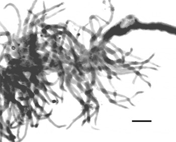
In a sample of 370 specimens of the hermit crab Pagurus proximus collected in September at Vostok Marine Biology Station, 77 specimens carried externae of P. reticulatus (20.8%), usually one externa on a host, rarely 2–3 or even 4 externae. A careful microdissection was performed to separate each interna of P. reticulatus with a single externa from host tissues as well as from the externa. The interna is emerald-green and comprises a few ‘lamp brushes’ of the morphology typical of the members of the genus Peltogaster, with numerous side branches arising from the axial trunk of each ‘brush’ (Figure 1).
A ‘lamp brush’ fragment from an interna of Peltogaster reticulatus. Scale bar: 50 µm.

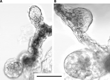
Short-term culture in vitro has made it possible to observe directly the living internae and to visualize the budding of stolon-like epithelial tubular structures filled with cells (Figure 2).
Budding of stolon-like structures in living internae of Peltogaster reticulatus. Scale bar: 50 µm.

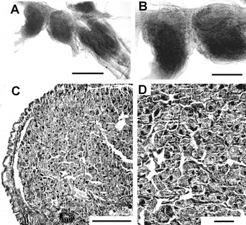
Histological study and histochemical detection of alkaline phosphatase activity showed that most of the studied specimens of P. reticulatus with a single mature externa had additional primordial externae or early buds; these rudiments often form groups of three or four primordial externae. Intense alkaline phosphatase activity was found in the primordial externae (Figure 3A, B).
Morphology of primordial externae of Peltogaster reticulatus: (A, B) histochemical detection of alkaline phosphatase activity; (C) a fragment of a primordial externa of P. reticulatus (histological section); (D) oocytes in the ovary (histological section). Scale bars: (A) 100 µm; (B, C) 50 µm; (D) 20 µm.

The intensity of cytochemical reaction of rhizocephalan cells in the primordia is comparable to that in embryonic stem cells of mice, used as a benchmark; the brick-red product of the cytochemical reaction is typical of the buds of P. reticulatus as well as cells of mouse embryonic stem cell (ESC) lines in vitro (Isaeva et al., Reference Isaeva, Shukaljuk and Kizilova2003; Shukalyuk et al., Reference Shukalyuk, Isaeva, Kizilova and Baiborodin2005).
Ovaries of the most developed primordial externae were filled with growing oocytes (Figure 3C, D), so the process of oogenesis begins in externa rudiments of P. reticulatus before the externae appear on the surface of host body.
DISCUSSION
Because an interna, connected to a mature externa, also integrates three or four additional primordial externae and several buds of earlier developmental stages in P. reticulatus, this species, which can be considered non-colonial based on the common presence of a single externa on each host, actually appears to have a hidden colonial organization. Primordial externae in the root system probably are capable of replacing the lost externa, particularly, as the result of host molting (Isaeva et al., Reference Isaeva, Dolganov and Shukalyuk2005).
The terms ‘externa’ and ‘interna’ were introduced by Yves Delage (Reference Delage1883) to distinguish the internal and external parts of Sacculina carcini. Høeg and coauthors prefer to use the term ‘root system’ instead of ‘interna’ (Bresciani & Høeg, Reference Bresciani and Høeg2001; Høeg et al., Reference Høeg, Glenner, Shields and Rohde2005). A dissection of the Peltogasterella sulcata (= Chlorogaster sulcatus) internae allowed researchers to visualize and schematize the system of several generations of multiple primordial externae (Pérez, Reference Pérez1931; Høeg & Lützen, Reference Høeg and Lützen1995). We distinguish in the root system (interna) of the colonial rhizocephalan Polyascus polygenea and Peltogasterella gracilis two differentiated systems, the trophic and the reproductive ones. We have observed inside each colonial interna of P. polygenea and P. gracilis numerous module elements of both reproductive and trophic systems at different stages of development. So the reproductive system manifests itself in each colony as multiple mature externae and more numerous primordial externae, several in P. polygenea and dozens in P. gracilis, belonging to successive generations (Isaeva et al., Reference Isaeva, Shukalyuk, Trofimova, Korn and Rybakov2001, Reference Isaeva, Shukalyuk, Korn and Rybakov2004; Shukalyuk et al., Reference Shukalyuk, Isaeva, Kizilova and Baiborodin2005, 2007). The trophic system fulfils the functions of absorption, digestion, storage and transport of nutrients. ‘Lamp brushes’ with the main trunk and side branches are typical for members of the family Peltogastridae (Høeg, Reference Høeg, Harrison and Humes1992; Høeg & Lützen, Reference Høeg and Lützen1995; Bresciani & Høeg, Reference Bresciani and Høeg2001), but the morphology of the root system varies in the genera Peltogaster, Peltogasterella and Septosaccus (Bresciani & Høeg, Reference Bresciani and Høeg2001). In colonial peltogastrids, each externa is supported by its own trophic module called ‘lamp brush’, or ‘bottled brush’ (Bresciani & Høeg, Reference Bresciani and Høeg2001). The relatively small, emerald-green interna of P. reticulatus also includes the trophic modules in the form of few lamp brushes. The green colour of the interna probably is dependent on the presence of dehydrobilirubin, similar to what is known about another peltogastrid species, Briarosaccus callosus (Meyers, Reference Meyers and Kinne1990). Thus, the organism of studied colonial rhizocephalans at the parasitic stage of the life cycle comprises a set of modular reproductive elements (mature and primordial externae) and trophic modules integrated into a colony.
The earliest primordia of future externae in the colonial rhizocephalans apparently arise as epithelial buds of stolon-like structures (Isaeva et al., Reference Isaeva, Shukalyuk, Korn and Rybakov2004; Shukalyuk et al., Reference Shukalyuk, Isaeva, Kizilova and Baiborodin2005). Budding stolons as long, slender epithelial tubes have been found in other colonial metazoans: Coelenterata, Bryozoa and Tunicata (Berrill, Reference Berrill1961). We consider the organization of colonial rhizocephalans using the same approach and the same terminology that has been adopted for other colonial invertebrates.
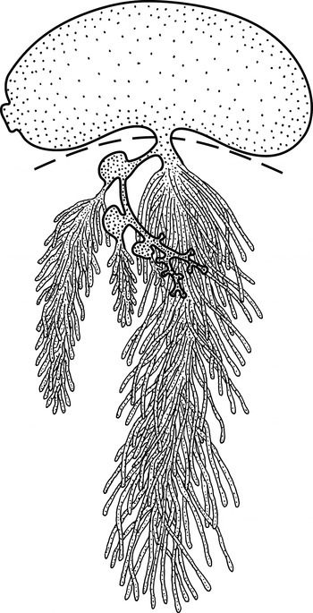
The hidden colonial organization of P. reticulatus, at parasitic stage of the life cycle is schematized in Figure 4.
A schematic sketch of hidden coloniality in Peltogaster reticulatus: the externa and the budding stolon are outlined by thick line and the trophic lamp brushes by thiner line; the host body wall as the boundary between externa and endoparasitic interna is shown by interrupted line.

The budding stolon including a few primordial externae and several early buds is evidence of asexual reproduction in P. reticulatus.
The budding of stolon-like epithelial stalks filled with migrating stem cells clearly takes place in the studied rhizocephalans (Isaeva et al., Reference Isaeva, Shukalyuk, Trofimova, Korn and Rybakov2001, Reference Isaeva, Shukalyuk, Korn and Rybakov2004; Rybakov & Shukalyuk, Reference Rybakov and Shukalyuk2004; Shulalyuk et al., Reference Shukalyuk, Isaeva, Kizilova and Baiborodin2005). The earliest primordia of future externae in the colonial rhizocephalans apparently arise as epithelial buds of stolon-like structures. Within each bud is a cluster of undifferentiated stem cells and such cells are also found inside the stolons (Isaeva et al., Reference Isaeva, Shukalyuk, Trofimova, Korn and Rybakov2001, Reference Isaeva, Shukaljuk and Kizilova2003, Reference Isaeva, Shukalyuk, Korn and Rybakov2004; Shukalyuk et al., Reference Shukalyuk, Isaeva, Kizilova and Baiborodin2005). An early bud has characteristic morphology: it is an epithelial bladder enveloping a mass of non-differentiated cells of the future visceral sac, the so-called ‘nucleus’. Early nuclei have been observed in non-colonial rhizocephalans (Bocquet-Védrine & Parent, Reference Bocquet-Védrine and Parent1972; Rubiliani et al., Reference Rubiliani, Turquier and Payen1982; Høeg, Reference Høeg1982; Høeg & Lützen, Reference Høeg and Lützen1995; Glenner, Reference Glenner2001). In the colonial P. polygenea, morphologically similar ‘nuclei have been described (Takahashi & Lützen, Reference Takahashi and Lützen1998; Isaeva et al., Reference Isaeva, Shukalyuk, Trofimova, Korn and Rybakov2001, Reference Isaeva, Shukalyuk, Korn and Rybakov2004; Lützen, Reference Lützen2002).
Smith (Reference Smith1906) and Potts (Reference Potts1915) were the first who suggested the ability of internae to bud in some rhizocephalan species forming multiple externae on a host. Thompsonia littoralis forms on one host up to 200 externae associated with a united root system; externae discarded during host moulting are able to reappear, apparently due to interna budding (Potts, Reference Potts1915; Lützen & Jespersen, Reference Lützen and Jespersen1992). The transitory appearance and following disappearance of multiple nuclei in early interna were described in the Sacculina carcini, which has just one adult externa (Rubiliani et al., Reference Rubiliani, Rubliani-Durozoi and Payen1980, 1982). As described for the non-colonial S. carcini, the cell mass of the early interna may be randomly divided into two parts, which leads to the formation of two externa sharing a common root system (Delage, Reference Delage1884; Smith, Reference Smith1906). Pérez (Reference Pérez1941) observed the appearance of ovaries in the root system of Peltogaster paguri following the discarding of the externa. There are additional primordial externae able to replace mature externa in case of loss (as a result of shedding or damage) in the interna of P. reticulatus with a single externa. It is likely that many or all members of such families such as Sacculinidae and Peltogastridae have a tendency to develop colonial organization. In species with one mature externa and several externa rudiments in the interna, the functioning externa, perhaps, has an inhibitory effect on the growth of other rudiments, and this effect can disappear in the case of loss of the dominant externa. We suppose the appearance of several additional primordial externae ensures the successful passage of the life cycle of the parasite.
The process of gonad differentiation was studied in only a few species of Rhizocephala; it was determined that the ovary develops from the cells of an early primordium (Bocquet-Védrine, Reference Bocquet-Védrine1961; Høeg, Reference Høeg1982). In the rhizocephalan Loxothylacus panopei, Glenner (Reference Glenner2001) traced cells in the primordial ovary of the future externa back to the vermigon, kentrogon and cypris larvae. Previously, we traced the differentiation of primordial externa in P. polygenea. It was shown that prior to the emergence of virgin externa of P. polygenea onto the body surface of the host crab it already has all differentiated tissue and organ systems including the ovary with oogonia and oocytes at all stages of oogenesis up to vitellogenesis (Isaeva et al., Reference Isaeva, Shukalyuk, Trofimova, Korn and Rybakov2001, Reference Isaeva, Shukalyuk, Korn and Rybakov2004). In P. reticulatus, an ovary in the primordial externa also contains growing oocytes; besides, the mantle, mantle cavity with mantle opening, male receptacles and oviduct are developed in each primordial externa.
Stem cells and whole buds in P. polygenea, P. gracilis and Th. isaevae selectively express alkaline phosphatase activity, the classical histochemical marker for mammalian embryonic stem and primary germ cells in vivo and in vitro. In stem cells of P. reticulatus the intensity of cytochemical reaction is comparable to that revealed under similar conditions in mouse embryonic stem cells in vitro, used as a benchmark. The brick-red product of the cytochemical reaction is typical for cells of mouse ESC lines as well as the stem cells of studied rhizocephalan species (Isaeva et al., Reference Isaeva, Shukaljuk and Kizilova2003, Reference Isaeva, Shukalyuk and Akhmadieva2008, Reference Isaeva, Akhmadieva, Aleksandrova and Shukalyuk2009; Shukalyuk et al., Reference Shukalyuk, Isaeva, Kizilova and Baiborodin2005). The cytochemical manifestation of alkaline phosphatase activity in stem cells of the colonial rhizocephalans was the first evidence of this activity in stem cells of invertebrates and a feature common to stem cells of animals belonging to such diverse taxa as mammals and crustaceans. So the classical method of identification of embryonic stem cells elaborated in mammals and other vertebrates is useful for identifying stem cells in different invertebrates (see Isaeva et al., Reference Isaeva, Shukaljuk and Kizilova2003, Reference Isaeva, Akhmadieva, Aleksandrova and Shukalyuk2009; Shukalyuk et al., Reference Shukalyuk, Isaeva, Kizilova and Baiborodin2005; Extavour, Reference Extavour, Minelli and Fusco2008; Sköld et al., Reference Sköld, Obst, Sköld, Åkesson, Rinkevich and Matranga2009).
The self-renewing pool of stem cells is the cellular basis of the reproductive strategy, including sexual and asexual reproduction, alternating in the life cycle of colonial rhizocephalans. Rhizocephalans are able to exploit a host for a long time (several years), controlling its morphology, physiology and behaviour, and using all resources of the host organism to produce a huge number of larvae. Blastogenesis and coloniality in the rhizocephalans involve a radical, evolutionary secondary change within ancestral reproductive biology due to the transition to parasitic life (Kasyanov et al., Reference Kasyanov, Korn and Rybakov1997). We believe that the longevity of the host hermit crab Pagurus proximus is between one and two years as well as in similar Pagurus species (McLay, Reference McLay1985; Mantelatto et al., Reference Mantelatto, Christofoletti and Valenti2005). The hosts had P. reticulatus externae during September, while during spring and summer we never observed the hosts with externae. Evidently, mature externae are lost in host moulting, and primordial externae are able to develop into externa ensuring successful reproduction of the parasitic barnacle during the following next year.
ACKNOWLEDGEMENT
This work was supported by the Russian Foundation for Basic Research (grant number 09-04-00019).