Introduction
After non-melanoma skin cancer, prostate cancer is the most commonly diagnosed cancer in New Zealand men, accounting for about a third of new diagnoses.Reference Ferlay, Soerjomataram and Ervik1 It is the leading cause of cancer death in this population, but if diagnosed and treated early it remains a curable disease. The role of radiotherapy in the treatment of prostate cancer is an established one. However, because of undesirable recurrence rates with conventional radiotherapy, there has been a move towards dose escalation over the years. Three-dimensional conformal radiotherapy and intensity-modulated radiotherapy (IMRT) are some of the techniques that have been used with good efficacy and acceptable toxicities.Reference Zelefsky, Yamada and Fuks2, Reference Zelefsky, Chan, Hunt, Yamada, Shippy and Amols3 However, recent clinical trials have continued supporting the use of high-dose-rate brachytherapy (HDRBT) as an effective alternative method of dose escalation.Reference Pollack, Zagars and Starkschall4–Reference Bachand, Martin, Beaulieu, Harel and Vigneault6 HDRBT has characteristics of highly accurate dose delivery and rapid dose fall-off, which allows for greater sparing of adjacent critical structures without significantly compromising the treatment effect. In addition, there is growing evidence of a radiobiological benefit based on the low α/β ratio of prostate cancer.Reference Williams, Taylor and Liu7 MortonReference Morton8 has declared prostate brachytherapy as the best method for dose escalation.
Despite its theoretical and practical advantages,Reference Morton8 there is a lack of clarity with regard to how HDRBT compares with other methods of dose escalation. Published randomised controlled trials have been criticised for using suboptimal doses in their external beam radiotherapy (EBRT) alone armsReference Hoskin, Motohashi, Bownes, Bryant and Ostler5, Reference Sathya, Davis and Julian9 and other studies have not used randomised cohorts.Reference Guix, Lacorte and Bartrina10
In 2006, Waikato Hospital became the first cancer centre in New Zealand to use HDRBT for men with prostate cancer as a form of dose escalation. In this study, we report data from a retrospective study of the first 137 patients who have been treated with EBRT and HDRBT boost between 2006 and 2010, outlining survival and late toxicity rates. Health-related quality of life and detailed sexual toxicity rates have been reported separately.Reference Conaglen, de Jong, Hartopeanu, Conaglen and Tyrie11
Methods and Materials
Waikato Hospital is a tertiary referral centre, including medical and radiation oncology services, serving a population of 360,000 people. All HDRBT treatment outcome and toxicity data were collected and recorded in patient and electronic charts. A specific prostate HDRBT database was also created. A total of 137 patients were included in the final analysis spanning from February 2006 to February 2010; follow-up was through to December 2013. All patients included in the analysis consented for data collection and research-related use of these data. Regional ethics committee approval was also obtained.
HDRBT patient selection and radiation technique
Eligibility criteria included adequate fitness for spinal anaesthesia and localised prostate cancer with intermediate- or high-risk features. Risk stratification was performed according to the National Comprehensive Cancer Network (NCCN)Reference Mohler, Bahnson and Boston12 guidelines. Patients with large prostates (>50mL) and/or significant lower urinary tract symptoms (International Prostate Symptom Score, IPSS, >12) were excluded. T stage was based on clinical and biopsy findings and upstaged if pelvic magnetic resonance imaging (MRI) showed more extensive disease. All patients who received HDRBT monotherapy or had inadequate follow-up (<9 months) were also excluded.
Some form of androgen-deprivation therapy (ADT) was administered to 59% (81/137) of the patients; this therapy was initiated in ten patients in the neoadjuvant setting for the purpose of cytoreduction to improve brachytherapy technical feasibility. Intermediate-risk patients were targeted to have 6 months and high-risk patients to have 36 months total duration of ADT.
HDRBT was delivered within 3 weeks of EBRT. All with the exception of two patients received 46Gy in 23 fractions of EBRT. The two exceptions received 35·75Gy in 13 fractionsReference Hoskin, Motohashi, Bownes, Bryant and Ostler5 for logistical reasons. A three-dimensional conformal radiotherapy technique was utilised with computed tomography (CT) planning. A standard five-field arrangement was typically used with 18MV beams.
All patients had brachytherapy delivered via one implant using the remote afterload method. Each patient had two fractions of 9·5Gy (total 19Gy) delivered at least 6 hours apart on the same day. Normal-tissue tolerances were set for the urethra and rectum as per the Radiation Therapy Oncology Group protocol.Reference Hsu, Bae and Shinohara13 The volume of urethra receiving 125% of the prescription dose was kept below 1mL (V125<1mL), and the volume of rectum receiving 75% of the prescription dose was kept below 1mL (V75<1mL). Patients were typically admitted to the hospital one day before the delivery of brachytherapy to undergo bowel preparation. All patients received a spinal anaesthetic and had indwelling urinary catheter (IDUC) insertions. Transperineal placement of catheters was performed with a reference template in situ. The patients then went for a radiation planning CT scan with subsequent target and critical organ delineation. Following dose delivery of the first fraction, CT scan and planning were repeated. Needle positions were adjusted as required before delivery of the second fraction. Following completion of dose delivery, bladder irrigation was performed, and the urinary catheter was removed once the patient was adequately mobilising and any haematuria settled. Patients were then typically discharged the next day with a course of oral antibiotics and α-blockers.
Equipment
The planning software used was Eclipse Brachyvision 11.0.42. Other equipment used included a Gammamed Plus IX afterloader (Varian Medical Systems, Inc., Palo Alto, CA, USA), Nucletron Proguide needles (Nucletron Corporation, Columbia, MD, USA) and a BK Medical Ultrasound unit (Analogic Corporation, MA, USA).
Follow-up and assessment
The plasma concentrations of post-brachytherapy prostate-specific antigen (PSA) in all patients were determined typically at 3- to 6-month intervals. Late gastrointestinal (GI) and genitourinary (GU) toxicities were assessed using the Common Terminology Criteria for Adverse Events v3·0, and sexual toxicity was assessed using the International Index of Erectile Function (IIEF-5) questionnaire. Late toxicity was defined as associated side-effects occurring 9 months or later following HDR implant. The highest-grade GI and GU toxicities were reported for each patient during their respective follow-up period. Phoenix PSA failure definition was used to define biochemical failure—that is, a PSA level of 2µg/L or more above nadir.Reference Roach, Hanks and Thames14 CT imaging and/or bone scintigraphy were used to look for evidence of regional or distant metastatic disease.
Database and statistical analysis
Information was obtained from patient medical and electronic charts and from the Waikato Hospital Prostate HDRBT Database. Statistical analysis was performed using GraphPad Prism (version 6.03; GraphPad Software, La Jolla, CA, USA) and SPSS (IBM SPSS Statistics for Windows, Version 20.0; IBM Corporation, Armonk, NY, USA). The Kaplan–Meier (KM) method was used to analyse biochemical progression-free survival (BPFS). This was defined as the time between the day of brachytherapy implant and the day of PSA failure and was censored on the day of the last PSA result if no failure had occurred. Distant Metastatic-Free Survival (DMFS), Overall Survival (OS), Prostate Cancer-Specific Survival (PCSS) and toxicity rates were also analysed via the KM method.
Results
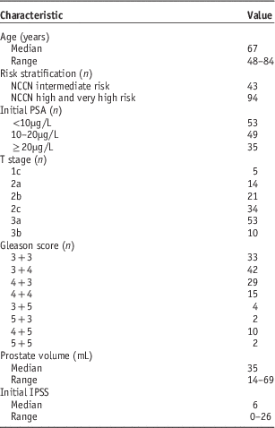
Table 1 summarises the patient cohort baseline characteristics. The median (range) age was 67 (48–84) years. The median (range) initial PSA was 12µg/L (2·3–113). A large proportion had T3a or T3b disease (46%), and nearly a quarter of patients had Gleason 8 or higher disease. Median prostate volume and initial IPSS were 35mL and 6, respectively. There were 43 (31%) intermediate-risk and 94 (69%) high-risk disease patients.
Patient characteristics
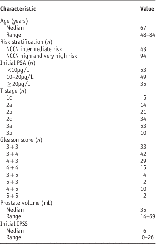
Abbreviations: n, number of patients; NCCN, National Comprehensive Cancer Network; IPSS, International Prostate Symptom Score; PSA, prostate-specific antigen.
The majority of patients had pre-treatment pelvic MRI staging. A total of 15 patients either did not have pelvic MRI done or a report was unable to be found.
Treatment outcomes
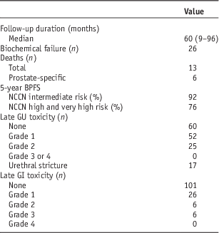
The majority of patients had at least 2 years of documented follow-up (93%, 127/137). All patients who had <2 years of follow-up were from different parts of the country or had re-located to another country shortly following completion of radiotherapy. Table 2 shows the summary of treatment outcomes. The median (range) follow-up period was 60 (9–96) months. A total of 26 patients failed biochemically. Figure 1 demonstrates the BPFS KM curve. The BPFS at 5 years was 92% for intermediate-risk and 76% for high-risk patients. Of the high-risk patients who failed biochemically, 6/21 had at least two high-risk features. Of the other 15 patients with only one high-risk feature, one had T3b disease and another had an initial plasma PSA concentration of 57µg/L. Another patient who had an initial PSA of 113µg/L remains biochemically free of disease after 7 years with undetectable PSA levels (<0·02µg/L). The DMFS at 5 years was 100% for intermediate-risk and 86% for high-risk patients. The OS at 5 years was 100% for intermediate-risk and 86% for high-risk patients. The PCSS for high-risk patients was 92% at 5 years. A total of 12 patients died with five of these due to prostate cancer. Five died from other types of malignancy (lung=2, rectum=1, multiple myeloma=1, liver=1), one died from a stroke and one death was of unknown cause (record not obtained).
Kaplan–Meier survival curve (very high-risk patients included in the high-risk group). Abbreviations: NCCN, National Comprehensive Cancer Network; BPFS, biochemical progression-free survival; HDRBT, high-dose-rate brachytherapy.

Follow-up and treatment outcomes
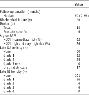
Abbreviations: n, number of patients; BPFS, biochemical progression-free survival; NCCN, National Comprehensive Cancer Network; GU, genitourinary; GI, gastrointestinal.
The median (range) time to PSA nadir was 18 (1–64) months.
Late toxicity outcomes
A total of 25 patients developed late grade 2 GU toxicity some time during the follow-up period; 17 patients developed a urethral stricture requiring non-surgical dilatation, and three of these patients required >1 dilatation. The 5-year rate of urethral stricture formation was 13%. Two patients had previous transurethral resection of the prostate (TURP), and one of these patients already had a urethral stricture at baseline that required dilatation before IDUC insertion on the day of brachytherapy. Of the patients who developed urethral strictures, 59% (10/17) were on treatment for hypertension. Median time to stricture formation was 30 months (range 13 to 82 months). Of the eight other patients who developed grade 2 GU toxicity, most had eventual improvement of their symptoms with conservative management to either grade 1 or 0. No patients developed late grade 3 or 4 GU toxicity.
The 5-year rate of late grade 2 and grade 3 GI toxicity was 4·7 and 4·6%, respectively. Six patients developed grade 3 rectal bleeding requiring surgical intervention. One of these patients was on long-term warfarin and four were on aspirin. Six patients developed late grade 2 toxicity, with three of these in the form of rectal bleeding. Seven out of the nine patients who developed late grade 2 or 3 rectal bleeding were on some form of long-term antiplatelet (aspirin, clopidogrel) or anticoagulation (warfarin) therapy. Most of these patients had eventual resolution of their symptoms.
Erectile dysfunction
A total of 100 patients had pre-HDRBT and post-HDRBT IIEF-5 scores recorded and were accessible; 45 of these patients had baseline severe dysfunction. Of the 39 patients who had normal erectile function or mild erectile dysfunction at baseline, 19 (49%) developed severe dysfunction at some point during the follow-up period.
Discussion
This is the first comprehensive prostate HDRBT series from a New Zealand centre to be reported. We analysed outcomes of the first 137 patients receiving prostate HDRBT as a form of dose escalation from 2006 to 2010. Our BPFS rates are comparable with other larger international centres.Reference Galalae, Martinez and Mate15–Reference Whalley, Patanjali, Jackson, Lovett, Chatfield and Hruby17 In particular, our high-risk cohort compared favourably with a relatively higher BPFS. Galalae et al. reported a 5-year freedom from biochemical failure rate of 69% for their pooled high-risk cohort, whereas Whalley et al. found a 4-year disease-free survival rate of 66%. Our cohort included a large proportion of patients with high-risk disease (69% according to NCCN stratification). Of the high-risk patients who failed biochemically, 8/21 either had at least two high-risk features or had a single high-risk feature that would place them at a very high risk of disease relapse.
Our 5-year urethral toxicity rate of 13% was marginally above the rate expected. Sullivan et al.Reference Sullivan, Williams, Tai, Foroudi, Cleeve and Duchesne18 analysed data from 474 patients undergoing HDRBT and reported a 6-year urethral stricture rate of 12%. It should be noted that our technical experience has increased over time and that a learning curve was evident at the beginning. All patients received a two-fraction regimen of brachytherapy via one implant, and each patient was scanned and the planning was carried out twice in order to maximise accuracy of radiation dose delivery. Urethral tolerance was met during brachytherapy planning for every patient. Two patients had previous TURP and one had a stricture preceding brachytherapy. The Sullivan study found that previous TURP and hypertension predicted the development of urethral stricture. Over half the patients in our cohort who developed strictures were on treatment for hypertension at baseline. Our CT target delineation of the prostate has since moved to a more urethral-sparing volume.Reference Mate, Gottesman, Hatton, Gribble and Hollebeke19 The reasoning behind this change in target delineation is the aim to reduce late urethral toxicity while maintaining an acceptable therapeutic index, given that the anterior lobe of the prostate is rarely involved with cancer.
Almost all the patients who developed late grade 2 or 3 rectal bleeding were on long-term aspirin or warfarin. Antiplatelet and anticoagulation therapy has been shown in various recent retrospective studiesReference Takeda, Ogawa and Ariga20–Reference Hamstra, Conlon and Daignault22 to increase the rate of late rectal bleeding toxicity. Given this, more caution may be required when defining the rectal tolerance of these patients or when selecting them for definitive radiotherapy.
Our study has limitations that are typical of a retrospective analysis. The follow-up period was relatively short and involved a small patient cohort. There was dependence on a variety of medical staff across different centres to grade follow-up toxicity levels. A large proportion of patients had their EBRT in different centres because they were geographically distant to our institution. The use of ADT was varied and a number of patients were on it purely for cytoreductive purposes. As with most prostate cancer series, erectile dysfunction secondary to radiotherapy was difficult to assess accurately, given the large proportion of patients on ADT.
HDRBT has evolved over time to be less resource intensive, and therefore more cost effective. Shah et al.Reference Shah, Lanni and Ghilezan23 reported comparable outcomes and improved cost-effectiveness with HDRBT over IMRT in patients with low- or intermediate-risk disease. In their analysis, low-dose-rate brachytherapy was the least costly treatment modality. However, over time, HDRBT has moved from multiple implants and fractions to single-implant and single-fraction regimens, and this should serve to improve its cost-effectiveness even further.
Since 2011, our centre has switched to a single-fraction technique as per the Toronto study by Morton et al.Reference Morton, Loblaw and Sankreacha24 An analysis of this cohort is planned for the future with direct comparison with the two-fraction cohort.
Conclusions
This study adds to the growing body of literature supporting the use of HDRBT as a method of dose escalation in localised prostate cancer. Survival rates in our high-risk patient cohort compared favourably with other centres. Rates of late toxicity, particularly grade 2 urethral stricture, were higher than expected. This raises the possibility that more careful patient selection is required or more conservative normal-tissue tolerances should be set in patients at higher risk for developing late GU or GI toxicity.
Acknowledgement
None.
Financial Support
None.
Conflicts of Interest
None.