RNA as a building material
While RNA is similar to DNA, just a few minor chemical differences between them cause RNA and DNA to behave in very distinct ways. The 2′-OH of the sugar-phosphate backbone forces the double helical structure of RNA into the A-form conformation with tilted base pairs that spiral along the perimeter of the helix ( Figure 1a), and the substitution of uracil for thymine allows base pairing with either adenine or guanine. Additionally, RNA folds upon itself to form branched tree-like structures Reference Tinoco and Bustamante1 and creates complicated tertiary motifs through nonstandard base pairs. Reference Leontis and Westhof2 RNA keeps its reactive backbone intact under acidic conditions and requires salty conditions with divalent cations to fold and function well. RNA helices have more mechanical stiffness than DNA helices, Reference Kebbekus, Draper and Hagerman3 but are less stable toward backbone hydrolysis and are rapidly broken down by ubiquitous RNA degrading enzymes.
RNA nanostructures built from helices and structural motifs. (a) Comparison of (left) DNA and (right) RNA helices in side- and end view. (b) Original RNA knot structure. Left schematics show the strand path and helices. Right schematics show the resulting topological knot, where
 $3_1^ -$
is the annotation for a negative trefoil knot. (c) RNA nanostructure created using the three-way junction (3WJ) of the pRNA motif. (d) RNA nanostructure created using the dimerizaton initiation site motif, where colors indicate different structural motifs.
Reference Chworos, Severcan, Koyfman, Weinkam, Oroudjev, Hansma and Jaeger12
(e) Crystal structure of RNA triangle composed RNA helices and three corner motifs.
Reference Boerneke, Dibrov and Hermann15
(f) RNA triangle composed of helices and kink-turn motifs bound to L7Ae proteins.
Reference Ohno, Kobayashi, Kabata, Endo, Iwasa, Yoshimura, Takeyasu, Inoue and Saito16
(g) RNA hexagon for delivery of siRNAs to cells.
Reference Afonin, Viard, Koyfman, Martins, Kasprzak, Panigaj, Desai, Santhanam, Grabow, Jaeger, Heldman, Reiser, Chiu, Freed and Shapiro17
(h) RNA square for attachment of multiple RNA functionalities: Fluorescent RNA aptamers, spinach and malachite green (MG), siRNA, and hepatitis B virus ribozyme (HBV rbz). Adapted with permission from Reference Reference Hartmann, Bindereif, Schon and Westhof47. © 2014 American Chemical Society. Note: tRNA, transfer RNA; siRNA, small interfering RNA; pRNA, packaging RNA or prohead RNA.

RNA structures found in nature exhibit multiple functional behaviors, such as small molecule and protein binding (by aptamers), conformational changes triggered by binding (by riboswitches), and catalytic activities (by ribozymes). These structural and functional properties make RNA molecules a central player in the function of the cell and an intriguing multipurpose material for nanoscale engineering.
Origins of RNA nanotechnology
N. Seeman originally proposed rational design of RNA nanostructures as a side note in the 1982 paper on nucleic acid junctions and lattices, Reference Seeman4 where he suggested that the principles for rational design of DNA nanostructures could be translated to RNA. The first rationally designed RNA nanostructure realized was a covalent RNA knot Reference Wang, Di Gate and Seeman5 (Figure 1b), which by being used as a substrate led to the discovery of the first RNA topoisomerase activity (E. coli DNA topo III). Topoisomerases are ubiquitous enzymes that regulate the topological state of DNA globally and locally, facilitating essential cellular functions, including replication, transcription, and DNA repair.
In 1996, another type of RNA nanostructure, called tecto-RNAs, was proposed to be constructed as a mosaic of recurrent RNA motifs found in natural RNA structures. Reference Westhof, Masquida and Jaeger6 Initial RNA nanostructures were designed using loop-receptor motifs, Reference Jaeger and Leontis7,Reference Jaeger, Westhof and Leontis8 but the use of sequence-specific RNA interaction elements, such as the dimerization initiation site (DIS) from human immunodeficiency virus Reference Skripkin, Paillart, Marquet, Ehresmann and Ehresmann9 and the RNA complex of the packaging motor of the phi29 bacteriophage (pRNA), Reference Guo, Zhang, Chen, Garver and Trottier10 allowed more complex RNA nanostructures to be constructed (Figure 1c–d). Reference Shu, Moll, Deng, Mao and Guo11,Reference Chworos, Severcan, Koyfman, Weinkam, Oroudjev, Hansma and Jaeger12
The library of structural RNA motifs that can be used in the tecto-RNA method has been expanding by discovery of several new motifs in, for example, the crystal structures of the ribosomal subunits. Reference Nissen, Hansen, Ban, Moore and Steitz13,Reference Wimberly, Brodersen, Clemons, Morgan-Warren, Carter, Vonrhein, Hartsch and Ramakrishnan14 Examples of unique RNA architectures that can be realized by the tecto-RNA method are nanotriangles composed of helices and RNA motifs with sharp bends Reference Boerneke, Dibrov and Hermann15 (Figure 1e) and RNA triangles where the binding of the ribosomal protein L7Ae to the kink-turn motif defines a 120 degree angle Reference Ohno, Kobayashi, Kabata, Endo, Iwasa, Yoshimura, Takeyasu, Inoue and Saito16 (Figure 1f). One of the main applications of RNA nanotechnology has been to develop nanoparticles for drug delivery, for example, by attaching small interfering RNAs (siRNAs) that can inhibit the expression of disease-causing genes by the RNA interference pathway (Figure 1g) Reference Afonin, Viard, Koyfman, Martins, Kasprzak, Panigaj, Desai, Santhanam, Grabow, Jaeger, Heldman, Reiser, Chiu, Freed and Shapiro17 or attaching several elements such as fluorescent RNA aptamers for imaging or ribozymes for target cleavage (Figure 1h). The progress of RNA nanotechnology for applications in medicine has been reviewed elsewhere. Reference Grabow and Jaeger18–Reference Myhrvold and Silver20
Cross-fertilization from DNA nanotechnology
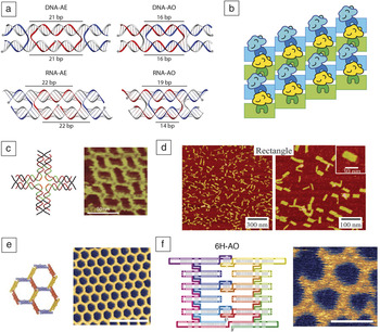
For several years, the research areas of DNA and RNA nanotechnology have developed rather independently, mainly because of the focus of DNA nanotechnology on using simple crossover motifs to create large extended structures and RNA nanotechnology’s focus on exploiting RNA tertiary motifs for creating smaller nanoparticles. Reference Stewar and Franco21 Some cross-fertilization has happened along the way that has primarily depended on the translation of DNA double crossover (DX) motifs to their RNA counterparts that are different due to the shape of the A-form helix of RNA Reference Geary, Andersen, Murata and Kobayashi22 ( Figure 2a) and the adaptation of the DNA strand displacement mechanism Reference Yurke, Turberfield, Mills, Simmeld and Neumann23 to DNA-RNA hybrids and RNA structures. Reference Afonin, Viard, Martins, Lockett, Maciag, Freed, Heldman, Jaeger, Blumenthal and Shapiro24,Reference Sulc, Ouldridge, Romano, Doye and Louis25
RNA nanostructures inspired by DNA nanotechnology. (a) Comparison of DNA and RNA double crossover motifs of the antiparallel even (AE) and odd (AO) type. The spacings in base pairs (bps) between crossovers are indicated on the top and bottom helix; 5′ ends are indicated with a dot. Graphics created by Steffen Sparvath. (b) Schematic of RNA tiles as scaffolds for enzymes. RNA tiles are shown in cyan and green. Enzymes are shown in cyan and yellow with a binding domain shown in dark blue and green. Graphics created by Guido Grossi. (c) Multistranded DNA-RNA hybrid four-way junction shown as structural model and atomic force microscopy (AFM) micrograph. Reference Ko, Su, Zhang, Ribbe, Jiang and Mao28 (d) RNA-DNA origami visualized by AFM. Reference Wang, Ko, Tian, Hao and Mao32 (e) Single-stranded RNA origami tiles of the two-helix AE type forming a honeycomb lattice. Model shown on left and AFM micrograph on right. Reference Geary, Rothemund and Andersen33 Scale bar = 50 nm. (f) Six-helix anti-parallel odd (6H-AO) RNA origami tile forming a honeycomb lattice. Strand path shown on left where color gradient indicates directionality of RNA strand from 5′ end (cyan) to 3′ end (green). AFM micrograph shown on right. Reference Geary, Rothemund and Andersen33 Scale bar = 50 nm.

As for DNA nanotechnology, one of the goals for RNA nanotechnology is to create large programmable scaffolds to arrange molecular components such as enzymes for optimal performance (Figure 2b). The benefit of scaffolding is evident from natural systems where protein scaffolds are often used to direct enzymatic pathways or cell signaling cascades. Reference Good, Zalatan and Lim26 Inspired by this, RNA nanostructures that form lattices have been expressed in E. coli cells, where the RNA lattices were decorated with binding sites for enzymes and used to bring enzymes involved in hydrogen production into close proximity to increase reaction yield. Reference Delebecque, Lindner, Silver and Aldaye27 The correct design of RNA lattices is of major importance to control the spatial organization of the bound components, Reference Geary, Andersen, Murata and Kobayashi22 and many different RNA lattices have been implemented with inspiration from DNA nanotechnology. Motifs like the DNA four-point star motif have been translated to A-form helices to create DNA-RNA hybrid lattices Reference Ko, Su, Zhang, Ribbe, Jiang and Mao28 (Figure 2c), and large DX lattices have been realized. Reference Stewart, Subramanian and Franco29 The DNA origami method Reference Rothemund30 also has been adopted to A-form helices to allow folding of DNA-RNA hybrid origami structures Reference Endo, Yamamoto, Tatsumi, Emura, Hidaka and Sugiyama31,Reference Wang, Ko, Tian, Hao and Mao32 (Figure 2d).
Recently the tecto-RNA method has been combined with RNA DX motifs with the goal of creating larger RNA nanostructures that fold from a single strand, named the single-stranded RNA (ssRNA) origami method Reference Geary, Rothemund and Andersen33 (Figure 2e–f). These structures are beneficial in that they can in some cases, be folded co-transcriptionally and thus can possibly be expressed and folded correctly in cells. More recently, the field of synthetic DNA topology has been applied to produce RNA knots using tensegrity motifs from DNA nanotechnology. Reference Liu, Shao, Chen, Tse-Dinh, Piccirilli and Weizmann34 Both of these new developments in the RNA nanotechnology field are described in the following.
Co-transcriptional assembly of RNA nanostructures
In nature, RNA structures such as ribosomes fold co-transcriptionally in a complex process involving formation of subdomains and binding of several protein components. Compared to the heat-annealing procedures used to self-assemble DNA nanostructures, the co-transcriptional self-assembly of RNA structures occurs progressively as the chain grows from the RNA polymerase. Reference Cruz and Westhof35,Reference Woodson36 It is of great interest to be able to design RNA nanostructures that assemble co-transcriptionally because of the improved speed, fidelity, and scalability of enzymatic synthesis and the opportunity for expression in cells for large-scale production or for manipulating intracellular processes.
Despite the fact that current sequence design methods for creating RNA nanostructures do not take the kinetics of co-transcriptional folding into account, examples of designed RNA nanostructures that fold into the desired structures directly from the transcription reaction have been demonstrated. Examples include the assembly of a nanocube from 12 ssRNA molecules when transcribed in vitro Reference Afonin, Bindewald, Yaghoubian, Voss, Jacovetty, Shapiro and Jaeger37 and filaments and tubes assembled from ssRNA molecules when transcribed from plasmids in E. coli. Reference Delebecque, Lindner, Silver and Aldaye27 Tecto-RNA nanostructures forming hexagonal RNA rings or RNA hearts have also been successfully assembled when transcribed in vitro. Reference Afonin, Desai, Viard, Kireeva, Bindewald, Case, Maciag, Kasprzak, Kim, Sappe, Stepler, Kewalramani, Kashlev, Blumenthal and Shapiro38–Reference Geary, Chworos, Verzemnieks, Voss and Jaeger40 Large RNA microsponges have been created by rolling circle transcription, which uses circular templates to create large and structurally heterogeneous assemblies. Reference Lee, Hong, Bonner, Poon and Hammond41
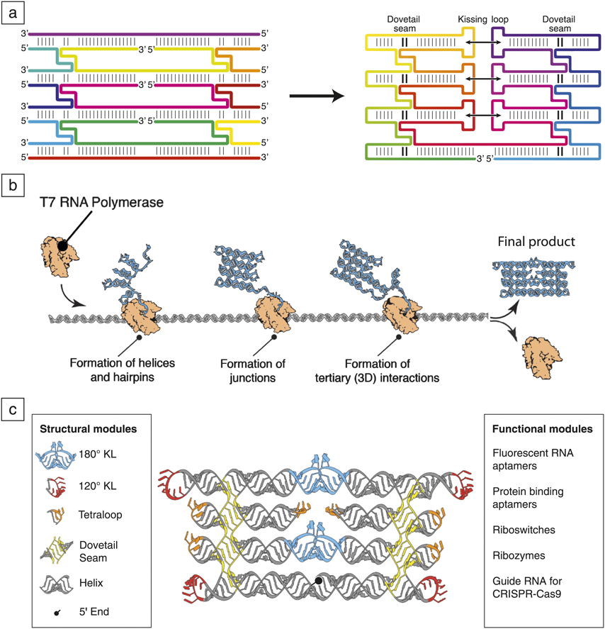
Recently Geary, Rothemund, and Andersen introduced the ssRNA origami method, which allows the design of RNA nanostructures that fold from a single strand during synthesis by RNA polymerase. Reference Geary, Rothemund and Andersen33 Conceptually, the method starts from an RNA nanostructure constructed as a multi-stranded DX lattice ( Figure 3a, left) and uses several loop motifs to concatenate the strands into one long strand (Figure 3a, right). Tetra loops are used to cap the ends of helices on the edges of the RNA nanostructure, while kissing loops are used to bridge helices interior to the RNA nanostructure. Junctions between helices arise at the crossover points between helices that have been named “dovetail seams.” The placement of the 5′ and 3′ ends of the strand affects the folding path of the RNA nanostructure.
Single-stranded RNA origami. (a) Left: RNA double crossover structure composed of multiple strands, where each strand is indicated with a different color, and 5′ and 3′ indicate directionality of the RNA strands. Right: The RNA double crossover structure is converted to one strand by inserting hairpin loops and kissing loops (KLs). Helix junctions form a dovetail seam. Reference Jasinski, Haque, Binzel and Guo19 Color gradient indicates directionality of RNA strand from 5′ end (cyan) to 3′ end (green). (b) Co-transcriptional folding by the T7 RNA polymerase (orange) produces RNA nanostructures (cyan) from the DNA template (gray). The RNA nanostructure forms in a series of steps: First, helices and hairpins are made, then, junctions are formed, and finally, tertiary interactions are made. Reference Geary, Rothemund and Andersen33 (c) Construction of RNA origami structures from structural modules allows further addition of functional modules on the external helices. Reference Good, Zalatan and Lim26 Note: CRISPR, clustered regularly interspaced short palindromic repeats.

Designing the order in which the RNA structure will fold depends on both the composition and thermodynamics of folding individual structural elements and the choice of folding path. In the initial study, it was possible to design several complex RNA nanostructures that fold co-transcriptionally by taking into consideration that secondary structures fold rapidly, and tertiary structures fold much more slowly. Using these basic rules, it was possible to scale the co-transcriptionally folded structure to 440 nucleotides, whereas a larger structure of 660 nucleotides failed to form by co-transcriptional folding. Reference Geary, Rothemund and Andersen33 The co-transcriptional folding process for the RNA origami structures was not investigated experimentally but likely occurs in a stepwise manner, where RNA helices and hairpins form first, junctions between helices form later, and tertiary contacts form last in order to produce the final product (Figure 3b).
RNA origami structures can be built following tutorials that describe the use of computer software to manually construct new architectures and corresponding sequence designs. Reference Sparvath, Geary and Andersen42 The current structures are built using only five structural modules that, in principle, can be extended to make any arbitrary 2D shape (Figure 3c, left and middle). The RNA origami scaffolds are intended to be used as pegboards for functional RNA modules (Figure 3c, right) that can be integrated on the structure at the computer design stage. Examples of RNA functional modules are fluorescent RNA aptamers, such as spinach or mango, which by binding a fluorescent dye, tag the RNA nanostructure; protein binding aptamers such as MS2 or PP7, as fusion proteins, can add a protein of choice to the RNA scaffold; riboswitches and ribozymes may be added to induce conformational changes or cleavage reactions; and guide RNA for CRISPR-Cas9 (clustered regularly interspaced short palindromic repeats) may be added to target the RNA scaffold to a genomic location of choice (e.g., a promoter site). With user-friendly software to add these modules to RNA origami scaffolds, it should become possible to easily create functional scaffolds for specific use as tools in synthetic biology or for cell biology studies.
Construction of topological RNA nanostructures
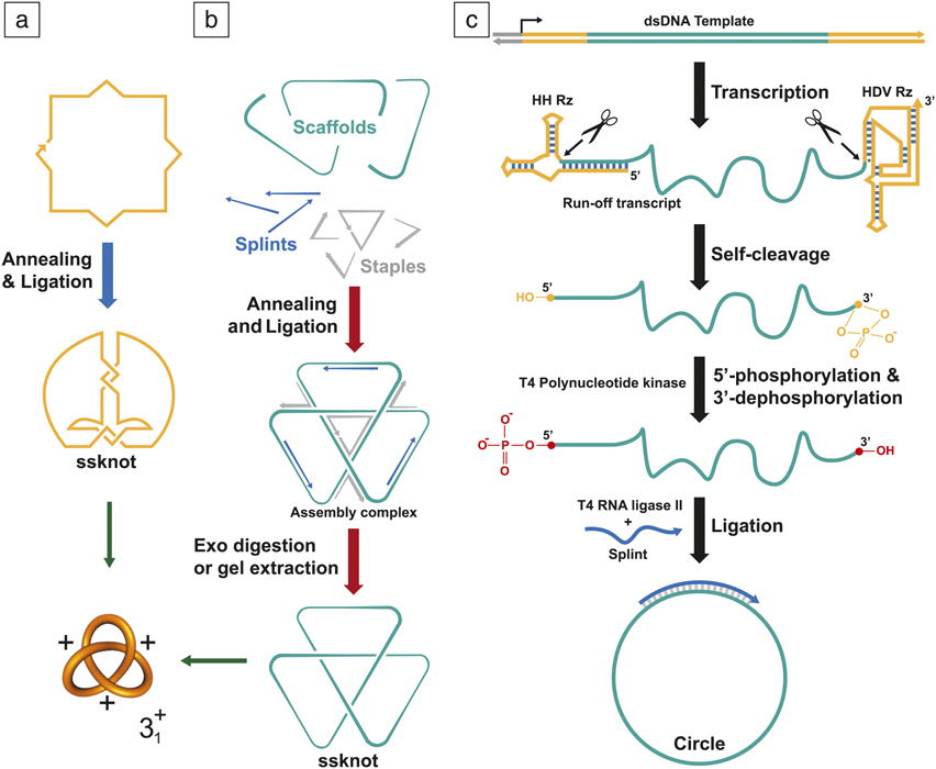
Relatively little progress has been made in RNA topology since the fabrication of the original RNA knot using the helix-based method developed by Seeman and co-workers Reference Wang, Di Gate and Seeman5 ( Figure 4a). Reference Du, Stollar and Seeman43 Recently, a more general and versatile strategy was developed to further push the field of nucleic acid topology. Reference Liu, Chen, Akhter, Cronin and Weizmann44 This strategy involves programming four-way junctions (4WJs), followed by ligation to create closed-loop DNA structures (Figure 4b). Removal of the accompanying staple and splint strands produces relaxed molecular topologies, which is in contrast to the helix-based method, where the topologies are fixed to a certain conformation by a strong intra-strand base pairing. Node signs (positive or negative) are controlled by changing the interjunction distance between the 4WJs in the structural tensegrity triangle motif. Reference Liu, Wang, Deng, Walulu and Mao45 The 4WJs strategy proved its utility, being critical to the successful construction of a total of nine distinct nontrivial single-stranded DNA (ssDNA) molecular topologies.
Construction of RNA topological structures. (a) Helix-based method for the preparation of positive single-stranded knot (ssknot): Reference Wang, Di Gate and Seeman5,Reference Du, Stollar and Seeman43 A single-stranded DNA is annealed, followed by ligation to obtain the ssknot. (b) Four-way junction method for the preparation of a positive ssknot. Reference Delebecque, Lindner, Silver and Aldaye27,Reference Cruz and Westhof35 The scaffold in (a) and (b) can be single-stranded DNA (ssDNA) or single-stranded RNA (ssRNA). For panel (b), the splints and staples are DNA strands. Strategy to construct single-stranded trefoil knot in (b): All the strands are annealed in a one-pot fashion. The ssknot is formed after ligation, then, exonuclease digestion or gel extraction removes the linear strands (splints and staples) producing the relaxed ssknot structure. (c) DNA-splinted RNA ligation preparation: dsDNA template is transcribed to generate the ssRNA product, including both ribozymes at the ends (HH Rz at 5′ and the HDV Rz at 3′), then self-cleavage processes create uniform ends. T4 kinase introduces a phosphate group at 5′, while removing the cyclic 2′-3′ phosphate on the sugar at 3′ makes the ssRNA product suitable for ligation. With the help of a splint strand and an annealing process, the ssRNA is circularized by T4 RNA ligase II. Note: HH Rz, hammerhead ribozyme; HDV Rz, hepatitis D virus ribozyme; dsDNA, double-stranded DNA; 5′ and 3′ indicate the carbon numbers in the sugar backbone.

To provide direct evidence for the formation of programmed nodes of the negative and positive knots, Weizmann and co-workers developed a procedure to install complex topological structures into a vector via a method named “topological grafting.” Using this approach, a small topological motif was ligated into a larger nucleic acid sequence (vector), resulting in the topology of the small motif integrating into the vector. By using RecA, a protein that binds tightly to DNA, the topology of the original topological motif become visible via atomic force microscopy. The 4WJs method also enables the realization of the more challenging double-stranded DNA (dsDNA) version of the knot using the ssDNA knot as a template. This has historically proven challenging, since to create knots with the more rigid dsDNA, precise design of curvature and torsion in 3D space is essential. Reference Liu, Chen, Akhter, Cronin and Weizmann44
Recently, using the same principles, it was shown that it is possible to create RNA and DNA-RNA hybrid topological structures. Reference Liu, Shao, Chen, Tse-Dinh, Piccirilli and Weizmann34 Using the 4WJs method, different topological composites have been formed containing only ssRNA (positive and negative knots), hybrid catenae (Borromean rings) of ssDNA and ssRNA, or DNA-RNA hybrid double-stranded knots. As previously discussed, the synthesis is flexible, can accommodate the formation of both node signs, and can be used to help solve unexplored problems within the field. There are broad implications for applicability of the resultant topological structures. The researchers investigated the actions of two kinds of nucleic acid-processing enzymes, and they studied the topological conversion catalyzed by DNA topoisomerase and the topological inhibition of DNA polymerase or reverse transcriptase, on DNA or RNA knots, respectively. Reference Liu, Shao, Chen, Tse-Dinh, Piccirilli and Weizmann34,Reference Liu, Chen, Akhter, Cronin and Weizmann44 Interestingly, both single-stranded knots were able to function as substrates. The polymerase/reverse transcriptase activity during rolling circle amplification (RCA), when polymerase runs on a circular template and results in amplification of sequence, was severely inhibited when using a knotted template instead of the more relaxed circle substrate. This finding was used to develop a RCA-amplification-based and electrophoresis-free detection method for topoisomerase activity. As a result, they envisioned developing a high-throughput screening method for topoisomerase inhibitors. For the topoisomerase studies, remarkably, DNA topoisomerase I has activity on the RNA knots, which, through mutation analysis, was confirmed to be unique from the DNA activity.
Since commercially available synthetic RNAs are expensive, using enzymatic methods can simplify the production of the desired RNA strands and promises higher yields at a lower cost. In the context of enzymatic synthesis, RNA faces two problems when compared to DNA. RNAs generated via transcription tend to show length heterogeneity of the 5′ and 3′ ends, making template-assisted ligation of the product strands difficult. Also, a suitable RNA ligase needs to be identified. It is likely that these difficulties in the ligation of in vitro transcribed RNA product are the reason for the low number of reported RNA topological structures.
To overcome these problems, two techniques have been employed. First, to ensure uniform ends to the transcribed RNA, researchers encoded self-cleaving ribozymes within the designed dsDNA template—a hammerhead ribozyme at the 5′ and hepatitis D virus ribozyme Reference Avis, Conn and Walker46 at the 3′ (Figure 4c). The ribozymes have a self-cleaving mechanism, form homogeneous termini for newly transcribed RNA strands, and prepare the strands for the templated assembly. Second, for the ligation process, a general T4 RNA ligase II is used to achieve ligation efficiency comparable to the routinely used DNA ligase enzyme. Reference Hartmann, Bindereif, Schon and Westhof47 By overcoming these two issues, drastic steps have been taken toward improving the feasibility of the synthesis of RNA topological structures.
Future directions for RNA nanotechnology
To advance the field of RNA nanotechnology, design methodology must be developed to allow larger and more complex RNA nanostructures to be created. When comparing the current artificially designed RNA nanostructures to the size and complexity of the ribosome, Reference Nissen, Hansen, Ban, Moore and Steitz13,Reference Wimberly, Brodersen, Clemons, Morgan-Warren, Carter, Vonrhein, Hartsch and Ramakrishnan14 this task seems daunting. To approach more complex RNA designs, several challenges need to be addressed: (1) improving sequence design algorithms to handle large RNA structures, for example, by parallelization to reduce computational time; (2) developing 3D modeling software for constructing new architectures by combining RNA structural motifs; (3) improving gene synthesis methods for long sequences with a large number of complementary sequences; and (4) developing simulation software to predict the folding path of large RNA nanostructures to avoid topological traps in heat-annealing or co-transcriptional folding. Inspired by DNA nanotechnology, further developments of RNA nanotechnology should focus on the creation of functional RNA devices. Compared to DNA nanotechnology, RNA nanostructures have the potential to be produced and used inside cells. One of the main challenges is to express and verify that the RNA nanostructures fold correctly in the cell. Verification can be done by indirect or direct characterization experiments and will be central to support the development of true applications of RNA nanoparticles and scaffolds for metabolic engineering, cell signaling, biosensor devices, and manipulation tools for cell biology.
Future progress in the field of RNA topology should be assisted by the development of strategies that will allow researchers to tackle the common problems associated with RNA. With the significant number of DNA topologies in existence, producing a number of analogous RNA topologies is the next logical step. However, the most topologically strained DNA topologies may not permit a full double-stranded structure, thereby preventing the use of this method in some cases. By generating monodisperse ssRNAs with the ribozymes approach, this alternative assembly method should allow more RNA topologies to be generated without relying on the presence of the DNA analogue. Beyond these advancements, investigations on how these RNA topologies interact with RNA binding enzymes (polymerases, RNases, and topoisomerase) will hopefully further the understanding of how organisms manage topological challenges in RNA, and lead to a new understanding of how such enzymes can become targets for treatments or antibiotics. Another potential outlet for future work uses the programmability of both DNA and RNA, with the potential of using nucleic acid topologies to program other materials through templated assembly. The inherent properties of the nucleic acids could lead to improved control over chirality as well as structure. Reference Niu, Hili and Liu48 Assemblies of nanoparticles, artificial biomolecules, and polymers could become more sophisticated from successes in nucleic acid topology.
Acknowledgments
This work was supported by the European Research Council (Grant Number 683305) funded to E.S.A. and the National Science Foundation CAREER Award (DMR-15,55,361) to Y.W.

Yossi Weizmann heads a research group in the Department of Chemistry at The University of Chicago. He obtained his PhD degree at The Hebrew University of Jerusalem, Israel, in 2007. He was subsequently a postdoctoral associate at the Massachusetts Institute of Technology. His research focuses on the design and construction of synthetic nucleic acid structure and topology for biomedical and biomimetic applications, and synthesis of metallic nanoparticles for plasmonic bioapplications. Weizmann can be reached by phone at 773-702-6107 or by email at yweizmann@uchicago.edu.

Ebbe Sloth Andersen heads a research group at the Interdisciplinary Nanoscience Center, Aarhus University, Denmark. He obtained his PhD degree at Aarhus University in 2006. He was a postdoctoral fellow at the Centre for DNA Nanotechnology and has been a visiting associate at the California Institute of Technology. His research focuses on nucleic acid design principles and applications in biosensing, drug delivery, and synthetic biology. Andersen can be reached by phone at +45 41178619 or by email at esa@inano.au.dk.