Introduction
In eukaryotic cells, 146 bps DNA is wrapped around two copies of each histone – H2A, H2B, H3, and H4 to form a nucleosome – the basic structural unit of chromatin (Luger et al., Reference Luger, Mäder, Richmondm, Sargent and Richmond1997; Kornberg & Lorch, Reference Kornberg and Lorch1999). Tremendous diversity in the histone/nucleosome structure is generated by a variety of post-translational modifications, such as acetylation, phosphorylation, methylation, ubiquitination and citrullination. Some modifications, including methylation, acetylation and phosphorylation, are reversible and dynamic and are often associated with inducible expression of individual genes. The citrullination which is catalyzed by peptidylarginine deiminases (PADs) results in the conversion of arginine residue of a protein into the non-conventional amino acid citrulline. This situation results in the decrease in the isoelectric point of the protein and affects its secondary and tertiary structure (Gyorgy et al., Reference Gyorgy, Toth, Tarcsa, Falus and Buzas2006). Protein citrullination has been shown to influence protein–protein interaction (Tarcsa et al., Reference Tarcsa, Marekov, Mei, Melino, Lee and Steinert1996), modulate signalling potency (Proost et al., Reference Proost, Loos, Mortier, Schutyser, Gouwy, Noppen, Dillen, Ronsse, Conings, Struyf, Opdenakker, Maudgal and Van2008; Loos et al., Reference Loos, Opdenakker, Van and Proost2009) and interfere with susceptibility to proteolytic degradation (Pritzker et al., Reference Pritzker, Joshi, Gowan, Harauz and Moscarello2000).
In growing oocytes genes are actively transcribed (Bachvarova, Reference Bachvarova1985) but the transcriptional activity is decreased to the marginal level when oocytes are fully grown. If any activity remains, it is at a very low level during meiotic maturation. Following fertilization, early embryos are still transcriptionally silent and the development is regulated by using maternally-derived transcripts (Johnson, Reference Johnson1981, Bolton et al., Reference Bolton, Oades and Johnson1984). Zygotic gene activation occurs according to species-specific timing and is known to occur in two phases: initially at a low level and then in a burst. Immediately after fertilization, genomic DNA is repressed to establish the regulatory mechanism for gene-specific expression in the preimplantation embryos. Gene-specific transcription factors and/or changes in chromatin structure are then required for the expression of specific genes to relieve repression. Histones are hyperacetylated in the active chromatin domain (Hebbes et al., Reference Hebbes, Thorne and Crane-Robinson1988) and hypoacetylated in the transcriptionally silent domain (Braunstein et al., Reference Braunstein, Rose, Holmes, Allis and Broach1993, Jeppesen & Turner, Reference Jeppesen and Turner1993).
Peptidylarginine deiminases (PADs) that catalyze citrullination are expressed in a variety of cells, targeting mainly structural proteins and are involved in development and cell differentiation. Among the five isomers, PAD4 has a relatively broad substrate specificity, and can deiminate multiple arginine sites on histones H3 (R2, R8, R17, and R26) and H4 (R3) (Wang et al., Reference Wang, Wysocka, Sayegh, Lee, Perlin, Leonelli, Sonbuchner, McDonald, Cook and Dou2004). In our previous study (Brahmajosyula & Miyake, Reference Brahmajosyula and Miyake2011,) we showed the expression of PAD4 in pig oocytes and parthenogenetic diploid embryos and that histone H3 was one of the targets of PAD4. It has been shown that PAD4 mediates gene expression by regulating arginine methylation and citrullination in histones, and plays a role in decondensation of chromatin in granulocytes and neutrophils via citrullination (Wang et al., Reference Wang, Wysocka, Sayegh, Lee, Perlin, Leonelli, Sonbuchner, McDonald, Cook and Dou2004, Reference Wang, Li, Stadler, Correll, Li, Wang, Hayama, Leonelli, Han, Grigoryev, Allis and Coonrod2009). Recent studies by Li et al. (Reference Li, Yao, Zhangm, Li, Luo, Thompson, Gilmourm and Wang2008, Reference Li, Wang, Yao, Doret, Hao, Shen, Qiu, Zhang, Wang, Chen and Wang2010) demonstrated the regulation of p53 target gene expression by PAD4 and the coordination of PAD4 and histone deacetylase (HDAC)2 in the regulation of p53 target gene expression. These data prompted us to study the role of PAD4 in embryonic development particularly in the pre-implantation stage owing to the unique status of the zygote being totipotent.
Materials and methods
This study was approved by the Institutional Animal Care and Use Committee (Permission numbers: 19–5-30, 19–5-31, 22–05-13 and 22–05-15) and carried out according to the Kobe University Animal Experimentation Regulations.
In vitro maturation (IVM) of pig oocytes and generation of diploid parthenotes
Prepubertal gilt (Sus scrofa domestica) ovaries were collected from a local abattoir and transported to the laboratory within 2 h. The ovaries were washed once with the cationic detergent, 0.2% (w/v) cetyltrimethyl ammonium bromide followed by thorough washing with Ca2+-free Dulbecco's phosphate-buffered saline (PBS) that contained 0.1% (w/v) polyvinyl alcohol (PVA; Sigma-Aldrich Chemical Co., St. Louis, MO, USA). Healthy antral follicles sized between 4–6 mm were collected and the oocyte–cumulus–granulosa cell complexes (OCGCs) were separated intact from the inner surface of these follicles and cultured for maturation in 2 ml of TCM-199 (Earl's salt; Nissui Pharmaceutical Co. Ltd, Tokyo, Japan) supplemented with 10% (v/v) heat-inactivated fetal calf serum (FCS; ICN Biomedicals, Inc., Aurora, Ohio, USA), 0.1 mg/ml sodium pyruvate, 0.08 mg/ml kanamycin sulphate (Sigma-Aldrich) and 0.1 IU/ml hMG (Pergonal; Teikokuzoki, Tokyo, Japan) along with two thecal shells freed from follicular fluid and granulosa cells. The culture was carried out in a CO2 incubator at 38.5°C under 5% CO2 in humidified air for 42–48 h with gentle agitation.
Mature pig oocytes after 42–48 h maturation culture were briefly exposed to 0.01% (w/v) hyaluronidase (Sigma-Aldrich) and gently pipetted to remove the cumulus cells. Denuded oocytes with first polar bodies in groups of 20 were selected and thoroughly washed in field solution composed of 0.3 mM mannitol, 0.1 mM MgSO4 and 0.05 mM CaCl2 (Zimmermann & Vienken, Reference Zimmermann and Vienken1982) supplemented with 0.01% (w/v) PVA before placing them in 100 μl of field solution between two parallel stainless steel electrodes in an activation chamber (FTC-03; Shimadzu Co, Ltd., Kyoto, Japan). A single direct current pulse of 1500 V/cm for 100 μs was supplied from an electrocell fusion device (LF101; Bex Co., Tokyo, Japan). The oocytes were then washed thoroughly in equilibrated porcine zygote medium (PZM3; Yoshioka et al., Reference Yoshioka, Suzuki, Tanaka, Anas and Iwamura2002) supplemented with 3.0 mg/ml bovine serum albumin (BSA; Wako Pure Chemical Industries, Osaka, Japan) before placing them in cytochalasin B (Sigma-Aldrich) supplemented PZM3 medium at 38.5°C in a humidified incubator with 5% CO2 in air for 4 h.
Designing PAD4 siRNA
Partial sequence (178 amino acids in length) of pig PAD was obtained from Ensembl genome browser. Three stealth siRNAs to target three different regions of PAD4 were designed using an online RNAi designer (Invitrogen Life Technologies, Carlsbad, CA, USA). Sequences of the siRNAs used are as follows. #1 – forward: CAAGGAGGUGUAUGUGUGCAGGAUA, reverse: UAUCCUGCACACAUACACCUCCUUG; #2 – forward: CCGAGGAACAGAGGCUUGAAGGAUU, reverse: AAUCCUUCAAGCCUCUGUUCCUCGG; #3 – forward: CAAACAUGGUGAACAUGCUGGUGCU, reverse: AGCACCAGCAUGUUCACCAUGUUUG.
Primary cell culture of pig cumulus cells
At first, the efficiency of siRNA induced PAD4 knockdown was examined in pig cumulus cells. Cumulus cells from pig metaphase II oocytes were collected during denudation, washed by centrifugation with 0.01% (v/v) PBS-PVA for 5 min at 1,000 rpm and grown in vitro in 2 ml of TCM-199 medium (Earl's salt; Nissui Pharmaceutical Co., Ltd, Tokyo, Japan) supplemented with 10% (v/v) heat-inactivated fetal calf serum (FCS; ICN Biomedicals, Inc., Aurora, Ohio, USA) in a 5% CO2 incubator at 38.5°C for 24–48 h. Then the cells were washed with PBS-PVA followed by lysing with 0.25% trypsin EDTA (T4049; Sigma-Aldrich) and seeded on a 35 mm cell culture dish (353001; Becton and Dickinson Falcon™ Co, Franklin Lakes, NJ, USA) allowing for confluence in the incubator for 24 h.
Transfection
Cumulus cells with 50–60% confluence were used for transfection with PAD4 targeted siRNAs. Before transfection, Lipofectamine 2000 (Invitrogen Life Technologies) was diluted by mixing 5 μl in 250 μl Opti-MEM medium without serum (31988; GIBCO-Invitrogen™, Auckland, New Zealand) and kept at room temperature for 5 min. Each siRNA (20 μM) was prepared by mixing 1 μl in 250 μl Opti-MEM. The diluted siRNA was combined with the diluted lipid (total volume 500 μl) and incubated for 20 min at room temperature. Each of these mixtures was added to each culture dish and gently rocked back and forth. The cells were incubated at 38.5°C in the CO2 incubator for 4–6 h and then the medium was changed to low serum DMEM (11885; GIBCO-Invitrogen™) and cultured for total 24–48 h. One of the dishes with cumulus cells was added with only diluted lipofectamine as a negative control.
RNA interference in parthenogenetic diploids at 1-cell stage
Electrostimulated metaphase II oocytes were injected with 5–10 pl of PAD4 siRNA#2 (20 μM) within 4 h post activation using PIEZO (PNAS-CT150; Prime Tech, Ibaraki, Japan) driven microinjector (Narishige Group, Tokyo, Japan). Then the activated oocytes were changed to fresh drops of PZM3 medium for in vitro development. Development was monitored at every stage; 2-cell (24 h), early 4-cell (48 h), late 4-cell (72 h), morula (96 h) and blastocyst (120 h) by taking bright field pictures under inverted microscope. A group of activated oocytes were mock injected with 0.1% diethylpyrocarbonate (DEPC) water as negative control. For confirming the PAD4 knockdown, control 2-cell embryos and both 1-cell and 2-cell embryos from siRNA injected group at 32 h and blastocysts at 120 h post activation were collected and subjected to indirect immunofluorescence study.
PAD4 inhibition with Cl-amidine
To counter check the results of siRNA experiments, the effect of PAD4 inhibition by a potent PAD4 inactivator Cl-amidine was studied. Initially, to check the toxicity of Cl-amidine in embryos, immediately after electro-stimulation, metaphase II oocytes were placed in cytochalasin B-containing PZM3 medium supplemented with 100 μM, 200 μM and 500 μM of Cl-amidine (Gift from Prof. P.R. Thompson, South Carolina University, USA). After 4 h, they were transferred to equilibrated drops of PZM3 medium supplemented with same concentrations of Cl-amidine respectively and cultured for 24 h totally. After that, embryos were thoroughly washed in fresh drops of PZM3 and kept for in vitro development up to blastocyst stage (120 h).
Next, to check the effect of Cl-amidine exposure for various durations, electrically activated oocytes were treated with 500 μM Cl-amidine from the time of activation for 6 h, 10 h, 12 h, 15 h and 24 h totally and subsequently cultured for 120 h, up to the blastocyst stage. In the next experiment, embryos at 0 h, 24 h, 48 h and 72 h post activation were treated with Cl-amidine (500 μM) for 6 h and 24 h to check the effect of PAD4 inhibition for short or long durations at various stages of development. After treatment with Cl-amidine, the embryos were washed thoroughly and cultured further up to blastocyst stage (120 h).
A group of activated oocytes were not treated with Cl-amidine as control in each experimental group. Cl-amidine treated and control samples were collected at various stages for indirect immunofluorescence analysis (IIF).
Immunocytology of embryos
Samples were fixed in 2% paraformaldehyde supplemented with 0.2% Triton X-100 (Nacalai Tesque, Inc., Kyoto, Japan) for 20 min. The fixed embryos were washed thoroughly twice in PBS–PVA for 5 min each and then stored in 1% BSA supplemented PBS–PVA at 4°C until use. They were blocked with 10% goat serum in 1% BSA–PBS–PVA for 1 h. Mock injected control and PAD4 siRNA injected embryos collected at 2-cell and blastocyst stage were incubated with rabbit polyclonal anti-human PAD4 antibody (1:25; AP1072C, Abgent Inc, San Diego, CA, USA) at 4°C overnight. Next day the samples were washed thoroughly with PBS–PVA and incubated with Alexa Fluor488-labelled donkey anti-rabbit IgG, (1:500; Invitrogen, Carlsbad, CA, USA) for 1 h at room temperature. After washing thrice with PBS-PVA for 5 min each, DNA was stained with 200 μg/ml propidium iodide (PI; Sigma-Aldrich) or 2 μg/ml 4′,6-diamidino-2–185 phenylindole (DAPI; Molecular Probes Inc, Eugene, OR, USA). The embryos were washed again before being mounted on Vectashield solution (Vector Laboratories, Burlingame, CA, USA) and observed under fluorescence microscope (BX51-Fl; Olympus Optical Company Ltd., Tokyo, Japan) at appropriate UV excitation. The slides were gently pressed so as to rupture the zona to show that the signal is from the embryo itself, not from the zona.
SDS PAGE and western blot analysis
The transfected cumulus cells were collected from culture dish by lysing followed by washing with PBS–PVA. Equal amount of 2× sample buffer containing 10% glycerol, 2% SDS, 80 mM Tris–HCl (pH 6.8) and 0.02% bromophenol blue was added to it and boiled for 5 min before storing at –20°C. Electrophoresis was done on 12.5% minigel SDS-PAGE (AE 6530; Atto Bioscience and Technology, Tokyo, Japan) and the proteins were transferred to polyvinylidene difluoride (PVDF) membranes (Immobilon P; Millipore, Bedford, MA, USA) on a semi-dry transfer cell (Trans-Blot SD; BioRad Laboratories, Hercules, CA, USA) for 1 h. Membranes were either cut into two, to process for two different antibodies or the same membrane was reprobed after probing for one antibody. The membranes were blocked for 1 h at room temperature with 5% FCS or fat-free milk in 0.1% Tween-20 in PBS (PBST) with gentle agitation and incubated overnight at 4°C with rabbit polyclonal anti-human PAD4 antibody (1:250; ab38772, Abcam) or mouse monoclonal anti-β-actin antibody (1:10 000; A2228, Sigma-Aldrich) in blocking solution. Subsequently, membranes were washed in PBST and incubated with horseradish peroxidase (HRP)-linked anti-rabbit IgG whole antibody from donkey (1:20000; GE healthcare UK. Ltd, Buckinghamshire, UK) or HRP-linked goat anti-mouse IgG (1:20000; Thermo fisher Scientific Inc., Rockford, IL, USA) for 2 h at room temperature. Membranes were washed three times with PBST and the HRP activity was detected by using enhanced chemiluminescence (ECL) super signal (Pierce, Thermo Fisher Scientific Inc., Rockford, IL, USA).
Statistical analysis
P-values were obtained using Fisher's Exact test in Table 1, by Bonferroni test in Figs. 3, 4 and 5 and by Mann–Whitney test in Table 2. All the experiments were repeated at least twice and for immunocytology each group contained at least three embryos.
Effect of RNAi-mediated PAD4 downregulation on the preimplantation development of parthenogenetic embryos in the pig

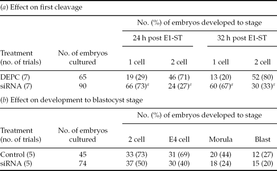
All eggs were observed every 24 h. Values of 2-cell, early 4-cell, morula and blastocyst (Blast) stages observed at 24 h, 48 h, 96 and 120 h respectively are shown.
a Values with asterisk ar significantly different from the control by Fisher's exact test (P < 0.01).
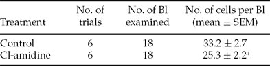
Effect of Cl-amidine treatment for 6 h from 0 h of electro-stimulation on total number of cells in the blastocyst
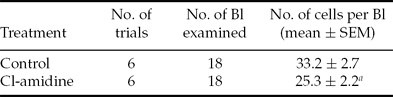
a Value with superscript is significantly different from the control by Mann–Whitney test (P < 0.05). Bl, blastocyst; SEM, standard error of the mean.
Results
RNAi efficiency by PAD4 targeted siRNA
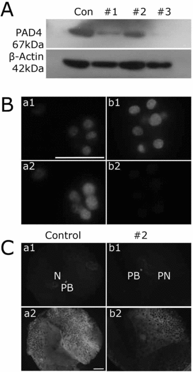
To check the efficiency of PAD4 targeted siRNAs, cumulus cells were transfected with all three siRNAs (#1, #2, #3). After 48 h of culture the cells were analyzed by western blotting. All the three siRNAs did show PAD4 downregulation compared with the control, which was treated with only lipofectamine (Fig. 1A). In activated oocytes, RNAi by injection showed evident PAD4 knockdown in the group treated with siRNA #2 (Fig. 1C-b2) in indirect immunofluorescence study. Therefore siRNA #2 was selected for the subsequent experiments. The high intensity V-shaped areas (in a2 and b2) are the zona ruptured areas. Cumulus cells collected from the metaphase II oocytes after in vitro maturation with or without siRNA#2 transfection were subjected to indirect immunofluorescence analysis. Cumulus cells from siRNA transfected oocytes showed evident PAD4 knockdown (Fig 1B-b2) compared with the control (Fig 1B-a2).
PAD4 expression in pig cumulus cells and parthenogenetic diploid embryos after RNAi with PAD4 siRNA. (A) In vitro cultured cumulus cells transfected with three different sequenced siRNAs (#1, #2, #3) and control cells (Con) treated with only lipofectamine were subjected to western blot analysis for PAD4 and β-actin 48 h after in vitro culture. The lower panel indicates loading control using β-actin. Cumulus cells transfected with all three siRNA sets exhibited effective PAD4 downregulation (upper panel). (B) Cumulus cells collected from transfected oocytes with siRNA #2 during maturation culture were subjected to indirect immunofluorescence analysis with anti-PAD4 antibody followed by secondary antibody labelled with Alexa Fluor 488 (green). Nuclei were stained with propidium iodide (PI). Cells treated with only lipofectamine are the control group (B-b). (C) Metaphase II oocytes were injected with 10–20 pl of siRNA #2 within 4 h of electro-stimulation. Control oocytes were injected with 0.1% diethylpyrocarbonate water (DEPC). After 32 h of in vitro culture embryos were collected, fixed, and subjected to indirect immunofluorescence analysis using anti-PAD4 antibody followed by secondary antibody labeled with Alexa fluor 488. Nuclei were stained with PI. siRNA #2 injected embryos (C-b2) showed obvious decrease of PAD4 expression compared to control. Note that the siRNA injected embryos were still at the pronuclear stage, while DEPC water injected embryos were at the 2-cell stage. High intensity V-shaped areas in a2 and b2 are the zona ruptured areas. Scale: 20 μm.
PAD4 reduction by PAD4 siRNA in pig parthenogenetic preimplantation embryos
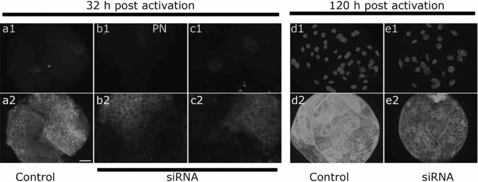
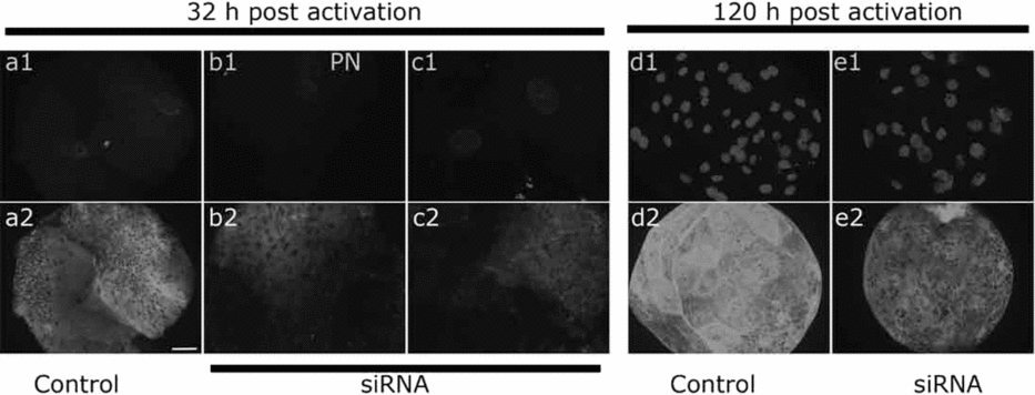
In order to check the role of PAD4 in preimplantation embryonic development, PAD4 protein synthesis was inhibited using RNAi. In vitro-matured metaphase II oocytes were injected with anti-PAD4 siRNA#2 within 4 h post electro-stimulation. At 24 h, the majority of the siRNA injected embryos were uncleaved. After an additional 8 h of culture, at 32 h post activation, the embryos were visualized again for the cleavage. But even at 32 h, many of the embryos were uncleaved. Then, at 32 h, both cleaved and uncleaved embryos from siRNA injected group and cleaved embryos from control group were collected and analyzed for PAD4 expression by immunofluorescence. The results revealed PAD4 downregulation manifested by reduced PAD4 signal, in both the 1-cell and 2-cell embryos that were injected with siRNA#2 (Fig 2-b2, c2) compared with the DEPC water injected control (Fig 2-a2). The rest of the embryos were cultured in vitro up to blastocyst stage. In the siRNA injected blastocysts at 120 h post activation PAD4 knockdown was clearly observed (Fig 2-e2) in comparison to the control blastocyst (Fig 2-d2). Strong intensified V-shaped areas (in a2, b2, c2 and e2) are the zona ruptured areas.
PAD4 expression in siRNA injected pig diploid parthenogenetic embryos. Electro-stimulated oocytes were injected with siRNA#2 within 4 h post activation. Control oocytes were injected with 0.1% diethylpyrocarbonate water (DEPC). At 32 h (a, b, c) and 120 h (d, e) post activation, embryos were subjected to indirect immunofluorescence analysis with anti-PAD4 antibody followed by Alexa fluor 488 labeled secondary antibody. Nuclei were stained with propidium iodide (PI) or DAPI. Both 1-cell (b2) and 2-cell embryos (c2) from siRNA injected group showed obvious PAD4 decrease compared with control 2-cell embryo (a2). Blastocysts that were developed from the siRNA injected activated oocytes showed reduced PAD4 labelling (e2), while control embryos showed bright fluorescence of PAD4 signal (d2). High intensity V-shaped areas in the lower panel pictures are the zona ruptured areas. Scale: 20 μm.
Effect of PAD4 inhibition by RNAi on pig parthenogenetic preimplantation embryonic development
To study the effect of PAD4 inhibition by RNAi on the developmental potential, activated oocytes were cultured after injection of siRNA and monitored for in vitro development along with control DEPC water injected activated oocytes. At 24 h post activation it was observed that the majority of the siRNA injected embryos did not cleave compared with DEPC-injected control embryos. The growth was monitored further and at 32 h the majority of siRNA injected embryos were still at 1-cell stage with significant delay in the development (Table 1A). Visual monitoring of the development up to blastocyst stage showed delay at each stage in the siRNA injected group compared to that of control group – although it was not significantly different (Table 1B). The total number of cells in the siRNA injected blastocysts (n = 15, 18.2 ± 2.0) was significantly lower than control blastocysts (n = 11, 29.6 ± 4.2; P < 0.05 by Fisher's exact test).
PAD4 inhibitor Cl-amidine toxicity check
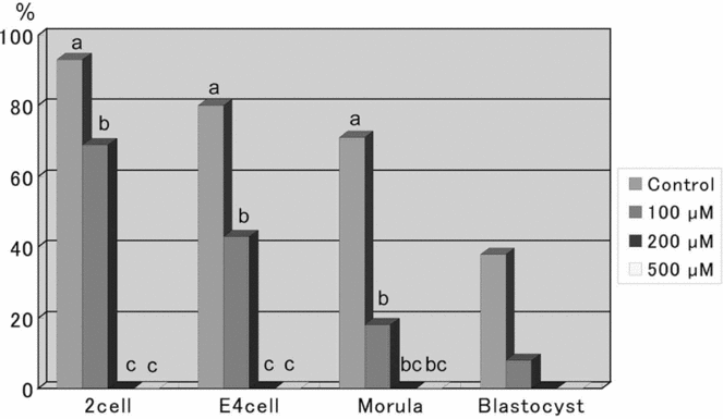
To confirm the results of RNAi experiments, similar experiments with PAD4 inhibitor Cl-amidine were performed. Prior to use, the toxicity of this inhibitor was checked on the embryonic survival. Cl-amidine at concentrations of 100 μM, 200 μM and 500 μM were added to the culture medium just after electro-stimulation. After 6 h and 24 h the embryos were washed and moved to fresh culture medium devoid of Cl-amidine and cultured up to 120 h post electro-stimulation. It was observed that even at a high concentration of 500 μM, development up to the blastocyst stage was not affected when the treatment was for short period of 6 h (data not shown). However, when the embryos were incubated in Cl-amidine for 24 h, all the embryos treated with 200 μM or more were arrested at 1-cell stage (Fig 3), but the embryos that were treated with 100 μM Cl-amidine developed up to blastocyst. To have the maximum inhibition effect, a dosage of 500 μM was chosen for the experiments.
Effect of different concentrations of Cl-amidine treatment on the development of pig diploid parthenogenetic embryos to blastocyst stage. The percentage of embryos developed to 2-cell, early 4-cell (E4 cell), morula and blastocyst stages were observed at 24 h, 48 h, 96 h and 120 h, post activation respectively. a, b,cValues with different superscripts at the same developmental stages are significantly different (p < 0.001) by Bonferroni test.
Effect of Cl-amidine treatment duration on the embryonic development
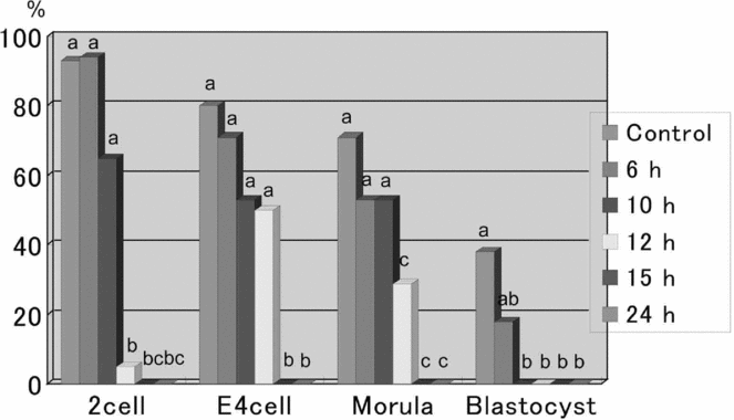
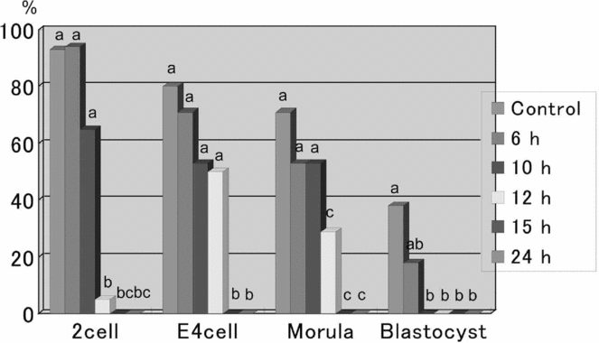
Activated embryos were treated with 500 μM of Cl-amidine for 6 h, 10 h, 12 h, 15 h and 24 h and checked for developmental competency. The embryos treated with Cl-amidine had a reduced rate of development in 6/10 h groups compared with the untreated control embryos. The embryos treated up to 12 h could develop, but slowly. Treatment beyond 12 h resulted in total developmental arrest at 1-cell stage (Fig 4). The blastomere number was significantly reduced in the Cl-amidine treated blastocysts compared with the control blastocysts (Table 2).
Effect of Cl-amidine treatment for different durations on the development of pig diploid parthenogenetic preimplantation embryos. Columns of embryos at the 2-cell, early 4-cell (E4 cell), morula and blastocyst stages show percentages at 24 h, 48 h, 96 h and 120 h post electro-stimulation, respectively. a, b, cValues with different superscripts at the same developmental stages are significantly different (p < 0.05) by Bonferroni test.
Effect of timing of Cl-amidine treatment on the development of parthenogenetic preimplantation embryos
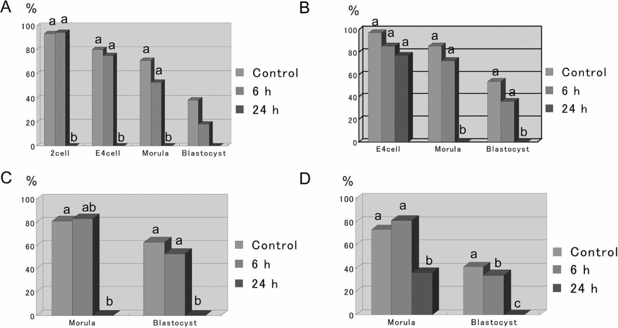
To investigate the effect of PAD4 inhibition at different stages of development, pig parthenogenetic embryos were treated with 500 μM Cl-amidine for 6 h or 24 h at 2 cell, early 4 cell, and late 4 cell stages (24 h, 48 h, and 72 h after electro-stimulation, respectively). Embryos treated for 6 h at any developmental stage could develop up to blastocyst (Fig 5A–D). There was no significant difference between control and 6 h treatment in all the groups. However, when the treatment was for a long duration (24 h), suppression at 1 cell stage, resulted in total arrest (Fig 5A). Embryos suppressed at 2-cell stage (Fig 5B), could cleave only once to 4-cell stage. Inhibition at early 4-cell stage (Fig 5C), resulted in arrest at early or late 4-cell stage, and inhibition at late 4-cell (Fig 5D) led to developmental arrest at morula. No blastocysts were formed in any of the long duration Cl-amidine treated groups.
Effect of Cl-amidine treatment at different developmental stages for 6 h or 24 h on the development of parthenogenetic embryos to blastocyst. Activated diploids were treated with 500 μM Cl-amidine at 1-cell (A; 0 h), 2-cell (B; 22–24 h), early 4-cell (C; 46–48 h), and late 4-cell (D; 70–72 h) and cultured upto blastocyst stage (120 h). a, b, cValues with different superscripts at the same developmental stages are significantly different (p < 0.05) by Bonferroni test.
Discussion
Several studies have found that PAD4 plays a repressive role in the expression of genes activated by estrogen and retinoic acid receptors (Cuthbert et al., Reference Cuthbert, Daujat, Snowden, Erdjument-Bromage, Hagiwara, Yamada, Schneider, Gregory, Tempst and Bannister2004, Wang et al., Reference Wang, Wysocka, Sayegh, Lee, Perlin, Leonelli, Sonbuchner, McDonald, Cook and Dou2004, Balint et al., Reference Balint, Szanto, Madi, Bauer, Gabor, Benko, Puskas, Davies and Nagy2005). In our previous study (Brahmajosyula & Miyake, Reference Brahmajosyula and Miyake2011) we analyzed the localization and expression of PAD4 in mammalian gametes and preimplantation embryos. In this study to investigate the role of PAD4 in embryonic development by inhibiting the synthesis of PAD4, at first we employed RNAi and then PAD4 inhibitor.
For studying RNAi in oocytes/preimplantation embryos, transfection cannot be a suitable mode as it has been shown that several commercially available transfection reagents are highly toxic to oocytes (mouse) even after dilution or a short incubation time (Svoboda et al. Reference Svoboda, Stein, Filipowicz and Schultz2004). Similarly plasmid transfection into preimplantation embryos has been inefficient (Stein et al. Reference Stein, Svoboda, Anger and Schultz2003). Therefore, the targeting RNA must be microinjected, which limits the timing of RNA introduction and the number of oocytes and embryos that can be analyzed for a phenotype in a single experiment. The most suitable stage for microinjection in embryos is 1-cell stage.
siRNAs target mRNA, degrade it and the corresponding protein product is depleted over a period of time, depending on the stability of the protein. Because RNAi often does not reduce protein levels to zero, the residual amount of targeted protein may be enough to perform the gene's function without resulting in a phenotypic change. Genes activated during early embryo development can be targeted, but the efficiency of targeting depends on the mRNA level, the role of the protein and its stability. Another problem is that the development of early embryos cannot be stopped or slowed down significantly to extend the exposure time to RNA.
Hence we chose to microinject siRNA into the activated oocytes. At the time of the experiment only partial sequence (178 amino acids) of Pig PAD was available when searched on Ensembl gene browser. This sequence was used to design stealth siRNA using Invitrogen RNAi designer. Among the various possible sequences, three were chosen. Initially the efficacy of siRNA was checked by transfection in cultured fresh cumulus cells. All the three sets of siRNA used effectively knocked down PAD4 synthesis which was evident as reduced PAD4 bands in western blotting results.
dsRNA microinjected into mouse one-cell embryos can be cultured up to the blastocyst stage and knockdown could still be seen at this stage (Stein, Reference Stein2009). In this study siRNA injected as little as approximately 5–10 pl could induce effective RNAi in the embryos both at 12 h post activation that is about 8 h post siRNA injection and up to 120 h post activation (116 h post injection). PAD4 protein reduction by siRNAs injected at 1-cell stage in this study indicates that PAD4 transcripts are maternal. Until the zygotic gene expression (which is at 4-cell stage in pig), there is very little transcription. After fertilization or artificial activation, the zygote or activated egg carries maternal transcripts and before the onset of ZGA this transcript pool is gradually depleted.
Visual examination of the developmental competency of these siRNA injected 1-cell diploids revealed delay in development at each stage, though it was not statistically significant perhaps due to the lower number of samples studied. However, at first cleavage the delay was significant. This situation could be due to intervention by p21 via p53 interacting with PAD4. It was shown that PAD4 interacts with p53 and represses the expression of p53 target gene p21 (Li et al., Reference Li, Yao, Zhangm, Li, Luo, Thompson, Gilmourm and Wang2008), in which p21 expression was increased post PAD4 depletion by RNAi. There are reports on the developmental delay in the embryos due to the up regulation of P21 via p53 pathway (Kawamura et al., Reference Kawamura, Uchijima, Horike, Tonami, Nishiyama, Amano, Asano, Kurihara and Kurihara2010).
The two potent PAD4 inhibitors invented so far, Cl-amidine and Fl-amidine, inactivate PAD4 by modifying an active site cysteine (Cys645) that is involved in nucleophilic catalysis (Causey & Thompson., Reference Causey and Thompson2008). Efficient PAD4 inhibition by Cl-amidine has been demonstrated by many groups in various cell types (Yao et al., Reference Yao, Li, Venters, Zheng, Thompson, Pugh and Wang2008, Wang et al., Reference Wang, Li, Stadler, Correll, Li, Wang, Hayama, Leonelli, Han, Grigoryev, Allis and Coonrod2009, Li et al., Reference Li, Wang, Yao, Doret, Hao, Shen, Qiu, Zhang, Wang, Chen and Wang2010). To substantiate the results of PAD4 suppression by RNAi, PAD4 inhibitor Cl-amidine was used. The present study showed the non-lethal effect of this inhibitor even at a higher concentration of 500 μM when the application period was 6 h, with no harm to the embryo or to the developmental ability of embryos except for the developmental delay and reduced cell number in blastocysts. The growth arrested embryos of the groups treated for 24 h were intact without degeneration even after leaving them in the culture medium for a couple more days. PAD4 suppression for 24 h at the 1-cell stage with 200 μM or more Cl-amidine resulted in complete growth arrest. Suppression at other stages of development for the same duration led to some growth, which was arrested later. This situation shows that PAD4 activity is crucial during the first cleavage and suppression of PAD4 is dose and duration dependent. At the 2-cell stage when the suppression was for 24 h, the embryos degenerated, showing that it was lethal at this stage. This emphasizes the critical condition of the embryo at 1-cell stage. Nevertheless, the factors involved in this growth arrest need to be investigated. The difference in the results of siRNA and Cl-amidine can be explained on the basis that the RNAi does not completely knock down the protein and the RNAi effect is short lived, whereas in the case of PAD4 inhibition by Cl-amidine there is total inactivation of the PAD4 enzyme at the effective level and duration of exposure.
Following fertilization, the synthesis of transcription factors starts. As these transcription factors are synthesized from maternal mRNAs, there needs to be a mechanism by which some particular mRNAs are translated only after fertilization.
Based on the results of the present study which showed developmental lag and arrest at certain periods, it can be assumed that PAD4 activity is essential for the normal development of preimplantation embryos. However, the factors involved and the pathway need to be studied.
The actual role of PAD4 can only be known when it is possible to completely deplete the target protein (PAD4) or in the absence of PAD4 gene by knocking out the gene. The current findings suggest PAD4 inhibition by Cl-amidine as a novel epigenetic approach for the control of embryonic development.
Acknowledgements
We are extremely grateful to Prof. P.R. Thompson and the University of South Carolina Research Foundation for providing the Cl-amidine.