Introduction
Bipolar disorder (BD) is a highly prevalent (Merikangas et al., Reference Merikangas, Jin, He, Kessler, Lee, Sampson, Viana, Andrade, Hu, Karam, Ladea, Medina-Mora, Ono, Posada-Villa, Sagar, Wells and Zarkov2011; Kessler et al., Reference Kessler, Petukhova, Sampson, Zaslavsky and Wittchen2012) and disabling (Bonnin et al., Reference Bonnin, Sanchez-Moreno, Martinez-Aran, Sole, Reinares, Rosa, Goikolea, Benabarre, Ayuso-Mateos, Ferrer, Vieta and Torrent2012) illness, characterized by recurrent episodes of mania/hypomania and depression (Anderson et al., Reference Anderson, Haddad and Scott2012). Although mood instability is the primary symptom, impairment of Executive Function (EF) has also been widely reported in patients with BD (Martinez-Aran et al., Reference Martinez-Aran, Vieta, Reinares, Colom, Torrent, Sanchez-Moreno, Benabarre, Goikolea, Comes and Salamero2004; Frangou et al., Reference Frangou, Donaldson, Hadjulis, Landau and Goldstein2005; Arts et al., Reference Arts, Jabben, Krabbendam and van Os2008; Schulze et al., Reference Schulze, Walshe, Stahl, Hall, Kravariti, Morris, Marshall, McDonald, Murray and Bramon2011; Torrent et al., Reference Torrent, Martinez-Aran, Daban, Sanchez-Moreno, Comes, Goikolea, Salamero and Vieta2006; Schulze et al., Reference Schulze, Walshe, Stahl, Hall, Kravariti, Morris, Marshall, McDonald, Murray and Bramon2011; Sole et al., Reference Sole, Bonnin, Torrent, Balanza-Martinez, Tabares-Seisdedos, Popovic, Martinez-Aran and Vieta2012; Dickinson et al., Reference Dickinson, Becerra and Coombes2017). Furthermore, the EF impairment may be a trait marker for BD, since it persists during remission (Ferrier et al., Reference Ferrier, Stanton, Kelly and Scott1999; Barbosa et al., Reference Barbosa, Rocha, Huguet, Ferreira, Salgado, Carvalho, Pariante and Teixeira2012), and can also be found in unaffected first-degree relatives of BD patients (Bora et al., Reference Bora, Vahip, Akdeniz, Ilerisoy, Aldemir and Alkan2008).
EF refers to non-intuitive behaviors which require concentration to perform (Diamond, Reference Diamond2013). Inhibition/interference, updating/Working Memory (WM) and flexibility/set-shifting are especially important sub-domains of EF (Miyake et al., Reference Miyake, Friedman, Emerson, Witzki, Howerter and Wager2000; Miyake and Friedman, Reference Miyake and Friedman2012) and have been shown to share a common pattern of brain activation (Friedman and Miyake, Reference Friedman and Miyake2017). This suggests that in performing a meta-analysis of EF in BD, it is not unreasonable to combine results from respective studies of the different sub-domains. Because of the impairment of EF, patients with BD face challenges during years of education, and in pursuing an occupation, as well as in interpersonal relationships (Altshuler et al., Reference Altshuler, Tekell, Biswas, Kilbourne, Evans, Tang and Bauer2007) which may further impede recovery following a period of illness (Robinson et al., Reference Robinson, Thompson, Gallagher, Goswami, Young, Ferrier and Moore2006). Obtaining an understanding of the neural basis of impairments in EF in patients with BD is likely to stimulate the development of new treatments.
Task-related functional magnetic resonance imaging (fMRI) has been widely used to determine the neural bases of cognition in healthy individuals (Niendam et al., Reference Niendam, Laird, Ray, Dean, Glahn and Carter2012) and in various psychiatric disorders (Honey et al., Reference Honey, Sharma, Suckling, Giampietro, Soni, Williams and Bullmore2003; Minzenberg et al., Reference Minzenberg, Laird, Thelen, Carter and Glahn2009; Chantiluke et al., Reference Chantiluke, Halari, Simic, Pariante, Papadopoulos, Giampietro and Rubia2012; Perkins et al., Reference Perkins, Bittar, McGillivray, Cox and Stokes2015). In healthy individuals, the so-called cortico-striatal cognitive control network has been shown to be consistently recruited during the performance of EF tasks (Niendam et al., Reference Niendam, Laird, Ray, Dean, Glahn and Carter2012). However, anomalies of the cortico-striatal system have been reported in BD patients during the performance of EF tasks (Blumberg et al., Reference Blumberg, Martin, Kaufman, Leung, Skudlarski, Lacadie, Fulbright, Gore, Charney, Krystal and Peterson2003; Gunning-Dixon et al., Reference Gunning-Dixon, Murphy, Alexopoulos, Majcher-Tascio and Young2008; Weathers et al., Reference Weathers, Stringaris, Deveney, Brotman, Zarate, Connolly, Fromm, LeBourdais, Pine and Leibenluft2012). Of the different structures comprising the cortico-striatal system, it is well-known that dorso-lateral prefrontal cortex (DLPFC) supports the performance of various EF tasks in both humans and non-human primates (O'Reilly et al., Reference O'Reilly, Herd and Pauli2010; Simpson et al., Reference Simpson, Kellendonk and Kandel2010), and for the striatal part, the activity of putamen was prominently involved in the EF task performance in BD patients (Bertocci et al., Reference Bertocci, Bebko, Mullin, Langenecker, Ladouceur, Almeida and Phillips2012). However, an fMRI study of the performance of EF tasks by patients with schizophrenia and BD found anomalies in cortico-striatal circuitry only in patients with schizophrenia (Badcock et al., Reference Badcock, Michiel and Rock2005). Thus, at the present time, there are no consistent findings regarding the neurofunctional abnormalities that occur during performance of EF tasks by patients with BD (Button et al., Reference Button, Ioannidis, Mokrysz, Nosek, Flint, Robinson and Munafo2013; Abe et al., Reference Abe, Rolstad, Petrovic, Ekman, Sparding, Ingvar and Landen2018), and which may be mainly due to the relatively small size of the cohorts studied.
Hajek et al. (Reference Hajek, Alda, Hajek and Ivanoff2013) conducted a meta-analysis of fMRI studies which focused on inhibition and included both patients with confirmed diagnosis and subjects at risk of developing BD with some subjects being younger than 18 years of age. More recently, Alustiza et al. (Reference Alustiza, Radua, Pla, Martin and Ortuno2017) performed a meta-analysis of fMRI studies of cognitive control in BD patients. Subjects in most of their included studies need to manipulate emotion while performing cognitive control tasks. Our meta-analysis is different from the previous two studies. First, we only included studies of adult patients with a confirmed diagnosis of BD. Furthermore, to obtain consistent results, we only included traditionally perceived EF tasks (i.e. EF evoked under affectively neutral situations), since EFs evoked under affective conditions are found closely associated with social and emotional process (Happaney et al., Reference Happaney, Zelazo and Stuss2004).
The primary aim of the present meta-analysis is to identify the most reliable and consistent pattern of alterations in functional neuroanatomy in adult BD patients compared with healthy controls (HC) during the performance of EF tasks. In addition, an analysis of within-group effects was performed. Furthermore, separate analyses were performed for the inhibition and WM sub-domains of EF, and also for patients with different sub-types of BD. Finally, heterogeneity, meta-regression, and sensitivity analyses were performed to test the robustness of findings.
Methods
Search strategies
A literature search of PubMed, Embase, ScienceDirect, and Web of Science databases up to 21 May 2018 was conducted to identify whole brain fMRI studies of BD patients performing EF tasks. The search strategy included the following terms (‘bipolar disorder’ OR ‘manic depressive psychos*’ OR mani* OR ‘bipolar depression’ OR ‘bipolar affective psychos*’) AND (‘executive function’ OR ‘executive control’ OR ‘cognitive control’ OR inhibit* OR ‘selective attention’ OR interference OR shifting OR flexibility OR ‘working memory’) AND (fMRI OR ‘functional magnetic resonance imaging’). These keywords were defined based on previous studies (Miyake et al., Reference Miyake, Friedman, Emerson, Witzki, Howerter and Wager2000; Minzenberg et al., Reference Minzenberg, Laird, Thelen, Carter and Glahn2009; Snyder, Reference Snyder2013). In addition, relevant review papers were also included (Chen et al., Reference Chen, Suckling, Lennox, Ooi and Bullmore2011; Hajek et al., Reference Hajek, Alda, Hajek and Ivanoff2013; Alustiza et al., Reference Alustiza, Radua, Pla, Martin and Ortuno2017). The search was restricted to publications written in the English language and published in peer-reviewed journals.
Selection criteria
fMRI studies were retained if (1) diagnosis of BD was confirmed according to the international classification of diseases-10 (ICD-10) and/or Diagnostic and Statistical Manual of Mental Disorders (DSM); (2) demographically matched HC were included; (3) the control condition contains all of the elements of the associated EF task condition other than the one element of interest (Raichle, Reference Raichle2015); (4) between-group contrasts based on whole-brain activation difference were reported; and (5) stereotactic 3D coordinates of brain activations were reported. Studies were excluded if (1) EF was evoked under affective or motivationally significant conditions; (2) only within-group results were reported; (3) results were based on the main effects of the group; (4) different extent thresholds were used within the same study; (5) BD patients were <18 years old. Non-original articles such as reviews, meta-analyses, conferences, and abstracts were excluded. If several studies reported overlapping samples, only the paper reporting the largest sample size was included.
Data extraction
The following information was compiled for all the included studies: first author, year of publication, cohort size, demographics [i.e. sex and age), illness variables (i.e. age at onset, illness duration, subtype, mood state, Young Manic Rating Scale (YMRS), medication, comorbidity], behavioral paradigm (i.e. type of EF, contrast of interest), imaging parameters (i.e. slice thickness, magnetic field strength, smoothing kernel, stereotactic template space, analysis software) and statistical threshold. The peak coordinates and corresponding t statistics of brain activation differences between BD patients and HC reported as significant were extracted into a txt file for each study. If in the original study 3D coordinates of brain activations were reported in Talairach space they were transformed into MNI space using the ‘tal2icbm_fsl’ transform (http://www.brainmap.org/icbm2tal/) as described by Lancaster et al. (Reference Lancaster, Tordesillas-Gutierrez, Martinez, Salinas, Evans, Zilles, Mazziotta and Fox2007). For studies in which data were analyzed using SPM or FSL brain activations were already reported in MNI space. The peak coordinates for within-group contrasts were also compiled. The above data were all extracted by two authors (Tian F.F. and Diao W.) and only contrasts which closely accorded with the aims of the present study were included (Muller et al., Reference Muller, Cieslik, Laird, Fox, Radua, Mataix-Cols, Tench, Yarkoni, Nichols, Turkeltaub, Wager and Eickhoff2018). When extra information was required, a request was made to the corresponding author by e-mail. In performing the meta-analysis, the guidelines for a Meta-analysis Of Observational Studies in Epidemiology (MOOSE) (online Supplementary Table S1) were followed (Stroup et al., Reference Stroup, Berlin, Morton, Olkin, Williamson, Rennie, Moher, Becker, Sipe and Thacker2000).
Voxel-wise meta-analysis
Anisotropic Effect-Size version of Seed-based Signed Differential Mapping (AES-SDM) software, version 5.14 (http://www.sdmproject.com) (Radua and Mataix-Cols, Reference Radua and Mataix-Cols2009) was used to identify brain regions which potentially show consistent significant differences in activation between BD patients and controls during performance of EF tasks. The description ‘Anisotropic’ kernel refers to the fact that neighboring voxels of a peak are assigned different values based on their spatial correlations with the peak (Radua et al., Reference Radua, Rubia, Canales-Rodriguez, Pomarol-Clotet, Fusar-Poli and Mataix-Cols2014). Since there are many sources of variability among the different studies which violate the assumptions for use of a fixed-effects model (Thompson and Higgins, Reference Thompson and Higgins2002), the meta-analysis was performed using a random-effects model. The setting of 20 mm for the Full Width at Half Maximum (FWHM) was found to provide the optimum balance between sensitivity and specificity for ES-SDM when a range of values was used in a simulation experiment (i.e. 10, 15, 20, 25, and 30 mm) (Radua et al., Reference Radua, Mataix-Cols, Phillips, El-Hage, Kronhaus, Cardoner and Surguladze2012). Effect-size brain maps were then recreated for each of the studies and to increase statistical stability, these brain maps randomly permuted 50 times. Results were considered statistically significant at uncorrected p < 0.005 (voxel level), with SDM-z > 1 (peak height) and cluster size ⩾10 voxels, which are all default settings in the SDM software (Radua and Mataix-Cols, Reference Radua and Mataix-Cols2012).
Terminology has been established to describe the findings from the application of the AES-SDM technique (Radua et al., Reference Radua, Mataix-Cols, Phillips, El-Hage, Kronhaus, Cardoner and Surguladze2012). In particular, hypo-activation refers to the circumstance when the t-statistic is a positive number and the change is above baseline and is greater in HC than in patients and which situation is frequently referred to as failure of activation, as compared with hyper-activation when the change is again below baseline but now is greater in patients than HC. Hypo-deactivation refers to the circumstance when the t-statistic is a positive number and the change is below baseline and is greater in HC than in patients and which situation is frequently referred to as failure of deactivation, as compared with hyper-deactivation when the change is again below baseline but now is greater in patients than HC.
Within-group analysis
Within-group analyses of the inhibition and WM tasks in patients and HC were performed to assist in the interpretation of the main findings of the study. No studies for set-shifting were retrieved.
Heterogeneity and publication bias analysis
Analysis of a map of QH statistics [i.e. χ2 distribution converted to z values and tested with a permutation approach (p < 0.005), uncorrected] was used to identify brain regions for which the activations showed significant heterogeneity and publication bias was evaluated using Egger's test (Egger et al., Reference Egger, Davey Smith, Schneider and Minder1997).
Sub-domain and sub-group analyses
Separate sub-domain meta-analyses were performed for studies that reported findings for inhibition and WM tasks in order to investigate whether there were specific anomalies in brain function associated with each type of task. Furthermore, a separate sub-group meta-analysis was performed for studies that reported findings for euthymic patients and BD-I patients (not sufficient studies included BD-II patients). In addition, separate sub-group sub-domain meta-analyses were performed to investigate whether specific abnormalities in brain function were associated with the inhibition and WM tasks in euthymic patients. Finally, a meta-analysis of the sub-group of patients free of psychiatric comorbidity was also performed. Since only one study (Penfold et al., Reference Penfold, Vizueta, Townsend, Bookheimer and Altshuler2015) included medication-free patients, the effect of medication could not be investigated.
Reliability analysis and quality assessment
In order to assess the effect that an individual study may have on the estimated pooled effect size, a jack-knife sensitivity analysis was performed in which a different individual study was discarded at successive repeat iterations of the meta-analysis. In addition, a quality assessment score was computed according to the criteria modified from studies by Sanderson et al. (Reference Sanderson, Tatt and Higgins2007); Shepherd et al., (Reference Shepherd, Matheson, Laurens, Carr and Green2012). The relevant checklist included 15 items relating to, for example, method of recruitment, demographics, EF task design, image acquisition and analysis, and consistency of the conclusions (see online Supplementary Table S2).
Meta-regression analysis
The potential effects of average age, male percentage in the cohort, age at onset, illness duration, YMRS score, medication, and study quality may have on the results was explored by means of meta-regression using a linear random-effects model. A threshold of p < 0.0005 was used and only brain regions also found in the main analysis were considered.
Results
Characteristics of included studies
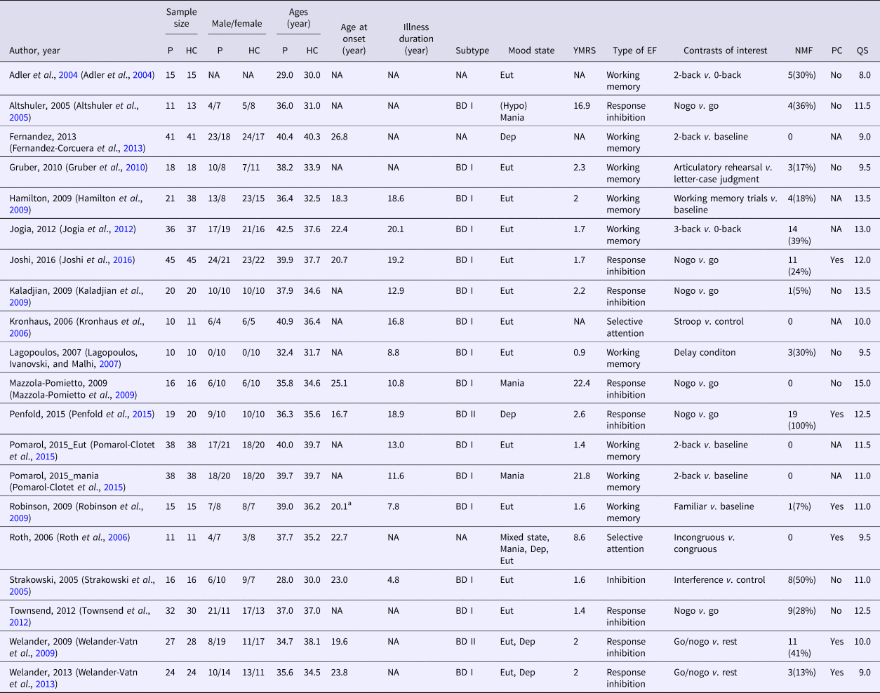
The main meta-analysis was based on 20 experiments which had been reported in 19 studies (Adler et al., Reference Adler, Holland, Schmithorst, Tuchfarber and Strakowski2004; Altshuler et al., Reference Altshuler, Bookheimer, Townsend, Proenza, Eisenberger, Sabb, Mintz and Cohen2005; Strakowski et al., Reference Strakowski, Adler, Holland, Mills, DelBello and Eliassen2005; Kronhaus et al., Reference Kronhaus, Lawrence, Williams, Frangou, Brammer, Williams, Andrew and Phillips2006; Roth et al., Reference Roth, Koven, Randolph, Flashman, Pixley, Ricketts, Wishart and Saykin2006; Lagopoulos et al., Reference Lagopoulos, Ivanovski and Malhi2007; Hamilton et al., Reference Hamilton, Altshuler, Townsend, Bookheimer, Phillips, Fischer, Woods, Mazziotta, Toga, Nuechterlein and Narr2009; Kaladjian et al., Reference Kaladjian, Jeanningros, Azorin, Nazarian, Roth and Mazzola-Pomietto2009; Mazzola-Pomietto et al., Reference Mazzola-Pomietto, Kaladjian, Azorin, Anton and Jeanningros2009; Robinson et al., Reference Robinson, Bearden, Monkul, Tordesillas-Gutierrez, Velligan, Frangou and Glahn2009; Welander-Vatn et al., Reference Welander-Vatn, Jensen, Lycke, Agartz, Server, Gadmar, Melle, Nakstad and Andreassen2009; Gruber et al., Reference Gruber, Tost, Henseler, Schmael, Scherk, Ende, Ruf, Falkai and Rietschel2010; Jogia et al., Reference Jogia, Dima, Kumari and Frangou2012; Townsend et al., Reference Townsend, Bookheimer, Foland-Ross, Moody, Eisenberger, Fischer, Cohen, Sugar and Altshuler2012; Fernandez-Corcuera et al., Reference Fernandez-Corcuera, Salvador, Monte, Salvador Sarro, Goikolea, Amann, Moro, Sans-Sansa, Ortiz-Gil, Vieta, Maristany, McKenna and Pomarol-Clotet2013; Welander-Vatn et al., Reference Welander-Vatn, Jensen, Otnaess, Agartz, Server, Melle and Andreassen2013; Penfold et al., Reference Penfold, Vizueta, Townsend, Bookheimer and Altshuler2015; Pomarol-Clotet et al., Reference Pomarol-Clotet, Alonso-Lana, Moro, Sarro, Bonnin, Goikolea, Fernandez-Corcuera, Amann, Romaguera, Vieta, Blanch, McKenna and Salvador2015; Joshi et al., Reference Joshi, Vizueta, Foland-Ross, Townsend, Bookheimer, Thompson, Narr and Altshuler2016) (see online Supplementary Fig. S1) and referred to 463 patients with BD [mean (s.d.) age, 36.87 (3.73) years] and 484 HC [mean (s.d.) age, 35.31 (3.13) years]. The main demographic and clinical characteristics of the participants and a summary description of the EF tasks performed in each study are presented in Table 1, whilst details of the imaging parameters, statistical threshold, and results of the between-group analysis for each study are presented in online Supplementary Table S3. One study (Adler et al., Reference Adler, Holland, Schmithorst, Tuchfarber and Strakowski2004) did not report the sex of patients, and for those studies which reported the ratio of males in the cohorts are 213/454 (46.9%) for patients with BD and 232/454 (48.7%) for HC. There were no significant differences in age (t = 1.43, p = 0.16) and sex (χ2 = 0.31, p = 0.58) between patients with BD and HC. A total of 96 patients (20.7%) were medication-free, being all patients of Penfold et al. (Reference Penfold, Vizueta, Townsend, Bookheimer and Altshuler2015) and an average of 26.7% patients of the 13 other datasets, and in which six datasets did not include any medication-free patients. The EF tasks to which the results of the meta-analysis refer include inhibition tasks (11 datasets, 236 patients, and 241 controls) and WM tasks (nine datasets, 232 patients, and 250 controls). Only 14 studies (Altshuler et al., Reference Altshuler, Bookheimer, Townsend, Proenza, Eisenberger, Sabb, Mintz and Cohen2005; Kronhaus et al., Reference Kronhaus, Lawrence, Williams, Frangou, Brammer, Williams, Andrew and Phillips2006; Roth et al., Reference Roth, Koven, Randolph, Flashman, Pixley, Ricketts, Wishart and Saykin2006; Lagopoulos et al., Reference Lagopoulos, Ivanovski and Malhi2007; Hamilton et al., Reference Hamilton, Altshuler, Townsend, Bookheimer, Phillips, Fischer, Woods, Mazziotta, Toga, Nuechterlein and Narr2009; Kaladjian et al., Reference Kaladjian, Jeanningros, Azorin, Nazarian, Roth and Mazzola-Pomietto2009; Mazzola-Pomietto et al., Reference Mazzola-Pomietto, Kaladjian, Azorin, Anton and Jeanningros2009; Welander-Vatn et al., Reference Welander-Vatn, Jensen, Lycke, Agartz, Server, Gadmar, Melle, Nakstad and Andreassen2009; Gruber et al., Reference Gruber, Tost, Henseler, Schmael, Scherk, Ende, Ruf, Falkai and Rietschel2010; Jogia et al., Reference Jogia, Dima, Kumari and Frangou2012; Townsend et al., Reference Townsend, Bookheimer, Foland-Ross, Moody, Eisenberger, Fischer, Cohen, Sugar and Altshuler2012; Fernandez-Corcuera et al., Reference Fernandez-Corcuera, Salvador, Monte, Salvador Sarro, Goikolea, Amann, Moro, Sans-Sansa, Ortiz-Gil, Vieta, Maristany, McKenna and Pomarol-Clotet2013; Welander-Vatn et al., Reference Welander-Vatn, Jensen, Otnaess, Agartz, Server, Melle and Andreassen2013; Penfold et al., Reference Penfold, Vizueta, Townsend, Bookheimer and Altshuler2015) reported results of within-group analysis.
Demographic and clinical characteristics of all subjects included in the meta-analysis

Altman Self-Rated Mania Scale, aage at the first episode
P, patients; HC, healthy controls; NA, not available; BD, bipolar disorder; Eut, euthymia; Dep, depression; YMRS, Young Manic Rating Scale; EF, Executive Function; NMF, number of patients of medication free; PC, psychiatric comorbidity; QS, quality score.
Meta-analysis of significant differences in brain activation between patients with BD and HC for EF tasks
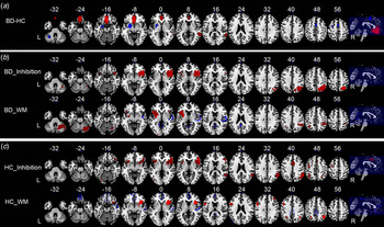
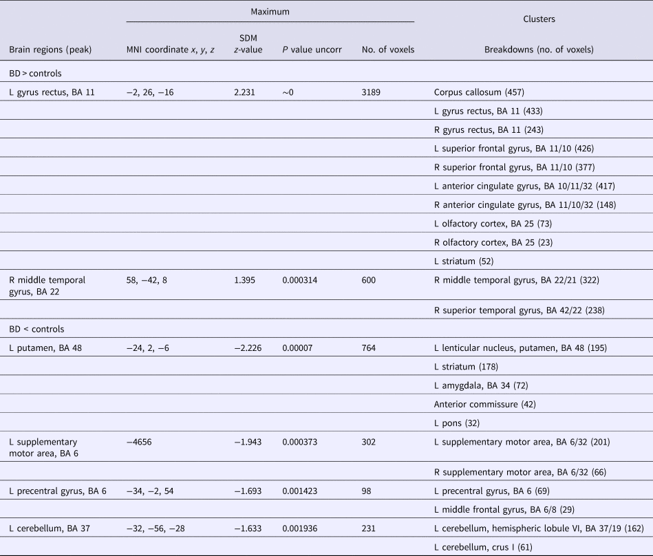
Compared with HC, patients with BD showed significant hypo-activation in the striatum (p = 0.00007), supplementary motor area (SMA, BA 6, p = 0.00037), and precentral gyrus (BA 6, p = 0.0014) of the left cerebral hemisphere together with left cerebellum (BA 37, p = 0.0019) (Fig. 1a and Table 2) and hyper-activation (i.e. failure of deactivation) in the left gyrus rectus (BA 11, p ≈ 0) and right middle temporal gyrus (BA 22, p = 0.00031). The hyper-activation (i.e. failure of deactivation) of left rectus gyrus (BA 11, MNI coordinates: x = −6, y = 34, z = −12; 442 voxels) was significantly heterogeneous between studies (p = 0.00003).
Results of meta-analysis using Anisotropic Effect Size Signed Differential Mapping showed that (a) BD patients showed significant hyper-activation (i.e. failure of deactivation) in right MTG and left gyrus rectus, and hypo-activation in left striatum, precentral gyrus and SMA and left cerebellum and left compared with the control group. Corresponding within-group analysis for patients with BD (b) showed significant activations right and left IFG, right SPG, ITG, SMA and ACC and right cerebellum, and left insula when performing inhibition tasks, and of right and left IFG, right insula and right cerebellum, and of left striatum, and MOG when performing WM tasks, whereas HC (c) showed significant activation of right and left IPG, right IFG, ACC, STG, MFG and striatum and left insula, fusiform gyrus, MFG, MOG, right striatum, when performing inhibition tasks, and of right and left IFG, right angular gyrus and MFG and of left SPG when performing WM tasks. BD, bipolar disorder; HC, healthy control; MTG, middle temporal gyrus; SMA, supplementary motor area; IFG, inferior frontal gyrus; SPG, superior parietal gyrus; ITG, inferior temporal gyrus; ACC, anterior cingulate cortex; WM, Working Memory; IPG, inferior parietal gyrus; MOG, middle occipital gyrus; MFG, middle frontal gyrus; STG, superior temporal gyrus.

Significant differences in brain activation between patients with BD and HCs

MNI, Montreal Neurological Institute; SDM, seed-based d mapping; BD, bipolar disorder; L, left; R, right; BA, Brodmann area.
Within-group brain activations
During performance of inhibition tasks, patients with BD showed activation in the left (BA 38, p = 0.0019) and right (BA 38, p = 0.0000042) inferior frontal gyrus, left insula (BA 48, p = 0.0017) and right SMA (BA 8, p = 0.0026), superior parietal gyrus (BA 7, p = 0.0000011), inferior temporal gyrus (BA 37, p = 0.00086), and anterior cingulate cortex (ACC, BA 24, p = 0.0033) as well as right cerebellum (BA 37, p = 0.0040). The right cerebellum activation (BA 37, MNI coordinates: x = 36, y = −60, z = −24) showed significant heterogeneity (p < 0.001). No brain region showed significant deactivation when BD patients performed the inhibition tasks.
During performance of WM tasks, patients with BD showed activation in the striatum (BA 48, p = 0.000017), inferior parietal gyri (BA 40, p = 0.00084), inferior parietal gyri (BA 3, p = 0.0013), and middle occipital gyrus (BA 18, p = 0.0037) in the left cerebral hemisphere (Fig. 1b and online Supplementary Table S4) and of insula (BA 47, p = 0.0033) and inferior parietal gyri (BA 40, p = 0.00028) of the right cerebral hemisphere together with right cerebellum (BA 19, p = 0.00086), and right inferior parietal gyri (BA 40, MNI coordinates: x = 38, y = −54, z = 46, p < 0.001) showed significant heterogeneity between studies. Patients with BD showed deactivation in the right superior temporal gyrus (BA 48) and right precuneus (BA 23), extending to posterior cingulate cortex.
During performance of inhibition tasks, HC showed activation in the insula (BA 48, p = 0.00027), middle frontal gyrus (BA 6, p = 0.00009), inferior parietal gyri (BA 40, p = 0.00015), middle occipital gyrus (BA 19, p = 0.0010), and fusiform gyrus (BA 19, p = 0.00072) of the left cerebral hemisphere and striatum (p = 0.0016), and inferior frontal gyrus (BA 45, p ≈ 0), inferior parietal gyri (BA 39, p = 0.00000030), ACC (BA 32, p = 0.000095), superior temporal gyrus (BA 42, p = 0.0026), and middle frontal gyrus (BA 46, p = 0.0032) of the right cerebral hemisphere. No brain region showed significant deactivation when HC performed the inhibition tasks.
During performance of WM tasks HC showed activation of left (BA 44, p = 0.00047) and right (BA 47, p = 0.00020) inferior frontal gyrus, left (BA 7, p = 0.0015) and right (BA 7, p = 0.00078) superior parietal gyrus and right angular gyrus (BA 40, p = 0.00018) and middle frontal gyrus (BA 46, p = 0.0023) (Fig. 1c and online Supplementary Table S4). No brain region showed significant heterogeneity in the within-group analysis for HC. No brain region showed significant deactivation when HC performed the inhibition tasks. However, for the WM tasks, HC showed significant deactivation in the left superior frontal gyrus (BA 11), ACC (BA 32), and posterior cingulate cortex (BA 23) and right superior temporal gyrus (BA 22) and precentral gyrus (BA 4).
Sub-domain meta-analyses
When inhibition and WM tasks are considered separately and brain activations compared between patients with BD and HC, for the inhibition tasks patients with BD showed significant hypo-activation in the left (BA 48, p = 0.0000002) and right (BA 48, p = 0.0021) putamen, left SMA (BA 32, p = 0.000042) and right inferior frontal gyrus (BA 47, p = 0.0010). No significant increases in activity were observed in patients with BD relative to HC. However, significant heterogeneity was observed for right inferior frontal gyrus (BA 47, MNI coordinates: x = 42, y = 28, z = −4) (p < 0.001). For the WM tasks, patients with BD showed significant hypo-activation in the left precentral gyrus (BA 6, p = 0.00012) and left cerebellum (BA 37, p = 0.0016) relative to HC (see online Supplementary Fig. S2 and online Supplementary Table S5) and hyper-activation (i.e. failure of deactivation) in the left gyrus rectus (BA 11, p = 0.00000018) and right middle (BA 22, p = 0.0013) and superior (BA 22, p = 0.0032) temporal gyrus, where significant heterogeneity was observed for right middle temporal gyrus (BA 22, MNI coordinates: x = 58, y = −46, z = 10) (p = 0.0014).
Sub-group analyses
Three sub-group analyses were performed.
Firstly, when the analysis was restricted to patients in the euthymic state, relative to HC, significant hypo-activation was observed in the bilateral striatum (BA 48), left SMA (BA 6), and right inferior parietal gyri (BA 2). Increased activation (i.e. failure of deactivation) was observed in the left gyrus rectus (BA 11), right middle temporal gyrus (BA 21), and right superior temporal gyrus (BA 22), which pattern is almost identical to that reported for the main analysis. Significant heterogeneity was observed for the left SMA (BA 6, MNI coordinates: x = −4, y = 6, z = 56, p = 0.02).
When patients in the euthymic state performed the inhibition tasks, only areas of decreased brain activation were found which included bilateral striatum, left SMA, and right ACC (BA 32) relative to HC, and when they performed the WM tasks, decreased activation was found in the left precuneus (BA 7) and right inferior occipital gyrus (BA 19) and DLPFC (BA 46), and increased activation was found in the left ventromedial prefrontal cortex (vmPFC, BA 11) and right superior temporal gyrus (BA 22). Furthermore, significant heterogeneity was observed for right DLPFC (BA 46, MNI coordinates: x = 42, y = 54, z = −4, p < 0.001).
Secondly, when the analysis was restricted to patients diagnosed as type I, significant hypo-activation was observed in putamen (BA 48) extending to insula and amygdala, SMA (BA 32), and caudate nucleus of the left cerebral hemisphere and significant hyper-activations in the right superior temporal gyrus (BA 22) and left superior frontal gyrus (BA 11) relative to HC. Significant heterogeneity was observed in the right superior temporal gyrus (BA 22, MNI coordinates: x = 58, y = −46, z = 12, p < 0.05).
Thirdly, when the analysis was restricted to patients free of current psychiatric comorbidity, significant hypo-activation was observed in the left and right putamen (BA 48) and left ACC (BA 24) extending to left SMA relative to HC. There were no areas of significant hyper-activation or failure of deactivation in patients with BD relative to HC (online Supplementary Table S6). No brain regions were observed to exhibit significant heterogeneity.
Reliability analyses and quality assessment
As shown in online Supplementary Table S7, whole-brain jack-knife sensitivity analysis confirmed that the findings reported for left striatum and gyrus rectus were highly reproducible. However, in one of the study combinations, the results for left middle temporal gyrus was not significant, whereas left SMA and precentral gyri results remained significant for all but two combinations of studies and the result for left cerebellum remained significant in all but three combinations of studies. The quality assessment score for each study included in the meta-analysis is presented in Table 1. The range of scores is 8–15 with an average score of 11 points.
Publication bias analysis
Application of Egger's test produced no significant findings for any brain region in the primary analysis (p > 0.05, Bonferroni corrected), indicating that no publication bias is present.
Meta-regression analyses
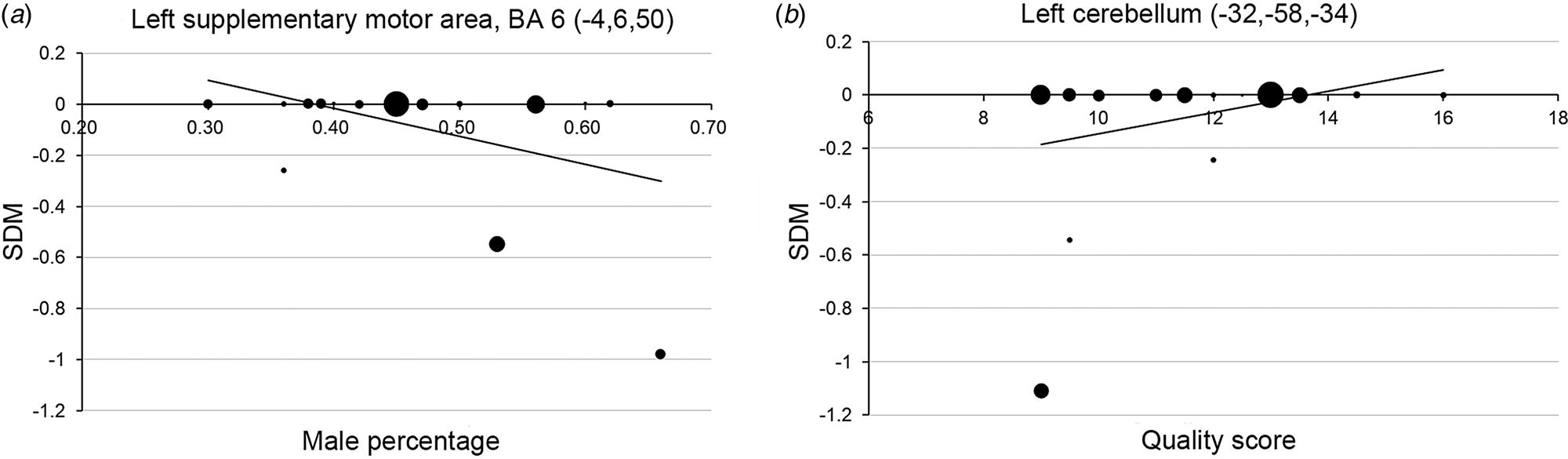
Meta-regression analysis of the effect of average age of the cohort, male percentage, age at onset, duration of illness, YMRS score, medication, and study quality on the difference in brain activation between patients with BD and HC showed that the greater the proportion of male patients in the cohort the more likely the observation of hypo-activation in the left SMA (MNI coordinates: x = −4, y = 6, z = 50; 38 voxels, SDM = −1.814, p = 0.0002, Fig. 2a). Furthermore, the lower the quality score of a study, the more likely the observation of hypo-activation of the cerebellum (MNI coordinates: x = −32, y = −58, z = −34; 282 voxels, SDM = 1.72, p = 0.00003, Fig. 2b). The meta-regression analysis produced no results relating to the average age of the cohort, age at onset, illness duration, YMRS score, or whether patients were receiving medication.
Results of meta-regression analysis. (a) higher proportion of males in the cohort is negatively associated with activation of left SMA, and (b) study quality score is positively associated with activation of left cerebellum.

Discussion
The fact that impairments in EF exist in patients with BD is well known, but the underlying alterations in brain function have yet to be established. In the present study, a meta-analysis has been performed of the results from all published task-related fMRI studies which compared brain activation in BD patients and HC performing EF tasks. Anomalous functioning of the striatum is the most significant finding. In particular, AES-SDM revealed hypo-activation (i.e. failure of activation) of the cortico-striatal system, including striatum, SMA, precentral gyrus, and cerebellum in BD patients relative to HC. Furthermore, hyper-activation (i.e. failure of deactivation) was found in the vmPFC in patients. In addition, activations of left SMA and left cerebellum were associated with the ratio of males in the study cohort and study quality, respectively.
The cortico-striatal system, namely striatum, SMA, precentral gyrus, and cerebellum, are commonly reported to be activated during the performance of EF tasks. For example, the striatum is mainly reported to be involved in the inhibitory control of motor behavior (Aron et al., Reference Aron, Schlaghecken, Fletcher, Bullmore, Eimer, Barker, Sahakian and Robbins2003; Chevrier et al., Reference Chevrier, Noseworthy and Schachar2007), and in a magnetic resonance spectroscopy study, it was reported that EF performance and NAA/Cr ratios of the striatum were both significantly reduced in BD patients relative to HC (Lai et al., Reference Lai, Zhong, Liao, Wang, Huang, Zhang, Sun, Zhao and Jia2018). SMA, on the other hand, plays a role in programming motor subroutines (Roland et al., Reference Roland, Larsen, Lassen and Skinhoj1980; Weinrich and Wise, Reference Weinrich and Wise1982) and inhibiting unwanted movements (Mostofsky et al., Reference Mostofsky, Schafer, Abrams, Goldberg, Flower, Boyce, Courtney, Calhoun, Kraut, Denckla and Pekar2003) and electrical stimulation of this area has been observed to inhibit actions (Fried et al., Reference Fried, Katz, McCarthy, Sass, Williamson, Spencer and Spencer1991). Patients with BD have been reported to show impaired performance of the Stroop color-word test and which was associated with decreased regional homogeneity (i.e. ReHo) in left SMA (Cui et al., Reference Cui, Gao, Jiao, Cao, Qi, Guo, Chen, Lu, Xiao, Su and Lu2016). The precentral gyrus has been reported to support planning and executing actions to achieve a behavioral goal (Nakayama et al., Reference Nakayama, Yamagata and Hoshi2016), and the finding of the present study is consistent with the report of hypo-activation of the precentral gyrus in BD patients relative to HC during the performance of EF tasks (Penfold et al., Reference Penfold, Vizueta, Townsend, Bookheimer and Altshuler2015). This anomalous function may be the result of reduced myelination which has previously been suggested based on the analysis of contrast between signals from grey and white-matter (Jorgensen et al., Reference Jorgensen, Nerland, Norbom, Doan, Nesvag, Morch-Johnsen, Haukvik, Melle, Andreassen, Westlye and Agartz2016). The cerebellum has been suggested to play a role in the formation of verbal WM traces (Ravizza et al., Reference Ravizza, McCormick, Schlerf, Justus, Ivry and Fiez2006; Boehringer et al., Reference Boehringer, Macher, Dukart, Villringer and Pleger2013; Tomlinson et al., Reference Tomlinson, Davis, Morgan and Bracewell2014). The striatum and the cerebellum both receive inputs from precentral gyrus and SMA (Hanakawa, Reference Hanakawa2011) which may significantly affect their functioning (Middleton and Strick, Reference Middleton and Strick2000).
The findings of the present study are consistent with a previous review in which anomalies in the function of the cortico-striatal system were reported in patients with BD (Green et al., Reference Green, Cahill and Malhi2007). Furthermore, using resting-state fMRI, Stoddard et al. (Reference Stoddard, Gotts, Brotman, Lever, Hsu, Zarate, Ernst, Pine and Leibenluft2016) reported that aberrant functional connectivity of the cortico-striatal system was correlated with cognitive control in BD. However, the findings of the present study partly differ from those reported in the meta-analysis of Alustiza et al. On the one hand, Alustiza et al. found the decreased activation areas were located mostly in the right hemisphere and the right inferior frontal gyrus, orbital part (BA 47) was the main hypoactivated area. These differences may be due to the emotion involvement during EF tasks in Alustiza et al. On the other hand, the hypoactivation of the striatum found in our study was consistent with that of Alustiza et al., which supports the opinion that the failure of activation in the striatum may be a core feature of BD.
The impairment of the striatum is significant and stable as reflected by the high p-value of the main result, the results of the heterogeneity and sensitivity analysis, and the sub-group analyses of euthymic and medication-free patients. However, abnormalities in the function of the striatum appear to be specifically related to the sub-domain of inhibition (p = 0.0000002, uncorrected) than WM. This is consistent with the demonstration by Vink et al. (Reference Vink, Kahn, Raemaekers, van den Heuvel, Boersma and Ramsey2005) that the striatum is critical for the control of execution of planned motor responses. Furthermore, at the molecular level, dopamine plays a crucial role in modulating motor readiness for inhibition (Ghahremani et al., Reference Ghahremani, Lee, Robertson, Tabibnia, Morgan, De Shetler, Brown, Monterosso, Aron, Mandelkern, Poldrack and London2012), and using PET imaging, Anand et al. (Reference Anand, Barkay, Dzemidzic, Albrecht, Karne, Zheng, Hutchins, Normandin and Yoder2011) reported that the dopamine transporter was significantly lower in the striatum in patients with BD relative to HC. In addition, in animal studies, the reaction time for the stop-signal was reported to be significantly slower in rats with lesions in medial striatum compared with normal rats (Eagle and Robbins, Reference Eagle and Robbins2003) and inhibition control was positively correlated with increased putamen volume in methamphetamine-exposed monkeys (Groman et al., Reference Groman, Morales, Lee, London and Jentsch2013). Finally, at the level of the gene, the concentration of the receptor TrkB mRNA for brain-derived neurotrophic factor was reported to be reduced in patients with BD (Reinhart et al., Reference Reinhart, Bove, Volfson, Lewis, Kleiman and Lanz2015), and Pacifico and Davis (Reference Pacifico and Davis2017) performed a transcriptomic analysis in the post-mortem BD patients and reported anomalies in the gene network specific to the dorsal striatum. Given the implications of associated findings at the level of the gene, molecule, and brain region, striatum appears to be a highly promising brain structure for further studies of the neural bases of impairments of inhibition in patients with BD.
In addition to the above findings of hypo-activation (i.e. failure of activation) of brain function in BD patients relative to HC, hyper-activation (i.e. failure of deactivation) was observed in vmPFC in BD patients relative to HC. There are at least two possible explanations for this finding. Firstly, the failure of deactivation may be related to abnormal function of the Default Mode Network (DMN) (Raichle and Mintun, Reference Raichle and Mintun2006) in patients with BD. This is supported by the fact that according to the definition of (Raichle et al., Reference Raichle, MacLeod, Snyder, Powers, Gusnard and Shulman2001), vmPFC lies within the DMN, activity of which is likely to be suppressed when attention-demanding, goal-directed EF tasks are performed (Broyd et al., Reference Broyd, Demanuele, Debener, Helps, James and Sonuga-Barke2009). Interestingly, the within-group analysis showed vmPFC to be deactivated in HC during the performance of WM tasks. Secondly, hyper-activation of vmPFC may indicate that patients with BD had to recruit more neurons than HC during the performance of EF tasks. This is supported by the fact that vmPFC is involved in performance monitoring, including detecting performance errors or conflicting response tendencies (Ridderinkhof et al., Reference Ridderinkhof, van den Wildenberg, Segalowitz and Carter2004; Alexander and Brown, Reference Alexander and Brown2010).
Relative to HC, euthymic patients showed abnormalities of brain function in left and right striatum, left SMA and right inferior parietal gyri when performing EF tasks and the finding is more significant than for the pooled results referring to all BD patients. Furthermore, the sub-domain, sub-group analyses revealed that the striatum and SMA were preferentially involved in inhibition and the parietal gyrus in WM. Taken together these findings are consistent with the interpretation that the greater hypo-activity of the striatum bilaterally in euthymic patients is related to their generally worse performance in performing inhibition tasks (Filip et al., Reference Filip, Linhartova, Hlavata, Sumec, Balaz, Bares and Kasparek2018). Furthermore, inferior parietal cortex is important for retaining temporal information in both verbal and visual–spatial WM tasks (Ravizza et al., Reference Ravizza, Delgado, Chein, Becker and Fiez2004) and right parietal cortex plays a significant role in spatial rehearsal (Owen et al., Reference Owen, McMillan, Laird and Bullmore2005) on the performance of both of which is compromised in euthymic patients.
Previously it has been reported that impairment of EF is greater in BD-I than BD-II (Hsiao et al., Reference Hsiao, Wu, Wu, Hsu, Chen, Lee, Lee, Yeh, Yang, Ko and Lu2009) and additional cognitive impairments have been reported in BD-I (Silveira et al., Reference Silveira, Kozicky, Muralidharan, Bucker, Torres, Bond, Kapczinski, Kauer-Sant'Anna, Lam and Yatham2014). In the present analysis, however, there were no sufficient studies which had recruited BD-II patients to make a comparison with BD-I. Interestingly, neither of the two studies which included BD-II patients reported activation of the caudate during the performance of EF tasks, and in future work, it may be found that this anomaly is specific to BD-I patients. This is consistent with findings from a structural MRI study in which increased volume of the caudate nucleus was reported in patients with BD-I (Kozicky et al., Reference Kozicky, Ha, Torres, Bond, Honer, Lam and Yatham2013). Additionally, in the present study, a significant anomaly of cortico-striatal function was observed in BD-I patients and this is consistent with a previous brain PET study of BD-I patients (Li et al., Reference Li, Hsieh, Wang, Yang, Bai, Lin, Lan and Su2012).
Unlike previous fMRI studies (Ranganath et al., Reference Ranganath, Johnson and D'Esposito2003; Moreno-Lopez et al., Reference Moreno-Lopez, Stamatakis, Fernandez-Serrano, Gomez-Rio, Rodriguez-Fernandez, Perez-Garcia and Verdejo-Garcia2012; Weinstein et al., Reference Weinstein, Voss, Prakash, Chaddock, Szabo, White, Wojcicki, Mailey, McAuley, Kramer and Erickson2012; Han et al., Reference Han, Han, Bolo, Kim, Kim and Renshaw2015; Hough et al., Reference Hough, Luks, Lai, Vigil, Guillory, Nongpiur, Fekri, Kupferman, Mathalon and Mathews2016), the present analysis did not reveal decreased activation of DLPFC during the performance of EF tasks by BP patients. However, this is perhaps not surprising. Although fMRI studies have shown that DLPFC is an important brain region for EF (Miller and Cohen, Reference Miller and Cohen2001) this is more so for WM (Curtis and D'Esposito, Reference Curtis and D'Esposito2003; Barbey et al., Reference Barbey, Koenigs and Grafman2013; Brunoni and Vanderhasselt, Reference Brunoni and Vanderhasselt2014) than inhibition (Aron et al., Reference Aron, Robbins and Poldrack2004) tasks and this was supported by the results of sub-domain analysis. Thus, pooling results for inhibition and WM tasks may have reduced the significance of the DLPFC finding in the main meta-analysis. In addition, perhaps the findings were influenced by the effect of the medication. The patients recruited to the vast majority of studies included in the present meta-analysis were receiving medication and perhaps there would have been greater activation of DLPFC during the performance of EF tasks if unmedicated patients had been studied (Adler et al., Reference Adler, Holland, Schmithorst, Tuchfarber and Strakowski2004; Lesh et al., Reference Lesh, Tanase, Geib, Niendam, Yoon, Minzenberg, Ragland, Solomon and Carter2015).
There are several limitations of the meta-analysis reported here that should be considered. Firstly, since fMRI studies employ correlational methods, it is not possible to use the approach to unequivocally confirm whether brain regions are necessary for the performance of EF tasks or simply have an accessory role (Sarter et al., Reference Sarter, Berntson and Cacioppo1996). Secondly, none of the studies that were available to incorporate in the meta-analysis had included the performance of a set-shifting task. Thus the results obtained do not represent the full range of EF tasks and it is notable that there were significant differences between the results of the sub-modal analysis of the inhibition and WM tasks. In future research, it will be interesting to discover whether a functional abnormality in a particular brain region may underlie impairment of EF in all three domains of inhibition, WM, and task-switching in patients with BD. Thirdly, the majority of the patients included in the present meta-analysis were in a euthymic state, and so may not reflect the results that would be obtained from a study of patients in a manic or depressed state. Finally, the possibility that medication may have contributed to the findings of the present meta-analysis cannot be ruled out. Of the three studies which included both medicated and non-medicated patients, in two it was reported that there was no influence of medication on the fMRI activations (Strakowski et al., Reference Strakowski, Adler, Holland, Mills, DelBello and Eliassen2005; Welander-Vatn et al., Reference Welander-Vatn, Jensen, Lycke, Agartz, Server, Gadmar, Melle, Nakstad and Andreassen2009) and in one that medication may have affected the activation of ACC and DLPFC (Adler et al., Reference Adler, Holland, Schmithorst, Tuchfarber and Strakowski2004). Furthermore, Philips et al. (Reference Phillips, Travis, Fagiolini and Kupfer2008) reviewed the available literature and reported that some psychotropic medications may have an effect on brain activity in patients with BD. Thus, further studies are required to determine the extent to which the results of the meta-analysis reported here are domain-specific, group-specific, and if they are potentially influenced by effects of medication.
In conclusion, patients with BD showed significant abnormalities of brain activation compared with HC during the performance of EF tasks. The anomalous function of the striatum might represent a biomarker for impairment in EF in patients with BD, especially with respect to inhibition tasks.
Supplementary material
The supplementary material for this article can be found at https://doi.org/10.1017/S0033291719000473
Acknowledgments
This study was supported by the National Natural Science Foundation of China (Grant Nos. 81771812 and 81571637), the Sichuan Science and Technology Program (2018SZ0391), the Fundamental Research Funds for the Central Universities (Grant No. 2012017yjsy190).
Conflict of interest
None.