Introduction
Within the last decade, a subtype of Fronto-temporal lobar degeneration (FTLD) has been described which stains positive for the Fused in Sarcoma (FUS) protein at autopsy. This pathology appears to be associated with a distinct clinical subtype and was associated with FTLD in 5/118 patients from two centers (Snowden et al., Reference Snowden2011). Given its relative rarity and questions as to whether it is associated with mutations on the FUS gene, it is important for clinicians to be aware of this entity. We report here a pathologically confirmed case of FTLD-FUS leading to Fronto-temporal dementia (FTD). The patient's widow gave written informed consent to the publication of this case report.
There were 15 patients initially reported to have FUS inclusions as a subtype of FTLD, associated with a distinct phenotype of sporadic, early-onset (mean age at onset = 38; range = 28–55 years) behavioral variant FTD (bvFTD) with severe progressive changes in behavior and personality (Neumann et al., Reference Neumann, Rademakers, Roeber, Baker, Kretzschmar and Mackenzie2009). Language was not affected until late in the disease when aspontaneity and other frontal features appeared. Mean disease duration was seven years (range 4– 15 years).
Several subsequent case series of patients with FUS inclusions have also described a characteristic syndrome of young onset bvFTD but additionally with caudate atrophy and absent family history (Munoz et al., Reference Munoz2009; Rohrer et al., Reference Rohrer2010; Seelaar et al., Reference Seelaar2010; Urwin et al., Reference Urwin2010; Snowden et al., Reference Snowden2011; Chare et al., Reference Chare2014).
Urwin et al. (Reference Urwin2010) reported a high prevalence (36%) of psychotic symptoms. Snowden et al. (Reference Snowden2011) also observed a “stereotypic” behavioral subtype with obsessionality, rituals, social withdrawal, hyperorality, and utilization behavior, possibly related to caudate nucleus atrophy.
Discovery of the FUS gene triggered speculation that mutations of this gene may cause FTLD-FUS, but to date mutations in the FUS gene have only been associated with amyotrophic lateral sclerosis (Kwiatkowski et al., Reference Kwiatkowski2009) with no reported cases of FTLD or even of a strong family history of dementia.
Case description
Our patient was a well-educated, married business professional. Medical history included biopsy-proven pulmonary sarcoidosis (with no iritis or neurological complications), hypertension, and prostatism.
His mother died suddenly aged 79 years from probable cardiac causes, and his father aged 81 years from multiple myeloma. He has an older sister who is well.
He was noted to decline gradually from about mid-2013. He repeated stories, forgot conversations, hesitated when speaking, seemed subtly confused, had difficulty in understanding complex explanations, increased anxiety, and subtle change in personality, such as paying many compliments, which was out of character.
MRI brain in October 2013 compared with previous MRI from December 2012 (performed to rule out neurosarcoidosis) noted prominence of the frontal and temporal lobe sulci, further dilation of the temporal horn of the lateral ventricle and slight dilation of the third ventricle. There were also a number of small rounded T2 hyperintense foci in the deep hemispheric white matter of bilateral frontal and parietal lobes.
Neurological examination in November 2013 was normal with no frontal release signs or extra-pyramidal signs, Mini-Mental Sate Examination (MMSE) score 27/30 with 0/3 for word recall and inaccurate hand placement on the clock drawing test. Neuropsychological examination in late 2013 revealed markedly reduced phonemic fluency and inefficient new learning, but no rapid forgetting or anomia.
FDG-PET in January 2014 showed hypometabolism in medial and orbital frontal lobes, left greater than right medial and anterior temporal lobes, and lesser hypometabolism in bilateral parietal and left lateral temporal cortex.
By mid-2014, he had ceased working and displayed adynamia, social withdrawal, weight gain associated with having developed a sweet tooth and obsession with photographs and painting. By this stage, he fulfilled criteria for probable bvFTD (Rascovsky et al., Reference Rascovsky2011). Donepezil 10 mg, Souvenaid and escitalopram 20 mg were each trialed with no clear benefit. By late 2014 he developed disinhibition, such as approaching and touching strangers. In November 2014, MMSE score was 25/30 with 1/3 for word recall and mild disorientation and Abbreviated Mental Test Score (AMTS) was 8/10.
Memantine was trialed from late 2014 with no clear benefit. MMSE in February 2015 was 25/30 with 0/3 for word recall and mild disorientation.
By April 2015, disinhibition and impulsivity evolved to include shoplifting sweets, asking his daughter-in-law if she was wearing a brassiere and insisting on taking walks at night.
Repeat FDG-PET in April 2015 showed hypometabolism in the frontal and anterior temporal poles, worse on the left and in the left caudate nucleus. Brain amyloid PET imaging in April 2015 was negative for amyloid plaques.
In May 2015, AMTS was 6/10 with confabulation, psychomotor agitation and utilization.
By July 2015, he was regularly spitting on the floor and was markedly agitated. Risperidone was commenced and titrated up to an eventual dose of 2 mg daily with marginal benefit.
Examination in July 2015 revealed two-way anomia with loss of conceptual meaning of words, phonemic and semantic paraphasic errors, but no prosopagnosia, consistent with semantic dementia.
He entered residential care in October 2015, at which stage he was largely mute, and died of pneumonia in February 2016.
Neuropathology
Brain weight was 1,560 g. External examination showed softening, loss of tissue, and discoloration of the temporal and inferior frontal poles. Serial coronal sectioning revealed severe loss of tissue from the temporal pole and inferior aspect of the frontal lobe. Basal ganglia, diencephalon, cerebellum, and brainstem appeared unremarkable. There was atrophy to the caudate nucleus and pallor to the substantia nigra.
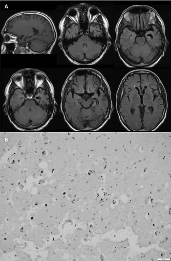
Sections from the atrophic temporal lobe showed subtotal neuronal loss and gliosis. There were numerous rounded FUS immunoreactive inclusions in the dentate and in residual pyramidal cells and in the adjacent cortical neurons, but in general there was severe loss of neurons in the hippocampus especially posteriorly (see Figure 1). Similar FUS-positive inclusions were seen in the frontal cortex, some with a ring-like structure that immunoreacted with FUS antibody. There was no evidence of granulomata or TDP43, beta amyloid or tau deposition. The morphologic features of basophilic inclusion body disease such as the rounded 7–30 µm basophilic bodies were not seen. There was demyelination of white matter secondary to wallerian degeneration underlying cortical areas with severe neuronal loss. Caudate nucleus showed loss of neurons. Brainstem sections showed substantia nigra with neuronal loss and pigment incontinence with no evidence of Lewy body formation. Examination was consistent with atypical FTLD with ubiquitin-positive inclusions.
(A) T1 sagittal and FLAIR axial MRI shows focal atrophy of the left temporal lobe with dilatation of the temporal horn of the lateral ventricle. There is also atrophy of the head of the caudate nucleus. (B) Pyramidal layer of the hippocampus (CA1) showing severe neuronal loss and gliosis with positive ring-like FUS inclusions within neurons and within the neutrophil. (FUS immunoperoxidase ×200).

Conclusion
FTLD-FUS has previously been associated with a distinct clinical syndrome of young age at onset, bvFTD with prominent changes in behavior and personality, absent family history and caudate atrophy (Neumann et al., Reference Neumann, Rademakers, Roeber, Baker, Kretzschmar and Mackenzie2009; Josephs et al., Reference Josephs2010; Seelaar et al., Reference Seelaar2010; Snowden et al., Reference Snowden2011). The present case was also a sporadic bvFTD with caudate atrophy, but was atypical in that he was older (59 years at symptom onset) and evolved to include language features. Moreover, the clinical course was quite rapid.
The main case series of FTLD-FUS report a mean age of onset of around 40 years with a range of 22–57 years (Neumann et al., Reference Neumann, Rademakers, Roeber, Baker, Kretzschmar and Mackenzie2009; Rohrer et al., Reference Rohrer2010; Seelaar et al., Reference Seelaar2010; Urwin et al., Reference Urwin2010; Snowden et al., Reference Snowden2011). Our case developed symptoms at 59 years, was diagnosed at 60 and died at 61. Thus our case was 2 years older than the known maximum age – the diagnosis should not be excluded if a patient presents in their late 50s.
All published cases of FTLD-FUS have presented with bvFTD (Neumann et al., Reference Neumann, Rademakers, Roeber, Baker, Kretzschmar and Mackenzie2009; Seelaar et al., Reference Seelaar2010; Urwin et al., Reference Urwin2010; Snowden et al., Reference Snowden2011; Chare et al., Reference Chare2014). While our case also initially presented with prominent changes in behavior and personality, his dementia did evolve to include features consistent with semantic dementia two years after symptom onset.
Conflict of interest
None.
Description of authors' roles
NI Bradfield drafted the manuscript. C McLean was the neuropathologist and assisted in revising the manuscript. J Drago, DG Darby and D Ames all saw the patient and assisted in revising the manuscript.
Acknowledgments
The authors would like to thank Dr Ben Harris, who conducted and provided the neuropsychological assessment and report, and the patient's family.