Introduction
The mutualism between fig trees (Ficus spp.) and their pollinating wasps is a classic model of coevolution, which has stimulated much research into the evolution, population genetics, biogeography, and ecology of the system (Janzen, Reference Janzen1979; Weiblen, Reference Weiblen2002; Jandér & Herre, Reference Jandér and Herre2010). However, the biology of the immature stages of the pollinating wasps has been relatively neglected (Galil & Eisikowitch, Reference Galil and Eisikowitch1968a ; Valdeyron & Lloyd, Reference Valdeyron and Lloyd1979). Even the location of egg deposition within the Ficus ovary has been revealed only recently by histological methods (Elias et al., Reference Elias, De Padua Teixeira, Kjellberg and Augusto Santinelo Pereira2012). The lack of information on the delimitation and description of pollinator larvae hinders the progress of various aspects of fig wasp research. Unlike most other wasps, pollinating and non-pollinating fig wasps are confined in their larval stages to the Ficus syconium, the enclosed inflorescence characteristic of the genus (Weiblen, Reference Weiblen2002). Pollinating fig wasps induce galls in Ficus ovaries, with one larva developing in each gall. Because the Ficus syconium is a closed system, it is not possible to observe live larvae at any point in time other than through destructive sampling.
Ficus hispida L. is a functionally dioecious (gynodioecious) species, in that the male and female reproductive functions are confined to different trees; seeds develop in the syconia of the female trees, and pollen (and pollinating wasps) in the syconia of the male trees. On Hainan Island, the pollinator species of F. hispida is Ceratosolen somlsi marchali Mayr. Female pollinator wasps (foundresses) enter a receptive syconium through the ostiole. When inside the central lumen of the syconium on a male tree, each foundress deposits her eggs individually into the ovaries by inserting her ovipositor through the styles, while simultaneously distributing the pollen that she has carried from her natal tree. The oviposited flowers are induced to become galls. After oviposition, each offspring grows within its gall as its natal syconium also grows to maturity, a process that takes approximately 30 days. Upon maturation, the male pollinators emerge from their galls first, mate with the females, and then chew holes in the syconium wall to enable the now pollen-laden females to disperse to other receptive F. hispida trees (Yang et al., Reference Yang, Peng, Song, Zhang, Wang, Zhao and Wang2002). Under natural condition, more than one (often 2 or 3) foundresses oviposit in a single syconium of F. hispida (Yang et al., Reference Yang, Peng, Song, Zhang, Wang, Zhao and Wang2002). F. hispida trees also harbor three non-pollinating fig wasp species (Peng et al., Reference Peng, Yang and Wang2005). Besides, there are abundant and diverse fig-associated insects, such as pulp eating insects (e.g., moths), sap feeders (e.g., aphids), predators (e.g., ants), and decomposers (e.g., fruit flies). Taken together with the pollinating fig wasps and non-pollinating fig wasps, they comprise a complex ecosystem (Yang et al., Reference Yang, Peng, Yang and Guan2008).
Lack of knowledge of the immature stages of pollinating fig wasps hinders progress in a variety of studies, such as wasp development and clarifying the ecological relationships between different wasp species. The purpose of this study was to delimitate and describe the immature stages of a pollinating fig wasp species, Ceratosolen solmsi marchali, the pollinator of F. hispida. We did this by monitoring wasp larvae of known ages over their development from egg to adult, a process that involved almost 1 month. Our data enabled us to identify the various key life-cycle stages, and to provide diagnostic characters of the pupal stage for both sexes.
Materials and methods
Study system and wasp life cycle
The material used for the study was collected from three male F. hispida trees (19°30′N, 109°31′E) growing near to the campus of the Chinese Academy of Tropical Agricultural Sciences in Danzhou, Hainan Island from June 12 to September 15.
Syconium sampling
Initially, we identified three male F. hispida trees that each displayed a large crop of pre-receptive syconia (A-phase) (Galil & Eisikowitch, Reference Galil and Eisikowitch1968b ). To prevent natural wasp oviposition and the interference of other insect species, we covered several fig branches with fine-mesh nylon bags, each displaying several syconia. All branches were inspected daily until the syconia within each bag showed signs of receptivity, with the entrance to the ostiole becoming visible. On detecting receptivity, the bag was removed, and a single female adult C. solmsi marchali (mated or virgin) that had been recently collected from a mature syconium of a nearby tree was placed at the entrance to the ostiole of each syconium. After wasp introduction, the figs were immediately re-covered with fine-mesh nylon bags to exclude other fig wasps and insects. After foundress entrance, we removed 3–6 syconia every 24 h and returned them to the laboratory for processing and measurement. A total of 521 syconia were checked. Average of daily maximum and minimum temperature was 34–25 °C, released by the National Meterological Administration.
Dissection, observation and measurement
Each syconium was dissected by a cross cut of the outside wall with a scalpel. With the aid of a stereomicroscope (SZ61, Olympus) and a pair of fine forceps, swollen galls were removed into a Petri dish containing 0.9% physiological saline. By the incremental removal of the ovary tissue, the contents of each gall could be exposed (egg or larva, depending on the age of the gall since foundress introduction). The ovaries from adult female wasps were also dissected to examine egg morphology prior to oviposition. Because fig wasp ovaries were pale and opaque (Dunn et al., Reference Dunn, Jansen-González, Cook, Yu and Pereira2011), the ovarian tissue was carmine dyed (Fagerström & Wiklund, Reference Fagerström and Wiklund1982). After dissection, each sample was photographed and measured with a SteREO Lumar.V12 imaging system (Carl Zeiss) using AxioVision (Carl Zeiss) image analysis software. Larvae for scanning electron microscope (SEM) observation were fixed in 4% glutaraldehyde and stored in 100% ethanol before dried in the Leica EM CPD300 Critical Point Dryer. They were then affixed to aluminum SEM stubs adhesive tabs. Stub-mounted specimens were sputter-coated with gold. The specimens were inspected under a Quanta200 SEM. Measurements recorded include body length, width, cephalic width, and mandible length. With respect to the measurements of curve bodies for certain stages, we measured several short straight parts along the midline of larval body, and then added them up to compute the total body length.
Sex identifying of immature C. solmsi marchali
PCRs of sex-specific transcripts of doublesex (dsx), an important gene in sex determination (Oliveira et al., Reference Oliveira, Werren, Verhulst, Giebel, Kamping, Beukeboom and Van De Zande2009), were used to confirm the sexes of larvae as early as 16 days post-oviposition (unpublished data). The results showed that all the offspring of virgin females were males, and most offspring of mated females were females (female/total offspring was 0.89±0.06, n=9). This was consistent with the previous reports that in one-foundress figs, offspring sex ratios of C. solmsi were highly female-biased (Hu et al., Reference Hu, Chen, Jiang, Huang, Niu and Fu2013). In addition, we did more than 20 sets (30 eggs or larvae per set) of morphologic comparisons on the eggs and larvae between the male offspring of virgin females and sex-unknown offspring of mated females. If there were any significant morphological difference between two sexes, it would be easily to distinguish.
Results
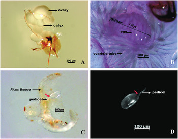
Eggs within ovaries (fig. 1A, B)
Pollinating fig wasps were proovigenic, with all eggs mature at eclosion. Approximately 100 eggs were in each calyx (fig. 1A, B).
Ovarian egg and a newly laid egg of C. solmsi marcahli: (A) Reproductive system of an adult female. (B) Inflated calyx and ovariole tubules after dissection. Calyxes were 401.23±0.02 μm (n=15) in length. (C) Egg adhering to a gall (4th day post-oviposition). (D) Egg separated from the Ficus tissue (4th day post-oviposition). Red arrows in C and D indicate cavities at the posterior end of eggs, emerged during embryo development.

Egg body elongate and pale in color (egg body: 111.62±0.41 μm long, range 97.85–123.24 μm; 39.62±0.07 μm wide, range 37.96–41.77 μm; n=17), with the posterior end connected to a pedicel (pedicel length: 52.03±0.09 μm, range 48.66–54.43 μm; n=17).
Eggs post-oviposition (fig. 1C, D)
The youngest eggs we could examine from galls were those on the second day post-oviposition. The egg was deposited inside the Ficus ovary with the pedicel attached to the style where it joined the Ficus ovary.
Egg smooth, without any tubercule or spine. No micropyle or sculpturing. The shape, color, and general appeared similar to ovarian eggs, except for the increase in size (body length: 146.67±0.39 μm, range 129.51–164.55 μm; body width: 68.61±0.15 μm, range 61–75.21 μm; pedicel length: 69.19±0.10 μm, range 64.26–73.42 μm; n=22). The developing embryo in the egg migrated to the anterior of the egg, leaving a cavity at the posterior end (fig. 1C, D).
After 5 days post-oviposition, a few larvae were inside the Ficus ovary, whereas others remained within eggs, which suggested that neonate larvae emerge about 5 days after oviposition (4.81±0.02 days, n=26).
Larval stages
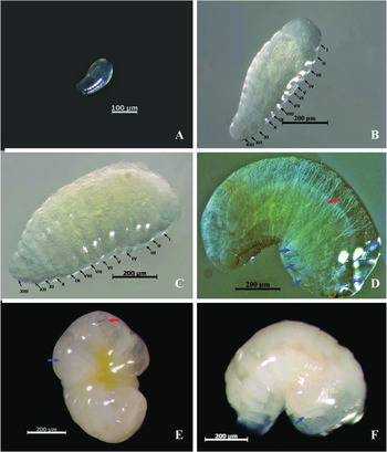
C. solmsi marchali had a typical chalcidiform larva, with a smooth, weakly sclerotized body surface, without any setae or tubercle (fig. 2A–F). The labium was not distinguishable. Mandibles were sclerotized and unidentate. Antennal tubercles were not present (Parker, Reference Parker1924; Chu & Cutkomp, Reference Chu and Cutkomp1992).
Larva and prepupa of C. solmsi marcahli: (A) First-instar larva (5th day post-oviposition). (B) Second-instar larva (10th day post-oviposition). (C) Third-instar larva (11th day post-oviposition). (D) Fourth-instar larva (16th day post-oviposition), showing presumed limb imaginal discs wrapped in each protrusion (blue arrows) and yellowish contents of alimentary tract (red arrow). (E) Fifth-instar larva from ventral view (19th day post-oviposition), showing lateral and latero-ventral protrusion (blue arrow) and red mouth parts (red arrow). (F) Prepupa (20th day post-oviposition) resembled the final-instar larva, lateral view, showing swollened mesosomal region of prepupa (blue arrow). Thirteen body segments (I–XIII) were illustrated in (B, C).

Changes in body form and mondibular structure indicate five instars prior to pupation. In first instar, the mandibles were indistinct even using scanning electron microscopy, width measurements of head capsules were made to check the instar delimitation. Exuviae from third and fourth instars that were casually found attached to larvae helped confirm the instars. We were not able to determine the precise timing of every larval instar by monitoring molting, because the larvae were often embedded in fragments of the Ficus ovary, which looked similar to exuviae. Larvae in instars 2–5 all had cranium and 13 body segments (I: head; II–IV: mesosoma; V–XIII: metasoma) (figs 2B, C, 3B and 4A).
Third- and fourth-instar larva (A) Mouthparts of third-instar larva (13th day post-oviposition), showing sharply edentate mandibles. (B) Ventral view of fourth-instar larva (17th day post-oviposition). (C) Enlarged view of mouthparts of fourth-instar larva (md: mandible; mp: maxillary palp; lp: labial palp). Thirteen body segments (I–XIII) were illustrated in B.

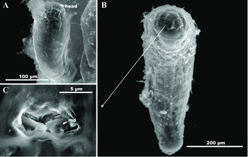
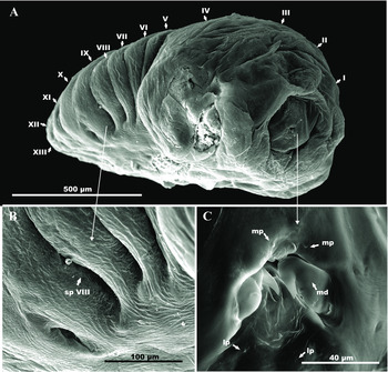
Final-instar larva under SEM: (A) Lateral view of fifth-instar larva (19th day post-oviposition). (B) Enlarged view of spiracle, showing the largest spiracle (spVIII). (C) Enlarged view of mouthparts (md: mandible; mp: maxillary palp; lp: labial palp). Thirteen body segments (I–XIII) were illustrated in A.

First-instar larva (figs 2A, 5A, B and 6A)
Length 240.21±2.6 μm, range 143.92–415.29 μm; width 106.90±1.2 μm range 77.21–115.92 μm; n=26. Head capsule width 59.10±0.5 μm, range 47.28–76.48 μm, n=17. Cylindrical and tapering apically. Translucent with faint body segmentation (figs 2A and 6A). Alimentary tract pale at the end of first instar. Gut contents gradually obvious as feeding ensued. Only modest segmental motion, unlike the vigorous first-instar larva of other hymenopteran species (Jackson, Reference Jackson1966; Jacas et al., Reference Jacas, Peña and Duncan2009; Gumovsky & Ramadan, Reference Gumovsky and Ramadan2011). Gall containing first-instar larva was filled with fluid and had a delicate outer membrane that was easily punctured during dissection (fig. 5A, B).
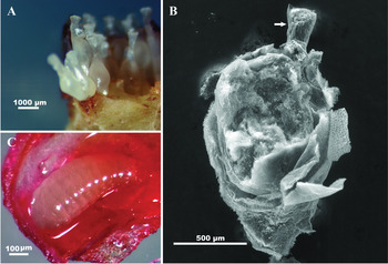
Ficus ovary and developing C. solmsi marchali larva in situ in a Ficus ovary: (A) Ficus ovaries under light microscope (6th day post-oviposition). (B) A dissected Ficus ovary under SEM (6th day post-oviposition). Arrow indicates style. (C) Larva of C. solmsi marchali in situ in a Ficus ovary, dyed with carmine (13th day post-oviposition).

First- and second-instar larvae under SEM: (A) First-instar larva (6th day post-oviposition), showing the head and faint body segmentation. (B) Second-instar larva (9th day post-oviposition). (C) Enlarged view of mouthparts of second instar, showing slender mandibles (md: mandible).

Second-instar larva (figs 2B and 6B, C)
Length 519.33±2.9 μm, range 427.54–648.55 μm; width 209.37±1.1 μm, range 162.84–279.65 μm; n=24. Head capsule width 124.56±0.7 μm, range 102.16–163.42 μm, n=21 (fig. 2B). Translucent, fusiform, and oval in cross-section (figs 2B and 6B). Cranium unsclerotized. Buccal region sclerotized of a light brown color. Mandibles concealed behind the labrum and small, slender and not protruding (fig. 6C). Mandible width 4.39±0.03 μm (n=6). Mid-gut filled with pale yellowish granules, occupied almost half of the body length. Larva sucked the fluid from the Ficus ovary by repeated contraction of the muscles surrounding its oesophagus.
Third-instar larva (figs 2C, 5C and 3A)
Molting was casually observed with exuviae attaching to the anal end of larvae after dissected from the Ficus ovary into physiological saline. Length 1016.77±7.2 μm, range 664.18–1354.93 μm; width 454.26±2.8 μm, range 281.60–526.89 μm; n=45. Head capsule width 192.78±0.4 μm, range 173.82–209.49 μm, n=22. Resembled second instar, except for the wider mesosomal segments (II–IV). Body wall semi-translucent (fig. 2C). Mandible length 29.19±0.5 μm (n=12), curved, with more sclerotized blades and well developed bases. Sharply edentate mandibles overlapped at the tips (fig. 3A). Active feeding, mid-gut packed with food filling almost the whole body (fig. 2C). Ficus ovaries took on the typical characters of wasp galls, with shriveled ovary integument enveloping the wasp larva (fig. 5C).
Fourth-instar larva (figs 2D and 3B, C)
Length 1553.75±8.5 μm, range 1522.39–1938.58 μm; width 905.35±4.7 μm, range 614.92–1104.67 μm; n=24. Less attenuated posteriorly and more arched, Body grayish white and less translucent than the larvae in preceding instars. Two protrusions emerged at the lateral and latero-ventral areas of the mesosomal segments (II–IV, figs 2C and 3B). Cranium sclerotized. Mandibles length 52.49±0.1 μm (n=16), heavily sclerotized (fig. 3C) and deeper red at the tips (fig. 2D). Alimentary tract extended and occupied larger area, appeared yellowish in color (fig. 2D).
Fifth-instar larva (figs 2E and 4)
Length 1891.71±8.6 μm, range 1617.15–2178.08 μm; width 1034.29±4.28 μm, range 911.01–1138.11 μm; n=17. Body strongly arched and turned white because of the accumulation of fat bodies. Mid-gut bright yellow, mandibles conspicuously red. Lateral and latero-ventral protruded, enveloping presumed developing legs and wing bud (fig. 2E). Mandibles well developed, broader and longer (64.0±0.4 μm in length, n=12), and sickle-form in shape, with darker pigmentation on the bases and margins (fig. 4B, C). Six spiracles presented ventrad on metasomal segments (VI–XI, fig. 4A). Spiracle VIII larger than other spiracles, external diameter 97.44±1.2 μm, n=6 (fig. 4A, B). Mid-gut moved rhythmically. Mobility limited to the mouthparts. Larva had consumed all the content of each Ficus ovary, leaving only the outer cast of the gall by the end of the final instar.
Prepupa (figs 2F and 7)
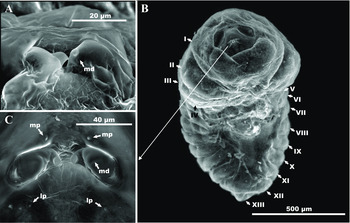
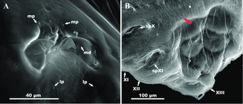
Retained the general morphology of the fifth larval instar. Abdominal region smoothly arched, a depressed area presented on the ventral region of abdomen, indicating an initial symptom of pupation (fig. 7B). Mesosomal region swollened (II–IV), approximate where the wing pads and legs emerged during pupal stage (fig. 2F). Mandibles resembled those in the final larval stage (fig. 7A). Pupal cuticle formed, with rapid internal changes taking place under it. Feeding completed and wasp was stationary. No peristalsis in the mid-gut. A period of approximately 1 day was required for this stage (n=27).
Prepupa of C. solmsi marchali under SEM: (A) Mandibles of prepupa (21th day post-oviposition), (md: mandible; mp: maxillary palp; lp: labial palp). (B) Ventral view of metasomal segmentation of pupa under larval skin, showing the depressed area on abdomen (red arrow), several metasomal segments (XI, XII, XIII), and the last two spiracles (spX and spXI).

Pupal stage
The pupae of C. solmsi marchali were exarate. Adult features were increasingly evident. The pupal stage could be divided into three different sub-stages (primary pupa, intermediate pupa or mature pupa) according to the body color and other characteristics. At the end of pupal stage, a yellow stain was found at the bottom of the inner wall of each gall, which was recognized as a meconium.
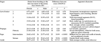
In the pupal stage, females could be recognized by the presence of two pink eye spots and three pink dots on the female head. They develop into compound eyes (Weiblen, Reference Weiblen2002) and three ocelli, respectively, that are absent in males. The developmental rate of male pupae appeared to be faster than in females, with males emerging earlier than females (table 1).
The time scale of C. somsi marchali from larva to adult and the significance of differences between sexes using ANOVA.

Primary pupa (fig. 8)
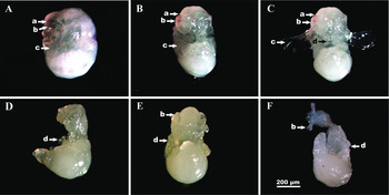
Body of both sexes white, divided into head, mesosoma, and metasoma. Pink compound eyes of females (fig. 8A–C) became increasingly more intense scarlet. Male pupa slenderer than female, with no wing buds. The telescopic male metasoma longer and curved. Antennae of the male (fig. 8F) shorter than the female (fig. 8B). All of these characters reflected the extreme sexual dimorphism of adult pollinating fig wasp (Jackson, Reference Jackson1966).
Primary pupa of both sexes, showing the compound eyes of female (a), antennae (b), wing buds of female (c), and legs (d): (A, B, C) Primary pupa of female (21st day post-oviposition) with pink eye spots and longer antennae. Wing buds in C were outspreaded artificially. (D, E, F) Primary pupa of male (21st day post-oviposition).

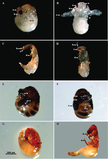
Approximately 24 days post-oviposition, three ocelli had emerged on the frons of female pupa, and turned into red, the same color of the female compound eyes (fig. 9B).
Intermediate pupa and mature pupa of both sexes, showing the compound eyes of female (a), antennae (b), wing buds of female (c), legs (d), ocelli of female (e), and mouthparts of male (f): (A, B) Lateral and ventral view of intermediate pupa of female (25th day post-oviposition). Wing buds in (B) were outspreaded artificially. (C, D) Intermediate pupa of male (25th day post-oviposition). (E, F) Mature pupa of female (27th day post-oviposition) with black wing pads. (G, H) Mature pupa of male (27th day post-oviposition).

Intermediate pupa (fig. 9A–D)
Female compound eyes and ocelli became brilliant red (on 24.42±0.02 day; n=26) and intensified gradually, finally became deep crimson in about 2 days (fig. 9B). Male sclerotized faster than female. Most parts of male's body turned yellow except for the reddish mouthparts on 23.77±0.02 day; n=22 (fig. 9C, D).
Mature pupa (fig. 9E–H)
Female pupa almost completed transitions, adult characteristics distinct, with black wings compressed in the pupal cuticle (on 26.65±0.02 day, n=20). Most parts of female body turned dark, with red eyes (fig. 9E, F). Males well sclerotized, turned orange with adult features visible (fig. 9G, H) on day 25.63±0.02 (n=24). Male pupa in this stage was able to twist its body slightly, whereas the female would remain stationary. Males eclosed in an average of 27.54±0.02 (n=26) days. Females eclosed in an average of 28.77±0.01 (n=31) days.
Discussion
Our data show that the entire immature period of C. solmsi marchali is approximately 28 days (male)–29 days (female) (table 1). Morphological differences between females and males before pupation are indistinct, and the duration of egg and larva development of both sexes was approximately equal. On average, eggs develop in 5 days; the growth of the five instars until prepupa lasts another 15 days. Consequently, the duration of postembryonic development from oviposition to prepupa is approximately 20 days. However, the time period since pupation (21st day post-oviposition, both sexes) to emergence is on average longer in females (8 days) than in males (7 days). The difference between sexes is significant since the intermediate-pupal stage. These results are consistent with the confirmed capacity for protandry in fig wasps (Joseph, Reference Joseph1984). Protandry means earlier male emergence (Fagerström & Wiklund, Reference Fagerström and Wiklund1982). There are several other documented cases of protandry in Hymenoptera (Bulmer, Reference Bulmer1983; Hastings, Reference Hastings1989).
The morphology of the ovaries of C. solmsi marchali was similar to that reported in other species of pollinators (Copland et al., Reference Copland, King and Hill1973; Ghara & Borges, Reference Ghara and Borges2010; Dunn et al., Reference Dunn, Jansen-González, Cook, Yu and Pereira2011). The egg load and morphological characteristics of ovarian eggs of C. solmi marchali concurred with those reported in Ceratosolen fusciceps, the pollinator of Ficus racemosa (Ghara & Borges, Reference Ghara and Borges2010).
Parker (Reference Parker1924) studied the egg and larval stages of Chalcidoidea. Although his description of Agaonid larva was not explicit, larva of C. solmsi marchali fit well into Parker's Group II Chalcidoid larvae (Parker, Reference Parker1924). The other chalcids in Parker's Group II include some species of Torymidae, Eurytomidae, Eulophidae, Pteromalidae, and Elasmidae (Parker, Reference Parker1924), phytophages occur in the first four families (Aeschlimann & Vitou, Reference Aeschlimann and Vitou1989; Askew, Reference Askew1998; LaSalle et al., Reference LaSalle, Raman, Schaefer, Withers, Raman, Schaefer and Withers2005). Larvae of C. solmsi marchali resemble those of other phytophagous chalcids in the early stages, such as Megastigmus nigrovariegatus Ashm. (Hymenoptera: Torymidae) (Milliron, Reference Milliron1949) and Eurytoma (Hymenoptera: Eurytomidae) (Williams, Reference Williams1960). The two lateral and ventro-lateral protrusions emerging in the fourth instar are believed to envelope the developing legs and wing pads, similar as described in ants (Brook et al., Reference Brook, Diaz-Benjumea and Cohen1996; Sameshima et al., Reference Sameshima, Miura and Matsumoto2004; Bowsher et al., Reference Bowsher, Wray and Abouheif2007). In Holometabola, wing primordium of Apocrita is set aside from the general larval epidermis before the last larval stage (Truman & Riddiford, Reference Truman and Riddiford1999), consistent with our results. The precise structure of the protrusions needs further investigation.
Our study presents the first detailed description of the immature stages of the pollinating fig wasp C. solmsi marchali, which can help us not only in further detailed studies on this species, but also to compare with the immature stages of other fig wasps.
Acknowledgements
This project was supported by the National Natural Science Foundation of China (NSFC grant numbers 31090253, 31172072, and 31210103912), partially by Major Innovation Program of Chinese Academy of Sciences (grant no. KSCX2-EW-Z-2), by Program of Ministry of Science and Technology of the Republic of China (grant no. 2012FY111100), by a grant (no. O529YX5105) from the Key Laboratory of the Zoological Systematics and Evolution of the Chinese Academy of Sciences, and by the National Science Fund for Fostering Talents in Basic Research (Special subjects in animal taxonomy, grant no. NSFC-J0930004).