Introduction
Carotid body tumours or chemodectomas are rare, slow-growing tumours usually manifesting in individuals in their fifth decade. In their sporadic form, these tumours are multifocal in less than 5 per cent of cases, with a male:female ratio of 0.7. The first case of a familial carotid body tumour was described by Chase in 1933,Reference Chase1 and it is now apparent that 10 per cent of cases are in fact familial. Lesions are more likely (>30 per cent) to be bilateral with an early age of onset, usually in the fourth decade or younger.Reference Grufferman, Gillman, Pasternak, Peterson and Young2 Whilst the mode of inheritance and the genes responsibleReference Heutink, van der Mey, Sandkuijl, van Gils, Bardoel and Breedveld3 have been established some time ago, predictive genetic testing for young family members has become available only recently. In this case report, we illustrate the presentation, diagnosis and management of such familial tumours, and make the case for predictive genetic screening.
Case report
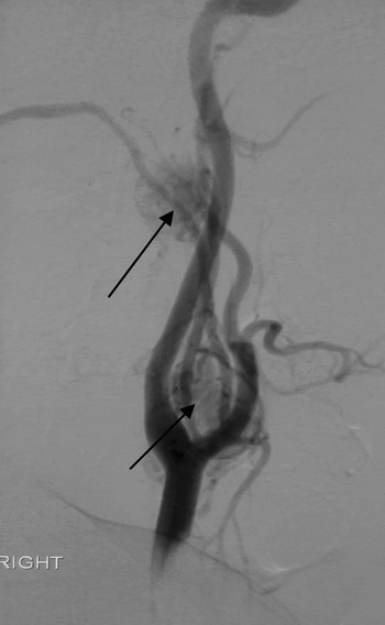
A 43-year-old man, AT, presented with headaches and a family history of carotid body tumours affecting his father and two paternal uncles. A magnetic resonance angiogram of the neck revealed two lesions in the right side of the neck, with an appearance in keeping with paragangliomas. Further angiography confirmed two lesions: a smaller, 18 mm lesion posterior to the right internal carotid artery at the level of C2; and a second, larger, 25 mm lesion medial to the right carotid bifurcation at the level of C4. The angiogram showing the two lesions is presented in Figure 1.
Angiogram showing two carotid body tumours (arrows), one at the carotid bifurcation and a smaller lesion posterior to the right internal carotid artery.

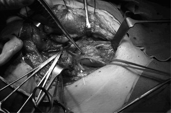
Subsequently, the patient underwent exploration of the neck. A mass was found to be encasing the common carotid artery, its bifurcation and internal and external branches. The vagus nerve and the surrounding structures were adherent to the lesion, as shown in Figure 2. The lesion was removed, preserving all surrounding structures. On exploration of the skull base, seeking the smaller, higher lesion, the lesion was found to be adjacent to the right internal carotid artery and surrounding the vagus nerve. Because removal of this lesion would have necessitated sacrifice of the vagus nerve, it was left in situ.
Intra-operative photograph, with forceps pointing to carotid body tumour at bifurcation and arrow pointing to the adjacent right vagus nerve.

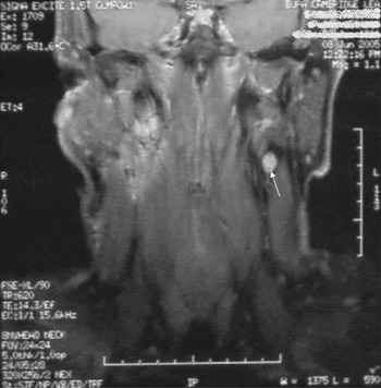
Post-operatively, AT made an uneventful recovery, with histology confirming carotid body paraganglioma. A magnetic resonance imaging (MRI) scan of the neck three months post-operatively confirmed the presence of the smaller lesion on the right seen at operation, but also showed an additional small lesion on the left at the carotid bifurcation (Figure 3).
Coronal magnetic resonance imaging scan of the neck performed 3 months post-operatively, with arrow pointing to additional carotid body tumour found on the left side.

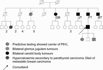
We then referred AT to the medical genetics department. His family tree is shown in Figure 4. His father, FT, along with his two uncles JT and BT and an aunt GB, all had bilateral carotid body tumours. The case of LF had been reported by Hamilton and Barros D'Sa in 1987.Reference Hamilton and Barros D'Sa4
AT's family tree, indicating distribution of carotid body tumours and results of predictive genetic testing.

Given the family history, it was thought likely that this family would have a mutation in succinate dehydrogenase subunit B or D. Genetic testing of AT confirmed a mutation in succinate dehydrogenase subunit D (P81L) causing familial phaeochromocytoma paraganglioma syndrome. However, a 24 hour urine collection from AT was not positive for raised catecholamines and abdominal imaging was also negative, thereby excluding phaeochromocytoma or secretory paraganglioma.
Following genetic counselling, AT's three children, two sons aged 11 and nine years and a daughter aged 13 years, underwent predictive testing for the same mutation. The daughter and the younger son tested positive.
Discussion
Paragangliomas are neuroendocrine tumours derived from the sympathetic or parasympathetic nervous system.Reference Benn, Gimenez-Roqueplo, Reilly, Bertherat, Burgess and Byth5 Of the parasympathetic paragangliomas, carotid body tumours are the most common and are usually physiologically inactive.Reference Pellitteri, Rinaldo, Myssiorek, Gary Jackson, Bradley and Devaney6 In contrast, paragangliomas from the sympathetic system, or phaeochromocytomas, are often active, with excess catecholamine secretion. Overall, 10–50 per cent of paragangliomas are familial, occurring either as part of tumour syndromes such as multiple endocrine neoplasia type two, von Hippel–Lindau disease, neurofibromatosis type one or due to hereditary paraganglioma genes (encoding succinate dehydrogenase subunits B, C or D).Reference Koch, Vortmeyer, Zhuang, Brouwers and Pacak7, Reference Young8 Succinate dehydrogenase is a mitochondrial enzyme important in the respiratory cycle. Mutations in both the succinate dehydrogenase subunit B and the succinate dehydrogenase subunit D genes predispose to familial phaeochromocytoma paraganglioma syndrome.Reference Astuti, Latif, Dallol, Dahia, Douglas and George9 However, succinate dehydrogenase subunit D mutation carriers are more likely to develop multifocal head and neck paragangliomas, whereas succinate dehydrogenase subunit B mutation carriers are more likely to present with phaeochromocytomas and to undergo malignant change.Reference Benn, Gimenez-Roqueplo, Reilly, Bertherat, Burgess and Byth5, Reference Suárez, Rodrigo, Ferlito, Cabanillas, Shaha and Rinaldo10 Patients with mutations in the succinate dehydrogenase subunit C gene are at risk of head and neck paragangliomas, which are seldom multiple and virtually exclusively benign.Reference Schiavi, Boedeker, Bausch, Peczkowska, Gomez and Strassburg11
Carotid body tumours most commonly present as an asymptomatic, lateral neck mass discovered by the patient or incidentally on neck examination. Characteristically, these tumours are pulsatile and can be moved in a horizontal plane only.Reference Knight, Gonzalez, Rary and Rush12 The diagnosis is usually confirmed by computed tomography (CT) or MRI scanning. Although generally slow-growing, carotid body tumours may be locally invasive or malignant in up to 3 per cent of cases.Reference Hamilton and Barros D'Sa4 Because of their malignant potential and greater surgical morbidity when large, it is important to excise them early. Before undertaking surgery, the specific vascular supply of the paraganglioma needs to be established. This helps reduce intra-operative bleeding, via either pre-operative embolisation or intra-operative ligation of the feeder vessel. Identification of the feeder vessel may be carried out with magnetic resonance angiography or intra-arterial digital subtraction angiography. In this regard, digital subtraction angiography is considered to be the gold standard.Reference Rosen, Palmer, Goldberg and Mustard13–Reference van den Berg, Wasser, van Gils, van der Mey, Hermans and van Buchem15
In patients with carotid body tumours who present young, have multifocal disease or have a positive family history, it is desirable to monitor the patient long term as well as to screen their close family members for similar lesions. As the present case illustrates, clinical examination has been found to be an unreliable screening tool, as it misses small lesionsReference Hamilton and Barros D'Sa4 and those near the skull base. Various screening techniques have been described for family members at risk. These include clinical examination and imaging techniques such as colour duplex,Reference Worsey, Laborde, Bower, Miller, Kresowik and Sharp16 CT,Reference Kohn, Raftery and Jewell17 MRI,Reference McCaffrey, Meyer, Michels, Piepgras and Marion18 metaiodobenzylguanidine scintigraphy,Reference Van Gils, van Erkel, Falke and Pauwels19 pentetreotide scanningReference Myssiorek and Palestro20 and Reference McCaffrey, Meyer, Michels, Piepgras and Marion18F-Dihydroxyphenylalanine (DOPA) whole-body positron emission tomography (PET).Reference Hoegerle, Ghanem, Altehoefer, Schipper, Brink and Moser21
• Carotid body tumours are familial in 10 per cent of cases
• Up to 3 per cent of carotid body tumours may be malignant, and early detection reduces subsequent surgical morbidity
• Previous screening modalities have relied upon detecting the presence of tumours in at-risk individuals, with concomitant health economic and welfare costs
• Genetic testing for familial paraganglioma syndromes has recently become available in Britain
• Predictive genetic testing allows selection of close family members for further screening to ensure tumours are detected early
Colour duplex is good at depicting carotid body tumours but cannot delineate higher vagal and jugular paragangliomas.Reference Alkadhi, Schuknecht, Stoeckli and Valavanis22 Magnetic resonance imaging allows better assessment of all paragangliomas but is problematic for claustrophobic patients. Compared with MRI, metaiodobenzylguanidine allows quicker whole body scanning and is also more tolerable for claustrophobic patients. However, this technique does expose patients to ionising radiation and is poor at detecting non-functioning paragangliomas.Reference Van Gils, van Erkel, Falke and Pauwels19 Pentetreotide scanning, using radio-labelled somatostatin analogue, does not require paragangliomas to be functional to enable detection and can be applied to the whole body.Reference Myssiorek and Palestro20 It is therefore a good screening tool for those at risk of multiple paragangliomas. Reference McCaffrey, Meyer, Michels, Piepgras and Marion18F-DOPA whole-body PET is yet another sensitive method of detecting multiple paragangliomas, and can identify lesions less than 1 cm in size. All screening modalities thus far described, irrespective of their strengths or weaknesses, require the presence of lesions for detection, implying that individuals at risk who have not yet developed tumours would need to undergo some form of life-long, regular screening. This has health economic implications as well as potential adverse effects on the individual from the screening procedure.
It was established that AT and his two children had a mutation in the succinate dehydrogenase subunit D gene which predisposed them to familial phaeochromocytoma paraganglioma syndrome. Germline mutations in the succinate dehydrogenase subunit D gene on chromosome 11q23 in familial paraganglioma were first identified by Baysal et al. Reference Baysal, Ferrell, Willett-Brozick, Lawrence, Myssiorek and Bosch23 and subsequently confirmed in cases of familial phaeochromocytomas by Astuti et al. Reference Astuti, Douglas, Lennard, Aligianis, Woodward and Evans24 At the time of writing, 66 unique mutations of succinate dehydrogenase subunit D associated with paragangliomas had been recorded on the relevant database (http://chromium.liacs.nl/lovd_sdh).Reference Ogawa, Shiga, Saijo, Ogawa, Kimura and Horii25 Mutations in this gene are inherited in an autosomal dominant fashion with genomic imprinting. Genomic imprinting means that expression of the mutated gene depends on the transmitting parent. For succinate dehydrogenase subunit D, the phenotype is seen only when the mutation is paternally inherited. Patients with germline mutations in succinate dehydrogenase subunit D are at risk of developing multiple head and neck paragangliomas at a young age (median age 28 years)Reference Benn, Gimenez-Roqueplo, Reilly, Bertherat, Burgess and Byth5 and can also develop phaeochromocytomas. Once susceptibility has been identified by genetic testing (in patients with family history of paragangliomas or those presenting young or harbouring multiple lesions), such patients need to be monitored over time in order to detect development or recurrence of these tumours. Patients as young as seven years have been detected with tumours;Reference Benn, Gimenez-Roqueplo, Reilly, Bertherat, Burgess and Byth5 in the present case report, the youngest mutation carrier was aged nine years. We would therefore advocate that predictive genetic screening of at-risk individuals commences in childhood. It has been suggested that genetically susceptible individuals could be monitored from the age of five to 10 years,Reference Suárez, Rodrigo, Ferlito, Cabanillas, Shaha and Rinaldo10 by annual blood pressure and urinary catecholamine measurements, plus an annual or biennial CT or MRI scanReference Benn, Gimenez-Roqueplo, Reilly, Bertherat, Burgess and Byth5 and a pentetreotide or Reference McCaffrey, Meyer, Michels, Piepgras and Marion18F-DOPA whole-body PET scan. Early detection of tumours means early intervention, reducing surgical morbidity and progression to malignancy.