Introduction
Increased prevalence of immune-mediated diseases in the West has been explained on the basis of the ‘hygiene hypothesis’ (Von Mutius, Reference Von Mutius2007), which proposes a direct link between the absence of appropriate priming of the immune response by infectious agents and the increased occurrence of immune-mediated disorders (Strachan, Reference Strachan1989). Several allergic and inflammatory disorders, such as type 1 diabetes (T1D), rheumatoid arthritis, inflammatory bowel disease and multiple sclerosis, are reported to have lower incidences in the areas where parasitic infections are more prevalent (Osada & Kanazawa, Reference Osada and Kanazawa2010). The rationale reached to account for this association is that the parasites might have modulated host immune responses while evolving exclusive and effective mechanisms to create an environment favouring their survival and longevity in the human host (Moller et al., Reference Moller, Gravenor, Roberts, Sun, Gao and Hopkin2007; Maizels, Reference Maizels2009). The results of experiments conducted using animal models of immune-mediated disorders are in support of the proposed immunomodulatory effect of helminth parasites (Zaccone & Hall, Reference Zaccone and Hall2012).
Type 1 diabetes is a multifactorial autoimmune disease characterized by selective destruction of insulin-producing beta cells of the islets of Langerhans by auto-antigen reactive inflammatory T cells (Esensten et al., Reference Esensten, Lee, Glimcher and Bluestone2009; Li et al., Reference Li, Song and Qin2014). Auto-reactive CD4+ (Cluster of Differentiation 4+) and CD8+ T cells are known to be actively involved in the tissue-damaging inflammatory T-helper cell 1 (Th1)-type immune response in T1D, characterized by elevation of interferon- γ (IFN-γ), interleukin (IL)-12, transcription factor T-bet and signal transducer and activator of transcription 1 (STAT1) (Pinkse et al., Reference Pinkse, Tysma and Bergen2005; Callewaert et al., Reference Callewaert, Gysemans, Ladrière, D'Hertog, Hagenbrock, Overbergh, Eizirik and Mathieu2007). Insulin is most widely used to treat T1D. However, insulin therapy is associated with the risk of mismatch among insulin levels, food intake and physical activity, often resulting in episodes of improper control of blood glucose (Alison, Reference Alison2014). Immune-suppressive drugs have also been explored in the treatment of T1D to control the destruction of beta-cells (Pescovitz et al., Reference Pescovitz, Greenbaum, Krause-Steinrauf, Becker, Gitelman, Goland and Raskin2009). However, these drugs are effective only in the short term and their long-term usage leads to serious toxic effects on the kidney, liver and nervous system (Wilson & Buckingham, Reference Wilson and Buckingham2001).
Helminth therapy has been explored as an alternative approach for the control of T1D (Zaccone & Hall, Reference Zaccone and Hall2012). Infection with the helminth parasites Schistosoma mansoni, Heligmosomoides polygyrus, Trichinella spiralis or Litomosoides sigmodontis has led to the prevention of the onset of T1D in non-obese diabetic mice (NOD) (Cooke et al., Reference Cooke, Tonks, Jones, O'Shea, Hutchings and Fulford1999; Saunders et al., Reference Saunders, Raine, Cooke and Lawrence2007; Hubner et al., Reference Hubner, Stoker and Mitre2009; Liu et al., Reference Liu, Saunders, Mishra, Mousavi, Liu and Gaydo2009; Mishra et al., Reference Mishra, Patel, Wu, Bleich and Gause2013; Osada et al., Reference Osada, Yamada, Nabeshima, Yamagishi, Ishiwata, Nakae and Kanazawa2013). However, helminth therapy using live worms is not appropriate as it would be ethically and practically difficult to implement it in preventive mode, and the sub-optimal doses expected to be used in the treatment may not be sufficient to control the pathology (McSorley & Maizels, Reference McSorley and Maizels2012). Homogenates of L. sigmodontis, adult worm and egg soluble antigens of S. mansoni, and secreted proteins from Fasciola hepatica have also been explored and found to be effective in preventing T1D in mice (El-Wakil et al., Reference El-Wakil, Aboushousha, El Haddad, Gamil, Mansour and El-Said2002; Zaccone et al., Reference Zaccone, Fehérvári, Jones, Sidobre, Kronenberg, Dunne and Cooke2003, Reference Zaccone, Burton, Miller, Jones, Dunne and Cooke2009; Hubner et al., Reference Hubner, Stoker and Mitre2009; Lund et al., Reference Lund, O'Brien, Hutchinson, Robinson, Simpson, Dalton and Donnelly2014). We reported earlier that the whole native filarial antigens, i.e. the somatic and excretory–secretory proteins, of B. malayi could attenuate the severity of streptozotocin (STZ)-induced T1D in mice (Amdare et al., Reference Amdare, Khatri, Yadav, Tarnekar, Goswami and Reddy2015). However, as the crude extracts of parasites are not appropriate for therapeutic use and may also contain other immunogenic substances that counter or inhibit protective components within the extract, it is more judicious to identify and use specific immune-modulatory molecules derived from helminth parasites.
By immunoscreening a Wuchereria bancrofti L3 cDNA library using microfilaraemic sera, a recombinant protein WbL2 (rWbL2) belonging to the SXP/RAL-2 family of proteins was identified in our laboratory (NCBI accession numbers: nucleotide sequence, EU183493; protein sequence, ABW38023). This showed 84% homology with other W. bancrofti proteins of the SXP/RAL-2 family, namely Wb-SXP1 and Wb14 (see supplementary fig. S1). rWbL2 could detect high levels of specific IgG4 antibodies in the sera of microfilarial carriers (Siva Prasad et al., unpublished observations). The elevation in IgG4 antibodies in these subjects is often associated with high levels of IL-10, which could play an important role in modulating the host immune response (Adjobimey & Hoerauf, Reference Adjobimey and Hoerauf2010). Another group of filarial molecules, i.e. abundant larval transcript (ALT) proteins, are produced in large amounts and are suggested to have a role in immune evasion by up-regulating the T-helper cell 2 (Th2) response in the host (Gomez-Escobar et al., Reference Gomez-Escobar, Bennett, Prieto-Lafuente, Aebischer, Blackburn and Maizels2005). Madhumathi et al. (Reference Madhumathi, Prince, Nageswara and Kaliraj2010) have shown that the T-cell epitopes of ALT2 from the filarial parasite Brugia malayi induce high levels of IL-10 secretions along with suppression of IFN-γ and IL-12 cytokines, suggesting their possible involvement in the modulation of the host immune system. In the earlier study from our laboratory, recombinant B. malayi abundant larval transcript 2 (rBmALT-2) with its effective immunomodulatory activity was shown to be a promising alternative therapeutic agent for T1D (Reddy et al., Reference Reddy, Reddy, Amdare, Khatri, Tarnekar, Goswami and Reddy2016). Hence, in the present study, we have assessed and compared the curative effects of rWbL2 and rBmALT-2 by administrating them individually and in combination to treat STZ-induced T1D in mice.
Materials and methods
Filarial proteins
rWbL2 and rBmALT-2 were prepared as described by Gnanasekar et al. (Reference Gnanasekar, Rao and He2004). In brief, pRSETA-WbL2 or pRSETA-BmALT-2 plasmids were constructed, transformed into BL21 (DE3) pLysS Escherichia coli expression host and expressed as His-tag proteins by inducing with 1 mm isopropyl β-d-1-thiogalactopyranoside (Merck Millipore, Bangalore, India). The recombinant proteins were purified using a nickel affinity chromatography column (Thermo Fisher Scientific, Mumbai, India), the protein content was estimated using a Bicinchoninic Acid Protein Assay kit (Thermo Fisher Scientific) and the endotoxin content was measured using Limulus amoebocyte lysate chromogenic quantitation kit (Thermo Fisher Scientific). The endotoxin content was found to be <0.2 EU/ml in both the recombinant protein preparations, which is well below the recommended cut-off level (Malyala & Singh, Reference Malyala and Singh2008).
Assessment of filarial proteins in vitro
Thioglycollate medium (1%; Sigma-Aldrich, Mumbai, India) was injected into the peritoneal cavity of BALB/c mice and the peritoneal exudate cells (PECs) were harvested on the fourth day by lavage with cold and sterile phosphate-buffered saline (PBS; 0.1 m, pH 7.2) containing 20 mm ethylenediaminetetraacetate (EDTA; Sigma-Aldrich). The cells were re-suspended in Roswell Park Memorial Institute (RPMI) 1640 medium supplemented with 10% heat-inactivated fetal calf serum (Thermo Fisher Scientific) and plated in triplicate (2 × 10⁵ cells/100 μl per well) in 96-well flat-bottom plates (Thermo Fisher Scientific). After 4 h of incubation (37°C in 5% CO2), the non-adherent cells were washed off and adherent cells in the wells were stimulated with E. coli lipopolysaccharide (LPS) (10 μg/100 μl per well; Sigma-Aldrich) in the presence or absence of rWbL2 or rBmALT-2 (10 μg/100 μl per well). In control wells, PECs were incubated with medium alone. After 24 h of incubation at 37°C in 5% CO2, the culture contents from triplicate wells were pooled separately, centrifuged and the supernatants were analysed for nitrite levels using Griess reagent (Taira et al., Reference Taira, Ohminea, Nanbub and Uedab2013). The culture supernatants were collected from parallel experiments after 72 h of incubation to analyse the levels of cytokines, including IL-10, IL-4, tumour necrosis factor-α (TNF-α) and IFN-γ.
Type 1 diabetes and treatment in mice
Female BALB/c mice (6–8 weeks of age) used in this study were bred in the animal house of our institute and maintained, under the supervision of a veterinarian, in a pathogen-free environment in standard laboratory conditions with free access to normal diet and drinking water ad libitum.
Multiple low-dose T1D was induced in BALB/c mice using streptozotocin (STZ; Sigma-Aldrich), as described by Santos-Junior et al. (Reference Santos-Junior, Sartori and Lima2009). The mice were fasted for 4 h then were administered freshly prepared STZ (40 mg/kg of STZ in 0.1 m sodium citrate buffer, pH 4.5) intraperitoneally (i.p.) for five consecutive days. Fasting blood glucose levels (obtained from a tail vein after 6 h of fasting) were monitored weekly using a glucometer (Gluco Check; Major Biochemical Corporation, Taiwan, China), starting from 7 days after the last dose of STZ. The mice with fasting blood glucose levels of ≥200 mg/dl for two consecutive weeks were considered to be diabetic and the incidence of diabetes was expressed as the percentage of mice in a group presenting with diabetes.
Mice (n = 6–8) were divided into different groups: STZ-rWbL2 group (diabetic mice treated with rWbL2), STZ-rBmALT-2 group (diabetic mice treated with rBmALT-2), STZ-rWbL2 + rBmALT-2 group (diabetic mice treated with combination of both rWbL2 and rBmALT-2), STZ-Alum group (diabetic mice treated with alum alone), STZ group (diabetic mice without any treatment) and Normal group (normal mice without any treatment). Initial experiments in diabetic mice on dose response with rBmALT-2 showed 25 μg/dose as the optimal quantity needed to achieve the significant therapeutic effect (data not shown).
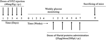
In this study the diabetic mice were treated by the administration of four doses of filarial proteins i.p. (each dose of 200 μl containing 25 μg of protein in 0.05 m PBS pH 7.2 and 1% alum) at intervals of 15 days (fig. 1). Starting from the initiation of the treatment, the mice were monitored for fasting blood glucose levels at weekly intervals until the end of experimental period.
Experimental protocol.

Histopathology and in vitro culture
At the end of the experimental period, the pancreas isolated from each sacrificed mouse was fixed in 10% forma-saline, embedded in paraffin and 5-μm cross-sections were cut and stained with haematoxylin and eosin (H&E). The total numbers of the pancreatic islet cells were counted and monitored for changes in morphology, degree of lymphocyte infiltration and inflammation. The degree of lymphocyte infiltration was scored and expressed as the percentage of examined islets with non-infiltrated (healthy islets), peri-insulitis (lymphocytes at the periphery of the islets) and intra-insulitis with ≤50% or ≥50% lymphocyte infiltration (Hubner et al., Reference Hubner, Stoker and Mitre2009).
For the in vitro culture of splenocytes, spleens were harvested aseptically from the mice, minced in RPMI 1640 medium, and single-cell suspensions were pelleted and re-suspended in erythrocyte lysis buffer (Invitrogen, Mumbai). The cells were washed three times in RPMI 1640 medium and plated in triplicate (2 × 10⁶ cells/ml per well) in 24-well flat-bottom plates (Thermo Fisher Scientific) in RPMI 1640 medium supplemented with 2 mm l-glutamine, 100 IU/ml penicillin, 100 μg/ml streptomycin, 25 mm HEPES buffer and 10% heat-inactivated fetal calf serum. The cells were stimulated separately with rWbL2 or rBmALT-2 (10 μg/well) or Concanavalin A (2 μg/well; positive control) (Sigma-Aldrich) or medium alone (negative control), and were incubated at 37°C in 5% CO2 atmosphere for 72 h. The final volume in each well was adjusted to 1 ml. The culture supernatants were collected after incubation and kept at −80°C until further used for the estimation of the cytokines (IL-10, IL-4, IL-5, TNF-α and IFN-γ) using enzyme-linked immunosorbent assay (ELISA) kits (Invitrogen).
Serology
Sera from experimental and control groups of mice were collected through the caudal vein and were used to analyse anti-insulin antibodies (IgG1 and IgG2a) using ELISA (Imai et al., Reference Imai, Tezuka and Fujita2001). Immuno plates (96-well; Thermo Fisher Scientific) were coated with human recombinant insulin (10 μg/100 μl per well; Sigma-Aldrich) in coating solution (100 mm carbonate–bicarbonate buffer, pH 9.5) at 4°C overnight. The wells were washed once with PBS/T (0.05 m PBS containing 0.05% of Tween 20, pH 7.2) and blocked by bovine serum albumin (2% in PBS, 300 μl/well) for 1 h at 37°C. After washing three times, the wells were incubated with the mouse sera (1:50 diluted in PBS) for 1 h at 37°C. The wells were then washed five times and the bound antibodies were detected by incubation with horseradish peroxidase (HRP)-conjugated goat anti-mouse IgG1 or IgG2a antibodies (1:5000 diluted in PBS; Invitrogen) for 1 h at 37°C. Finally, wells were washed five times and colour was developed by addition of 3,3ʹ,5,5ʹ-tetramethylbenzidine/ hydrogen peroxide (TMB/H2O2) substrate (100 μl/well). After 10 min of incubation, the reaction was stopped by addition of sulphuric acid (2 n; 50 μl) and the optical density was measured at 450 nm using a spectrophotometer (Biotek India, Mumbai).
Serum IgE levels were analysed using ELISA, as described by Boitelle et al. (Reference Boitelle, Di Lorenzo, Scales, Devaney, Kennedy, Garside and Lawrence2005), using optimal concentrations of rWbL2 or rBmALT-2 (10 μg/100 μl per well), 50-fold diluted sera and optimally diluted (1:5000) HRP-conjugated goat anti-mouse IgE antibodies as the probe (Southern Biotech, Birmingham, Alabama, USA).
Data analysis
The data were analysed using SPSS version 16.0 (SPSS Inc., Chicago, Illinois, USA). Results are presented as mean ± standard error of the mean (SEM) (n = 6–8 mice in each group) and are representative of two independent experiments. The statistical difference between the means of multiple groups for the normally distributed data was tested for significance using one-way analysis of variance (ANOVA) followed by Tukey's post-hoc test for multiple comparisons. For the data which were not normally distributed, the Kruskal–Wallis test was used. P values ≤ 0.05 were considered to be significant.
Results
Filarial proteins suppress inflammatory activity
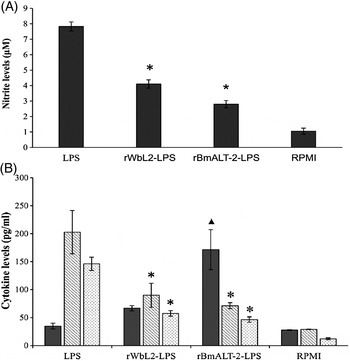
The nitrite levels were significantly lower (P ≤ 0.05) in the culture supernatants of the mouse PECs co-stimulated with LPS and filarial proteins rWbL2 or rBmALT-2, compared to the levels in the culture supernatants of the PECs stimulated with LPS only (fig. 2A). This anti-inflammatory effect was found to be associated with the significantly (P ≤ 0.05) reduced release of TNF-α and IFN-γ cytokines by the peritoneal macrophages following co-stimulation with the filarial proteins and LPS (fig. 2B). On the other hand, these macrophages released IL-10 at higher levels, although the increase was not significant from the macrophages co-stimulated with rWbL2 and LPS.
Mean levels (± SEM) of (A) nitrites (μm) in the supernatant of peritoneal exudate cells and (B) cytokines (pg/ml), including interleukin (IL)-10 (solid shading), interferon-γ (IFN-γ), (diagonal shading) and tumour necrosis factor-α (TNF-α) (stippled shading). *Level of significance with P ≤ 0.05.

Filarial proteins reduce the severity of STZ-induced T1D
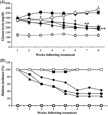
Treatment with rWbL2 or rBmALT-2 proteins (either individually or in combination) after the onset of clinically evident STZ-induced T1D in BALB/c mice, reduced the severity of the disease condition. A progressive reduction in the fasting blood glucose levels was observed in the diabetic mice following treatment with the filarial proteins, leading to a significant reduction at the end of the experimental period (fig. 3A). As many as 57.2% of diabetic mice treated with rWbL2 (8 of 14) and 66.6% of diabetic mice treated with rBmALT-2 (10 of 15) became non-diabetic by the end of the experimental period (fig. 3B and table 1). Treatment with the combination of rWbL2 and rBmALT2 resulted in 73.3% (11 of 15) diabetic mice becoming non-diabetic by the end of the experimental period (fig. 3B and table 1). In contrast, almost all the diabetic mice treated with alum alone (STZ-Alum) remained diabetic throughout the experimental period (fig. 3B and table 1). Neither the difference between the results of these two proteins, nor the difference between the results of their combination or any of the two individual proteins, was statistically significant in terms of reducing the blood glucose levels.
Mean levels (± SEM) of (A) fasting blood glucose levels (mg/dl) and (B) incidence of diabetes (%) in different groups of mice: STZ-rWbL2 (closed diamonds), STZ-rBmALT-2 (closed circles), STZ-rWbL2 + rBmALT-2 (closed triangles), STZ-Alum (closed squares), STZ (open squares) and Normal (open circles). *P ≤ 0.005 and **P ≤ 0.0001 in comparison with streptozotocin (STZ)-Alum and STZ groups, respectively.

The incidence of diabetes (%) and histological changes (%) in the pancreatic islets of mice treated with recombinant Wuchereria bancrofti L2 (rWbL2) and Brugia malayi abundant larval transcript 2 (rBmALT-2).

* Level of significance with P ≤ 0.005. STZ, streptozotocin.
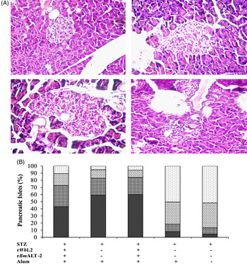
Histopathological examination of the pancreatic tissue of the mice in the control groups (STZ and STZ-Alum groups) showed cell destruction and severe insulitis with a higher degree of lymphocyte infiltration and inflammation (fig. 4A, B, table 1). In diabetic mice treated with rWbL2 or rBmALT-2, or with their combination, there was decrease in the number of damaged islets and also a reduction in the degree of lymphocyte infiltration and the severity of inflammation (fig. 4A, B, table 1). Accordingly, the scores as measures of the severity of inflammatory infiltration were significantly reduced in all the groups of diabetic mice treated with filarial proteins (table 1).
(A) Representative photographs of the H&E-stained sections of pancreas (original magnification, ×40) classified as (i) non-infiltrated, (ii) peri-insulitis, (iii) intra-insulitis with ≤50% lymphocyte infiltration and (iv) intra-insulitis with ≥50% lymphocyte infiltration (scale bar = 50 μm). (B) Percentage of pancreatic islet cells classified as non-infiltrated (solid shading), peri-insulitis (square shading), intra insulitis with ≤ 50% infiltrated lymphocytes (diagonal shading) and intra-insulitis with ≥50% infiltrated lymphocytes (stippled shading).

Effect on cytokine responses
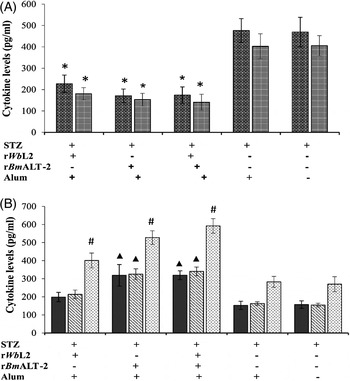
Pro-inflammatory cytokines IFN-γ and TNF-α are responsible for the destruction of pancreatic β cells in T1D. The splenocytes of mice treated with individual rWbL2 and rBmALT-2, or their combination, released significantly reduced levels of these two cytokines (P ≤ 0.005) compared to the splenocytes of the untreated mice or the mice treated with alum alone (fig. 5A). In contrast, there were significantly elevated (P ≤ 0.001) releases of IL-4 and IL-5 cytokines by the splenocytes of rBmALT-2 or rWbL2 + rBmALT-2 treated groups of diabetic mice (fig. 5B). Although the mice treated with rWbL2 also showed increased levels of IL-4 and IL-5 cytokines, the increase was not significant (fig. 5B). Further, significantly increased levels of IL-10 were observed in all the groups of the diabetic mice treated with filarial proteins (P ≤ 0.0001; fig. 5B). These results indicate that the suppression of the disease condition of T1D by the treatment with the filarial proteins is associated with polarization of the immune response towards the Th2 type.
Mean levels (± SEM) of cytokines (pg/ml) in the culture supernatants of splenocytes. (A) Interferon-γ (IFN-γ) (square shading) and tumour necrosis factor-α (TNF-α) (horizontal shading); (B) interleukin (IL)-4 (solid shading), IL-5 (diagonal shading) and IL-10 (stippled shading). *P ≤ 0.005; ▲, P ≤ 0.001 and #, P ≤ 0.0001 in comparison with STZ-Alum and STZ groups.

Effect on anti-insulin and IgE antibodies
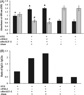
To investigate whether the observed Th2 polarization of the immune response was also reflected at the antibody levels against diabetes-associated auto-antigens, we measured the levels of auto-antibodies against insulin (auto-antigen) in the sera of the diabetic mice treated with the filarial proteins. Significantly increased levels of IgG1 (P ≤ 0.0001) and decreased levels of IgG2a (P ≤ 0.0005) antibodies were noted in the diabetic mice treated with rBmALT-2 or rWbL2 + rBmALT-2 proteins, which resulted in a significant increase in the ratio of IgG1:IgG2a (fig. 6A, B).
Mean levels (± SEM) of (A) IgG1 (solid shading) and IgG2a antibodies (diagonal shading) in the sera. (B) Ratio of IgG1/IgG2a. *P ≤ 0.01 in comparison with the STZ group; ▲, P ≤ 0.0001 in comparison with streptozotocin (STZ)-Alum and STZ groups; #, P ≤ 0.0005 in comparison with the STZ-Alum group.

The serum levels of antigen-specific IgE antibodies in treated mice were also investigated. The rBmALT-2-specific IgE antibodies were significantly increased (P ≤ 0.0001) in the diabetic mice treated with rBmALT-2 alone (mean ± SEM absorbance values of 0.62 ± 0.05) or with the combination of rWbL2 + rBmALT-2 (0.6 ± 0.04) compared to the levels in the control group of diabetic mice treated with alum (0.21 ± 0.06). The IgE antibodies against rWbL2 were also higher in the diabetic mice treated with rWbL2 (0.38 ± 0.04) or with rWbL2 + rBmALT-2 (0.51 ± 0.04) compared to the control group of mice treated with alum (0.25 ± 0.02), although the increase was significant (P ≤ 0.0001) only in the rWbL2 + rBmALT-2 group.
Discussion
Helminths, which are known to cause a variety of diseases in humans, have developed mechanisms to support their long-term survival in the host. They trigger profound regulatory effects on the host immune system to avoid exaggerated inflammatory responses directed against them (Flohr et al., Reference Flohr, Quinnell and Britton2009). Such helminth-mediated immunomodulation is shown to be involved in inducing protection against the development of autoimmune and allergic diseases. Helminths or their derived products are thus potentially useful alternative therapeutic agents for these diseases. T1D, a chronic autoimmune disorder, with several limitations in the currently available treatment methods, is one such disease targeted for helminth therapy.
Multiple low-dose administrations of STZ induce inflammation characterized by the infiltration of immune cells, such as lymphocytes, macrophages and mononuclear cells, into the islets of Langerhan, with subsequent destruction of β cells and progressive hyperglycaemia, which is the hallmark of T1D pathology (Tisch & McDevitt, Reference Tisch and McDevitt1996; Denis et al., Reference Denis, Mahmood, Benoist, Mathis and Weissleder2004; Espinoza-Jim'enez et al., Reference Espinoza-Jiménez, Rivera-Montoya, C'ardenas-Arreola, Moran and Terrazas2010). The clinical and pathological features of this model are similar to those observed in humans with T1D (Muller et al., Reference Muller, Schott-Ohly, Dohle and Gleichmann2002). Additionally, being an inducible model, it is possible to adjust the timing of onset of diabetes depending on the experimental protocol.
Initially, filarial proteins were screened in vitro for an anti-inflammatory effect against LPS-activated macrophages. LPS is a known endotoxin and, upon stimulation with LPS, macrophages trigger signals for the release of diverse inflammatory mediators, such as nitric oxide, and inflammatory cytokines, such as IFN-γ and TNF-α (Decker, Reference Decker1990; Alexandra et al., Reference Alexandra, Müller and Vollmar2002). Filarial proteins rWbL2 and rBmALT-2 significantly suppressed LPS-induced nitric oxide synthesis and also reduced expression of pro-inflammatory cytokines IFN-γ and TNF-α by PECs of mice. These results suggested that both rWbL2 and rBmALT-2 have the potential to modulate the immune response and thus are suitable for investigation as alternative therapeutic agents in T1D.
Treatment with individual rWbL2 and rBmALT-2, or their combination, could suppress the severity of established T1D in mice. Blood glucose levels were significantly reduced in all the groups of diabetic mice treated with these proteins. There was no significant difference between rBmALT-2 and rWbL2, or between any of these two proteins and their combination, in their effect on the reduction of blood glucose levels. At the end of the experimental period, there was marked reduction in the incidence of diabetes in all the groups of diabetic mice treated with filarial proteins. The treatment with the combination of rBmALT-2 and rWbL2 resulted in the highest reduction (73.3%) in the incidence of diabetes, which was not substantially different from the results obtained by treatment with individual proteins.
The anti-diabetic effect of these proteins is also reflected in histopathological observations of the pancreatic tissue of treated diabetic mice, which showed a higher number of healthy islets with normal architecture and reduced inflammation and lymphocyte infiltration. Taken together, these results imply that filarial proteins rWbL2 and rBmALT-2 could induce a robust and reproducible protection against STZ-induced diabetes.
In our earlier study (Reddy et al., Reference Reddy, Reddy, Amdare, Khatri, Tarnekar, Goswami and Reddy2016) rBmALT-2 was found to be more effective in ameliorating T1D. By the end of the fifth week after initiating treatment, all the diabetic mice had turned to normal. The difference in the therapeutic effect of rBmALT-2 in these two studies is possibly due to differences in the treatment regimens used. While the mice were treated with four doses of rBmALT-2 at weekly intervals in the earlier study, the protein was administered at intervals of 15 days in the present study. Brugia malayi somatic and excretory–secretory proteins were also able to significantly control hyperglycaemia and bring down the incidence of STZ-induced T1D by 71.5% in mice (Amdare et al., Reference Amdare, Khatri, Yadav, Tarnekar, Goswami and Reddy2015). Treatment with recombinant Dirofilaria immitis antigen (Imai et al., Reference Imai, Tezuka and Fujita2001) or with the secretory proteins of F. hepatica (Lund et al., Reference Lund, O'Brien, Hutchinson, Robinson, Simpson, Dalton and Donnelly2014) also resulted in the prevention of the development of diabetes in NOD mice.
Although the exact immunological mechanism responsible for the aetiology of T1D is not clearly understood, dysregulation of IFN-γ, TNF-α, IL-10, IL-4 and IL-5 cytokines and some auto-antigen-specific antibodies, such as IgG1 and IgG2a, have been shown to play a crucial role in the disease progression (Gillan et al., Reference Gillan, Lawrence and Devaney2005; Lund et al., Reference Lund, O'Brien, Hutchinson, Robinson, Simpson, Dalton and Donnelly2014; Bashi et al., Reference Bashi, Bizzaro, Shor, Blank and Shoenfeld2015). Th1-type inflammatory responses have been shown to be involved in the progression of autoimmune reactions in type 1 diabetes mediated through the secretion of TNF-α and IFN-γ by the differentiated Th1 CD4+ T cells, which in turn promote the CD8+ cytotoxic T cell-mediated destruction of insulin-producing β cells (Yoon & Jun, Reference Yoon and Jun2005; Wilson et al., Reference Wilson, Taylor, O'Gorman, Balic and Barr2010). Consistent with this supposition, in the current study we have observed the suppression in the production of TNF-α and IFN-γ cytokines by splenocytes of diabetic mice treated with filarial proteins. Besides the suppression of these pro-inflammatory cytokines, we observed up-regulation of IL-4 and IL-5 cytokines, which are recognized as strong inducers of the Th2 type of immune response in the spleen cells of treated mice. Up-regulation of IL-10, an anti-inflammatory immunological phenotype, is a general characteristic of helminth infection and hence this cytokine could play a vital role in the amelioration of autoimmune diabetes (Hussain & Delovitch, Reference Hussain and Delovitch2007; Amdare et al., Reference Amdare, Khatri, Yadav, Tarnekar, Goswami and Reddy2015; Bashi et al., Reference Bashi, Bizzaro, Shor, Blank and Shoenfeld2015). In tune with this phenomenon, in the current study, the production of IL-10 was elevated in the splenocytes of diabetic mice following treatment with filarial proteins. However, contrary to expectations, there were no significant changes in the production of the above cytokines from the splenocytes of treated diabetic mice in response to stimulation with auto-antigen insulin (data not shown). In an earlier study, Hubner et al. (Reference Hubner, Stoker and Mitre2009) also reported that there were no significant changes in the auto-antigen (insulin) specific cytokine responses from the splenocytes of filarial-infected NOD mice.
In the present study, though we have used alum to deliver filarial proteins to the mice, it is possible that these proteins alone may induce a similar immunomodulatory effect, which needs to be confirmed. The possibility of alum alone influencing the immunomodulatory effect in the treated mice can be ruled out as there were no significant changes in the clinical status or histopathological scores, or immune responses, in the diabetic mice treated with alum alone (STZ-Alum group). In the pilot experiments, we had also treated a group of normal mice with alum alone and they did not show any noticeable changes (data not shown here).
Proteins secreted from F. hepatica were reported to confer a protective effect against diabetes, associated with the suppression of IFN-γ and with the generation of IL-10-secreting regulatory B cells (Lund et al., Reference Lund, O'Brien, Hutchinson, Robinson, Simpson, Dalton and Donnelly2014). Earlier studies have also shown that infection with S. mansoni, T. spiralis and H. polygyrus can significantly inhibit or delay the development of diabetes in NOD mice by skewing of the Th1 immune response towards the protective Th2 type associated with the production of IL-4, IL-5 and IL-10 cytokines (Saunders et al., Reference Saunders, Raine, Cooke and Lawrence2007; Mishra et al., Reference Mishra, Patel, Wu, Bleich and Gause2013). IFN-γ secretion preferentially promotes switching of activated B cells towards the IgG2a isotype (Th1 type), and the cytokines IL-4 and IL-10 stimulate the production of the IgG1 isotype (Th2 type) (Espinoza-Jim'enez et al., Reference Espinoza-Jiménez, Rivera-Montoya, C'ardenas-Arreola, Moran and Terrazas2010). In the present study, treatment with filarial proteins resulted in significantly elevated insulin-specific IgG1 and reduced IgG2a levels. Hence, the protection induced by rWbL2 and rBmALT-2 against T1D might be associated with the generalized Th2 shift mediated by both cytokines and insulin-specific antibody production. We have also observed higher levels of antigen-specific IgE antibodies in the diabetic mice treated with filarial proteins, indicating that the antigen-specific IgE antibodies, in correlation with other immune parameters, might also have played a crucial role in reducing the severity of STZ-induced T1D. In an earlier study, the IgE antibodies induced by filarial parasites were reported to play a vital role in preventing the development of diabetes in NOD mice (Imai et al., Reference Imai, Tezuka and Fujita2001).
The results of the current study provide a valid proof in favour of exploring filarial proteins as effective alternative therapeutics for T1D. Further investigations into revealing the exact mechanism involved, determining their optimal dose and assessing the longevity of their curative effect, along with understanding the safety aspects of their long-term use, can pave the way to development of these proteins as effective anti-diabetic drugs.
Supplementary material
To view supplementary material for this article, please visit http://dx.doi.org/10.1017/S0022149X1600064X
Financial support
This research work was supported by the funding received from the Department of Biotechnology (DBT) (Project Id. BT/PR/4988/INF/22/155/2012) and Department of Science and Technology (DST-FIST) (Project Id. SR/FST/LSI-470/2010) of the Ministry of Science and Technology, Government of India.
Conflict of interest
None.
Ethical standards
The authors assert that all procedures contributing to this work comply with the ethical standards of the national regulatory body, the Committee for the Purpose of Control and Supervision of Experiments on Animals (CPCSEA), Government of India, and has been approved by the Institutional Animal Ethics Committee (IAEC).