The excellent long-term haemodynamic outcomes following surgical repair of tetralogy of Fallot are marred by a significant incidence of late post-operative arrhythmias. Traditionally, the majority of interest has been on the incidence and treatment of ventricular arrhythmias in this context, due to concerns about risk of sudden cardiac death. However, the prevalence of atrial tachyarrhythmias in these patients has recently been reported to be as high as 34%.Reference Roos-Hesselink, Perlroth, McGhie and Spitaels1 Increased realisation of the morbidity and symptom burden caused by atrial arrhythmias in surgically repaired congenital cardiac disease has highlighted the inadequacies of medical management and driven efforts to improve outcomes by other means, including catheter ablation.
Many factors contribute to the development of atrial tachyarrhythmias in this patient group including the legacy of pre-operative atrial pressure and volume overload, surgical scars, sinus node dysfunction, the development of pulmonary and tricuspid regurgitation, and progressive fibrosis with increasing age at the time of surgery.Reference Gatzoulis, Balaji and Webber2 Acting together, these factors create a complex arrhythmogenic milieu in which multiple macro- and micro-reentrant circuits co-exist to provide both the substrate and trigger for initiating and sustaining atrial tachyarrhythmias.
Epicardial ablation to achieve a right-sided or bi-atrial Cox maze procedure at the time of pulmonary valve replacement has been shown to reduce the risk of recurrent atrial arrhythmias following surgery in patients with previously repaired tetralogy of Fallot.Reference Karamlou, Silber and Lao3, Reference Deal, Mavroudis and Backer4 However, this option can really only be justified in patients, who require repeat cardiac surgery for haemodynamic indications late after repair – such as for pulmonary regurgitation.
The role of potent anti-arrhythmic drug therapy is limited in this context by concerns about ventricular pro-arrhythmia and aggravation of sinus node disease.
Their use is limited to drugs such a beta-blockers or rate-slowing calcium channel blockers, which are relatively ineffective or amiodarone, with its potential for toxicity when deployed in the long term in these predominantly young patients.Reference Etheridge, Craig and Compton5–Reference Cochrane, Siddins and Rosenfeldt8 Although routinely deployed, the overall effect of anti-arrhythmic therapy has been disappointing and often does not control arrhythmias sufficiently to improve quality of life.
The development of radiofrequency catheter ablation techniques and their success in other arrhythmias originally raised hopes for patients with arrhythmias following surgical repair of congenital cardiac disease. However, the complexity of the arrhythmia substrate made it extremely difficult to localise and ablate all atrial arrhythmia circuits/foci using conventional mapping techniques. Procedures were long and frustrating and the results disappointing, despite reports of acute procedural success.Reference Anne, van Rensburg, Adams, Ector, Van de Werf and Heidbuchel9
This analysis had three aims – first, to describe the outcome for patients whose main clinical problem was atrial tachyarrhythmia occurring late after surgical repair of tetralogy of Fallot; second, to define the nature and location of circuits/foci responsible for these atrial tachyarrhythmias; and third, to evaluate the outcome of computer-assisted mapping and catheter ablation in this patient group.
Methods
Patients
All patients with repaired tetralogy of Fallot are routinely followed in the adult congenital cardiac clinics and we currently have 293 patients under regular review in our centre. Symptomatic patients are assessed with exercise testing, echocardiography, and magnetic resonance imaging and if pulmonary regurgitation is deemed the primary haemodynamic problem, the patients are referred for pulmonary valve replacement. In this study, we describe a group of patients who did not meet the standard criteria for pulmonary valve replacement and whose main symptom was atrial tachyarrhythmia. The arrhythmia(s) were not satisfactorily controlled with medical therapy and, therefore, the patients were referred for electrophysiological study. The choice of anti-arrhythmic medication in this group was not standardised and the study period was from January, 2001 to June, 2007.
Electrophysiological study and ablation procedure
Computer-assisted mapping of atrial tachyarrhythmias was performed in all using either EnSite® (St Jude Medical Inc.) arrhythmia mapping and intra-cardiac catheter guidance or CARTO™ (Biosense Webster Inc.) electroanatomical mapping systems. The ablation procedure was carried out under conscious sedation in 12 and under general anaesthesia in four procedures. A 4-millimetre irrigation-tip catheter was used for radiofrequency ablation in 15 procedures and a cryo-ablation catheter in one procedure. Standard catheter configurations were used with the EnSite® multi-electrode non-contact catheter sited in the right atrium during EnSite®-guided procedures and a body surface reference catheter for CARTO™-guided procedures.
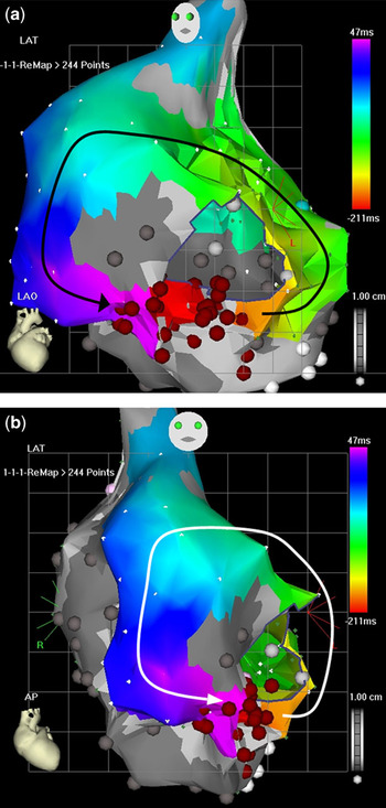
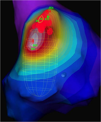
Routine pacing techniques including induction, overdrive termination, and entrainment were used in conjunction with computer-assisted mapping to identify the arrhythmia mechanism(s) and suitable target sites for ablation. Examples of electro-anatomical maps depicting macro-reentry and focal/micro-reentrant tachyarrhythmias are illustrated in Figures 1a and b and 2.
(a and b) (Carto): Macro-reentrant atrial tachyarrhythmia. The two electroanatomical maps represent an LAO and AP view of a macro-reentrant circuit around a central obstacle sited in the right atrium. Ablation sites are indicated by the red spheres and the direction of wavefront propagation is shown by the black and white arrows. LAO = left anterior oblique; AP = anterior posterior.

EnSite® (ESI): Focal/micro-reentrant atrial tachyarrhythmia. The activation map seen in the right anterior oblique projection illustrates wavefronts originating from a focus of myocardial tissue (red star) and spreading out in a centrifugal pattern. Ablation sites are indicated by the red spheres.

Classification of atrial tachyarrhythmias
For the purpose of this study, arrhythmias were defined as focal or micro-reentrant, if they were characterised by rapid repetitive electrical activations originating from a small area of myocardial tissue with activation wave fronts radiating centrifugally from the source. Macro-reentrant atrial tachyarrhythmias were defined as those characterised by waveform reentry around a central obstacle, typically around fixed obstructions or areas of scarring.
Clinical outcome
Patient status following ablation was classified based on symptoms, 12-lead electrocardiograph, 24-hour Holter monitoring, anti-arrhythmic medication, and subsequent need for surgical procedures.
Results
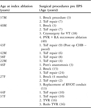
Ten consecutive patients (four males) of median age 39 plus or minus 8 years with atrial tachyarrhythmias following surgically repaired tetralogy of Fallot, were identified and included in the analysis. Median age at surgical repair was 7.5 plus or minus 6 years and all underwent one or more ablation procedures. The details of the surgical procedures performed are shown in Table 1. All patients had an atriotomy for atrial septal defect and/or transatrial ventricular septal defect closure at the time of surgery.
Surgical procedures pre electrophysiological studies.

CHB = complete heart block; EPS = electrophysiology study; PVR = pulmonary valve replacement; RA = right atrial; RAA = right atrial appendage; RVOT = right ventricular outflow tract; TVR = tricuspid valve replacement; VT = ventricular tachycardia
Brock procedure = Trans-ventricular resection of part of the right ventricular outflow tract to increase pulmonary blood flow; ToF repair = Patch closure of the ventricular septal defect, resection of thickened muscle from the right ventricular outflow tract, pulmonary valve repair, and augmentation of the pulmonary arteries (if necessary)
Out of the 10 patients, five underwent only one operation, one had two procedures, and four had three or more separate surgical procedures. The mean interval between reparative surgery and the onset of atrial tachyarrhythmia was 24 plus or minus 9 years.
Of the 22 arrhythmias identified, 21 were right atrial in origin. Only two patients had a single circuit with multiple separate arrhythmia foci/circuits identified in eight (80%) patients.
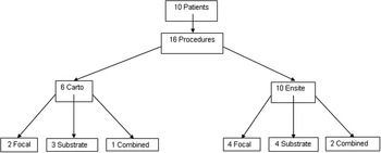
A total of 16 separate ablation procedures were performed on the 10 patients with 10 procedures guided by EnSite® and six by CARTO™ mapping systems (Fig 3). Mean procedure duration was 240 minutes with a range of 180–240 minutes and mean fluoroscopy dosage (dose area product) 829 plus or minus 733 centigray per square centimetre. Atrial tachyarrhythmias were incessant at the start of two procedures and were induced for mapping in the remaining 14 by programmed atrial stimulation (six), by programmed stimulation and isoprenaline (four), and spontaneously during catheter manipulation (four). EnSite® had difficulty in locating the arrhythmia origin/circuit for ablation in four procedures, three in the same patient, as it was unable to distinguish regions of electrical silence, irrelevant to the arrhythmia, from very low-voltage scar sites critical to the maintenance of the arrhythmia. There were no procedure-related complications.
Study design.

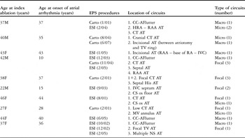
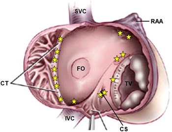
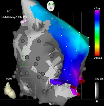
We identified 22 separate atrial tachyarrhythmia foci/circuits in these 10 patients (six macro-reentrant, 16 micro-reentrant/focal; Table 2). Two circuits were identified in five patients, three in two and four separate circuits in one patient. Figure 4 illustrates the location of the 15 micro-reentrant/focal right atrial tachyarrhythmias identified – the other micro-reentrant site was in the left atrium on the mitral valve annulus. Figure 5 is an example of an electro-anatomical map highlighting the typical extent of scar tissue in the right atrium of patients following surgical repair of tetralogy of Fallot.
Electrophysiological studies and arrhythmia foci ± circuits identified.

AF = atrial fibrillation; AFlutter = atrial flutter, AT = atrial tachycardia; CC = counter-clockwise; CS = coronary sinus; CT = crista terminalis; EPS = electrophysiology study; ESI = EnSite®; NS = non-sustained; TV = tricuspid valve
Location of 15 right atrial focal/micro-reentrant atrial tachyarrhythmias. CS = coronary sinus; CT = crista terminalis; FO = fossa ovalis; IVC = inferior caval vein; RAA = right atrial appendage; SVC = superior caval vein; TV = tricuspid valve.

(Carto): Scar tissue in the right atrium (shown in grey on the electroanatomical map).

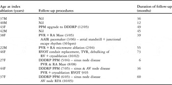
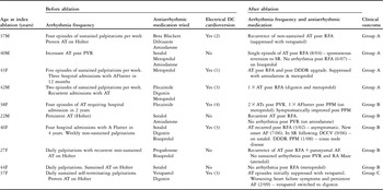
The mean duration of follow-up following the last ablation procedure was 41 plus or minus 20 months (Table 3). As part of their anti-arrhythmic management, five patients were paced for pre-existing sinus plus or minus atrioventricular nodal disease. Details of the arrhythmia burden before and after ablation and clinical outcome are included in Table 4.
Follow-up procedures and duration.

DDDRP = dual-chamber paced, dual-chamber sensed, dual response, rate modulated pacemaker; PPM = permanent pacemaker; PVR = pulmonary valve replacement; RAA = right atrial appendage; RFA = radiofrequency ablation; RVOT = right ventricular outflow tract; SR = sinus rhythm; TVR = tricuspid valve replacement
Arrhythmia burden before and after ablation and clinical outcome.

AAIR = atrial rate adaptive; AAx = antiarrhythmic; AF = atrial fibrillation; AFlutter = atrial flutter, AT = atrial tachycardia; CC = counter-clockwise; CHB = complete heart block; CS = coronary sinus; CT = crista terminalis; DC = direct current; EPS = electrophysiology study; ESI = EnSite®; HRA = high right atrium; IVC = inferior vena cava; MV = mitral valve; NS = non sustained; PPM = permanent pacemaker; PVR = pulmonary valve replacement; RAA = right atrial appendage; RFA = radiofrequency ablation; RVOT = right ventricular outflow tract; SR = sinus rhythm; TVR = tricuspid valve replacement; VT = ventricular tachycardia
Clinical outcome following the ablation procedure(s) could be grouped as follows:
• Group A: Improved symptoms following catheter ablation but remained on anti-arrhythmic drug therapy (four patients).
• Group B: Patients with no significant improvement in symptoms following catheter ablation who were referred for pulmonary valve replacement (plus or minus tricuspid valve repair) and surgical ablation (five patients).
• Group C: Patient with no significant improvement in symptoms but not suitable for further surgical intervention (one patient).
In the five patients who underwent further surgery, empiric radiofrequency or cryo-ablation lesions were sited and previous catheter linear ablation lesions were consolidated by the placement of three linear cut-and-sew or radiofrequency lesions in the right atrium intra-operatively, that is, inter-caval line in the lateral atrium linking superior to inferior caval vein; a cavo-tricuspid isthmus line between tricuspid valve and inferior caval vein; an anterior septal line between superior and inferior caval vein through the fossa ovalis.
Ablation success, defined as no recurrence of arrhythmia at 12-month follow-up was higher in patients who underwent CARTO™-guided (83%) versus EnSite®-guided (30%) ablation procedures. In addition, ablation success was higher in substrate-guided (57%) versus focal (33%) ablation procedures with a combined success rate of 67% when both focal and substrate ablation were performed during the same procedure.
The overall ablation success rate at 12-month follow-up was 50%, which is similar to other published seriesReference Papagiannis, Maounis, Laskari, Theodorakis and Rammos10; however, using the combined catheter and surgical ablation strategy, symptomatic improvement was achieved in nine out of the 10 patients in this study.
Discussion
This series provides important information on a small but clinically important group of patients after tetralogy of Fallot repair, whose primary problem was atrial tachyarrhythmia as opposed to pulmonary regurgitation. Although it is accepted that almost all have some degree of pulmonary regurgitation, the patients whose regurgitation was deemed significant enough to warrant valve replacement in the first instance were not considered in this series.
There are a number of important lessons from our experience. These arrhythmias are complex with both macro and micro-circuits and most patients have more than one substrate. The electrophysiological information included in this study will be of use to cardiologists and electrophysiologists managing similar patients. It is worth emphasising that the arrhythmia substrate is virtually always in the right atrium and, using the versions of the computerised mapping systems available at the time, CARTO™ provided more consistently useful guidance for these procedures than EnSite®. Our hypothesis for this is that contact mapping (CARTO™) seems superior to non-contact mapping (EnSite®) in terms of a defining scar and conduction channels more accurately and in interpreting very low voltage signals from the far field signals at high gain settings in these patients with significantly scarred atria. However, it is no surprise that benefits accrued incrementally from successive procedures. Information is also provided on typical arrhythmia locations.
The importance for the overall management of correcting sinus node disease is also clear from this series. In five patients, sinus node disease limited anti-arrhythmic drug doses and may have contributed directly to the frequency of tachyarrhythmia episodes. Although permanent pacemaker implantation can improve a patient’s symptoms – particularly, if there is chronotropic incompetence – and allow trials of more potent anti-arrhythmic therapy, this was not sufficient to obviate the need for ablation in several patients in this series.
The data also allow some speculation on the relative value of various approaches to mapping arrhythmias in this context. In the patients treated earlier in this series, activation mapping was performed with the help of the EnSite® multi-electrode non-contact balloon catheter to guide ablation. However, increasingly aware that multiple arrhythmias co-existed in these patients, we changed for later procedures to a strategy of scar substrate mapping and ablation. Substrate ablation aims to block multiple potential arrhythmia circuits by linking scarred areas to fixed obstructions by linear ablation and has been reported to improve overall success by allowing non-sustained or non-inducible arrhythmias to be addressed without the need to map each individually.Reference Nakagawa, Shah and Matsudaira11 Mapping was performed during sinus rhythm, guided by either NavX (EnSite®) or CARTO™ point-by-point generated maps. At regular intervals, attempts were made to induce arrhythmia to determine the efficacy of the lesions already identified or determine the need for further linear lesions. The need for multiple procedures in this series lends indirect support to the superiority of this strategy by confirming the inadequacies of the earlier approach – heavily reliant on the ability to induce sustained forms of each arrhythmia. However, although substrate ablation would be expected to abolish all macro-reentrant circuits – clinical and potential – it is less suited to addressing focal tachyarrhythmias.
The main finding from this analysis is that no single management strategy, whether catheter or surgically based, can be guaranteed to achieve complete arrhythmia abolition in these patients. An initial electrophysiological assessment should be recommended early in patients not controlled on oral anti-arrhythmic therapy. Those with focal right atrial tachycardias (5 of these 10 patients) are better treated by mapping-guided catheter ablation since an empiric surgically delivered ablation procedure lacks the necessary precision to be successful. In those patients with significant pulmonary regurgitation, pulmonary valve replacement can be combined with anti-arrhythmic linear lesions sited in the right atrium and this usually provides further symptomatic improvements. However, even then most patients will still require maintenance anti-arrhythmic drug therapy. There was only one patient in our series in whom we achieved little symptomatic improvement with catheter ablation and the degree of pulmonary regurgitation was mild and therefore not suitable for pulmonary valve replacement. Our assessment was that the arrhythmia in this patient did not justify isolated surgical ablation.
It is likely that atrial tachyarrhythmias will continue to pose a challenging problem in adults with surgically corrected congenital cardiac disease. The complex nature of arrhythmia circuits makes conventional mapping impossible and taxes existing mapping systems to their limits. The range of arrhythmia substrates likely to be encountered is described and should enable refinement of ablation strategies to optimise outcomes. However, currently, a hybrid-staged approach seems most effective. In this regard, it is probably more useful clinically to look at the degree of symptomatic improvement and the incremental benefit of repeat procedures rather than absolute ablation success or failure. These anti-arrhythmic procedures are currently best seen as palliative rather than as curative. In the longer term, understanding where and why arrhythmogenic scars evolve should allow modifications in the surgical technique so as to minimise their development. Prevention of these atrial arrhythmias, which currently impair quality of life so significantly in these patients, should be one of the ultimate goals of management.
Acknowledgements
The authors declare no conflicts of interest and relationship with industry.